basic anatomy and physiology of cervix to understand physiological changes in transformation zone during reproductive years, types of transformation zones and their importance
Cervical cancer arises from the transformation zone of the cervix. Risk factors include HPV infection, early age of first intercourse, multiple sexual partners, and smoking. It typically spreads locally first through direct extension, then can metastasize via lymph nodes or hematogenously to distant sites like lungs and liver. Screening via Pap smears can detect pre-cancerous changes and has reduced cervical cancer rates in developed nations by 75% over 50 years. Vaccination against HPV also prevents infection and future cancer development. Treatment and prognosis depends on the stage, with early localized disease having the best outcomes.
1) The breast is composed of adipose tissue, lactiferous ducts and tubules, blood vessels, nerves and lymphatics. It extends from the 2nd to 6th ribs and medially from the lateral border of the sternum to the mid-axillary line.
2) The breast parenchyma contains 15-20 lobes made up of lobules that empty into lactiferous ducts. It also contains stromal connective and fatty tissue.
3) Lymphatic drainage of the breast primarily follows the blood supply to the axillary lymph nodes but may also drain to internal mammary or intercostal nodes. Sentinel lymph node biopsy is used to help determine cancer stage
This document provides guidance on examining male external genitalia. It outlines relevant history to obtain, areas to inspect and palpate including the penis, scrotum, hernia, lymph nodes, and instructions for testicular self-exam. Considerations for different age groups are also discussed. The practice exam question addresses how to respond if a patient becomes sexually aroused during the exam.
The breast lies in the superficial fascia of the pectoral region and is divided into four quadrants. It extends from the 2nd to 6th ribs horizontally and from the sternum to the midaxillary line vertically. The breast contains lobules that produce milk via lactiferous ducts which drain at the nipple. Lymphatic drainage is primarily to the axillary lymph nodes but also occurs to internal mammary and intercostal nodes. The breast develops from the mammary ridge in utero.
The document discusses the anatomy of the breast. It covers topics such as location and extent of the breast, layers and structures within the breast like skin, parenchyma, ducts and lobes. It also discusses blood supply, lymphatic drainage including lymph node stations, nerve supply and radiological anatomy of the breast.
The document discusses the Pap smear screening test for cervical cancer. It describes how Pap smears have reduced cervical cancer incidence by 80% and mortality by 70% by allowing for treatment of pre-cancerous lesions. Screening should begin within 3 years of becoming sexually active and can typically decrease in frequency to every 2-3 years after 3 normal annual tests. Screening may stop at age 70 after recent negative tests or hysterectomy. The document outlines the anatomy of the cervix and squamo-columnar junction, techniques for Pap smear collection, abnormal findings, screening guidelines, and accuracy of Pap smears.
Fallopian tube radiology - Dr. Sumit SharmaSumit Sharma
The document discusses the fallopian tubes (also known as uterine tubes), including their anatomy, development, histology, diseases, and radiological evaluation. Some key points:
- The fallopian tubes connect the ovaries to the uterus and allow the passage of eggs. They have distinct segments including fimbriae, infundibulum, ampulla, and isthmus.
- A hysterosalpingogram is commonly used to evaluate the uterus and fallopian tubes for infertility or recurrent miscarriage. It can identify conditions like tubal blockage, polyps, or hydrosalpinx.
- Diseases of the fallopian tubes include congenital abnormalities, infections which
The document provides an overview of the breast anatomy and lymphatic drainage. It discusses:
- The breast's location, extent, and lymphatic system. Lymph flows primarily to axillary nodes but also to internal mammary and intercostal nodes.
- Sentinel lymph nodes are the first nodes draining a cancer, important for sentinel lymph node biopsy.
- The upper outer breast quadrant contains most glandular tissue and most cancers originate here.
- Lymphatic obstruction by cancer can cause lymphedema, peau d'orange skin, and nipple retraction. Mammography is used to detect breast masses.
The document provides information on breast anatomy and physiology. It discusses:
- The external anatomy of the breast including the nipple, areola, skin, and internal anatomy such as lobes, lobules, ducts and fatty tissue.
- Breast development from fetal stage through puberty and changes that occur during pregnancy, menopause and with age.
- Composition of the breast including glandular and fatty tissues.
- Radiographic appearance and changes in breast density with age.
Carcinoma Buccal Mucosa- Anatomy to ManagementDrAyush Garg
This document provides information on carcinoma of the buccal mucosa, including its anatomy, epidemiology, risk factors, clinical features, diagnosis, staging, treatment, and prognosis. Carcinoma of the buccal mucosa is most common in India, Malaysia, and Taiwan, where it affects men more than women. Risk factors include tobacco and betel nut chewing. Treatment depends on the stage, with surgery or radiation typically used for early stages and chemoradiation for advanced stages. Prognosis depends on factors like tumor size and nodal involvement.
Radical hysterectomy is a surgical procedure for treating cervical cancer. It involves removing the uterus, cervix, part of the vagina, and nearby lymph nodes and tissue. There are different classifications of radical hysterectomy based on the extent of tissue removed. Complications can include bleeding, infection, injury to nearby organs like the bladder or ureters. Radical hysterectomy is indicated for early stage cervical cancer and may provide better survival outcomes than radiation alone for some patients.
Brachytherapy involves implanting radioactive sources directly into tumors for short-range radiation treatment. It delivers high radiation doses to tumors while sparing surrounding tissues. Teletherapy uses external radiation beams from a source placed away from the body. For cervical cancer, brachytherapy treats the central tumor while teletherapy controls peripheral growth and metastases. The relative proportion of teletherapy increases with tumor stage. Brachytherapy is followed by teletherapy 4-6 weeks later, and chemotherapy may enhance radiation effects. Complications can include nausea, irritation, and late effects like fibrosis and osteoporosis.
The vagina is a musculomembranous tube extending from the vulva to the uterus. It is located between the bladder and rectum and is curved posteriorly. The vaginal canal is H-shaped with the lower end narrower than the upper end. It has anterior, posterior, and lateral fornices. The vagina has relations anteriorly to the bladder and urethra, posteriorly to the pouch of Douglas and rectum, and laterally to the levator ani muscles. Common clinical manifestations of vaginal conditions include discharge, vaginitis, prolapse, and cancer. Bacterial vaginosis, candidiasis, and trichomoniasis are treated with antibiotics like
Clinical presentation and investigations for breast carcinomaViswa Kumar
This document provides an overview of breast carcinoma, including:
1) The embryology, functional anatomy, blood supply, innervation, and lymphatics of the breast.
2) The epidemiology of breast cancer, noting it is the most common cancer in women worldwide.
3) Clinical presentations like palpable masses, pain, nipple discharge, and skin changes.
4) Recommendations for diagnostic tools like mammography, ultrasound, and MRI to evaluate symptoms based on patient age and risk factors.
5) The BI-RADS assessment system to categorize imaging findings and guide next steps.
Sentinal lymph node biopsy is a staging procedure used to detect occult metastases in early stage cancers. The sentinel lymph node is the first lymph node to receive drainage from a tumor. During the procedure, a radioactive tracer is injected around the tumor and followed using a gamma probe to identify the sentinel lymph node, which is then surgically removed and examined for cancer cells. Identifying metastases in the sentinel lymph node can help determine the need for further treatment such as neck dissection while avoiding unnecessary procedures in node-negative patients. While controversial in oral cancers due to variability in lymphatic drainage, sentinel lymph node biopsy may help improve staging and individualize treatment when performed by an experienced team.
The cervix is the lowermost part of the uterus that extends into the vagina. It is cylindrical in shape and divided into portions above and within the vagina. The endocervical canal contains ridges and glands lined by columnar epithelium. The ectocervix is covered by squamous epithelium. The cervix secretes mucus to aid sperm transport and acts as a barrier during pregnancy. The vagina is a fibro muscular tube extending from the vulva to the uterus. It has anterior, posterior and lateral walls lined by stratified squamous epithelium and contains glands, muscles and connective tissue. The vagina allows for sexual intercourse, birth and menstrual flow.
This document discusses the history and anatomy of neck dissections for head and neck cancer. It traces developments from the late 19th century of increasingly conservative neck dissections to preserve important structures. It describes the levels and boundaries of lymph node groups involved in the neck dissection staging system, including levels I-V. Key structures like the hypoglossal nerve and spinal accessory nerve are discussed in relation to the lymph node groups.
The perineum is the region between the thighs that contains the external genitalia and anal opening. It is bounded by the pelvic floor muscles superiorly and the thighs laterally. The perineum can be divided into the urogenital triangle anteriorly, containing the genital structures, and the anal triangle posteriorly, containing the anus. Each triangle contains superficial and deep pouches separated by fascial layers. The pouches contain muscles, nerves, and vessels related to the genital and anal structures. The perineal body is an important structure that provides support to the pelvic floor.
History of radical hysterectomy for cancer cervixSakshi Mundra
The document provides a history of radical hysterectomy for cervical cancer treatment over 2500 years. It discusses key pioneers and developments including Hippocrates attempting trachelectomy in 460-370 BC, Wilhelm Freund developing the first standardized radical hysterectomy technique in 1878, and Ernst Wertheim performing the first full radical abdominal hysterectomy in 1898. The modern radical hysterectomy was developed in the late 19th/early 20th century by surgeons including John Clark, Joseph Meigs, and Hidekazu Okabayashi. Classification systems for radical hysterectomy procedures including the Piver-Rutledge-Smith and Querleu and Morrow systems are also summarized.
FEMALE PELVIC APPLIED ANATOMY BY DR SHASHWAT JANIDR SHASHWAT JANI
This document provides an overview of female pelvic anatomy including the bones of the pelvis, pelvic spaces and landmarks, vasculature, lymphatic drainage, and innervation. Key points include the sacrum and coccyx forming the back of the pelvis, the true and false pelvis divided by the linea terminalis, important ligaments such as the sacrotuberous and sacrospinous, branches of the internal iliac artery including the uterine artery, lymphatic drainage pathways, and the four main pelvic spaces - retro pubic, para vesical, pouch of Douglas, and para rectal. A comprehensive understanding of pelvic anatomy is important for avoiding injuries during gynecological
Contracted pelvis, also known as pelvic disproportion, occurs when the essential diameters of the pelvis are shortened, altering the normal mechanism of labor. It can be caused by developmental, metabolic, traumatic or other factors. Pelvises are classified based on degree of contraction and pelvic architecture. Diagnosis involves history, physical exam including internal and external pelvimetry, and sometimes radiological imaging. An internal pelvimetry exam evaluates the inlet, cavity, and outlet to determine pelvic adequacy for vaginal delivery.
The prostate is a walnut-sized gland located below the bladder and above the urethra. It has an inverted cone shape with a base above and apex below. The prostate surrounds the urethra and produces fluid that comprises part of semen. It has four lobes - anterior, median, and two lateral lobes. The prostate contains the prostatic urethra, two ejaculatory ducts, and prostatic utricle. Blood supply comes from the inferior vesical artery and drains into the internal iliac and sacral lymph nodes.
Vasectomy is a surgical procedure for male sterilization that involves cutting, blocking, or tying the vas deferens tubes to prevent sperm from entering seminal fluid and causing pregnancy. The document discusses the history, types, procedure, risks, and role of nurses in vasectomy. It notes the procedure can be done with scalpels or non-scalpel methods like using ring forceps and dissectors to access the vas deferens without incisions. After the procedure, patients should rest and avoid heavy lifting for a week while waiting for follow up sperm tests to confirm effectiveness.
The breast is composed of lobes, lobules, and ducts. It receives its blood supply from the internal and external mammary arteries. Lymph drainage is primarily to the axillary lymph nodes.
Breast anatomy and development can vary between individuals. Common benign breast conditions include fibroadenomas, cysts, and fibrocystic changes.
Breast cancer originates in the breast ducts or lobules. HER2-positive breast cancer is a type where cancer cells overexpress the HER2 receptor, causing rapid growth. Physical signs may include a painless breast mass, nipple retraction, and enlarged lymph nodes.
This document outlines the procedures for performing a Pap smear test. It describes obtaining a cervical cell sample, the timing of the test in relation to a woman's menstrual cycle and sexual activity. It lists the necessary equipment and supplies. It provides instructions for preparing the patient, performing the test, and following up with the patient based on the test results.
Imaging in obstetrics & gynaecology (part 1- Gynaecological scans in benign c...drmcbansal
Ultrasonography is a type of medical imaging that uses high-frequency sound waves to visualize structures within the body. It is a non-invasive technique that provides real-time images and does not use radiation. Common applications of ultrasound in gynecology include evaluating the uterus, ovaries, and fallopian tubes. A transvaginal probe is often used to obtain detailed images of the pelvic organs. Normal ultrasound appearances of the ovaries include scattered antral follicles that develop during the menstrual cycle. Ovulation is identified by a decrease in size of the dominant follicle. The corpus luteum that forms after ovulation can be seen as a cyst or echogenic area on ultrasound.
lecture 5b The breast and pectoral region.pdfNatungaRonald1
The breast lies in the pectoral region and develops during puberty under the influence of female sex hormones. It is composed of lobules that radiate from the nipple. The breast undergoes changes during pregnancy and menopause. Cancer is a major health concern and can spread via the lymphatic drainage pathways.
The pectoral region contains muscles that act on the arm including the pectoralis major and minor. The serratus anterior muscle rotates the scapula. All three muscles receive nerve innervation from branches of the brachial plexus. Knowledge of the anatomical structures of the breast and pectoral region is important for clinical diagnosis and management.
The abdominal wall has 9 layers and develops from the lateral plate mesoderm. It closes by the end of the third month except at the umbilical ring. There are 4 muscles of the abdominal wall along with fascia layers. The rectus sheath surrounds the rectus abdominis muscle. Blood vessels and nerves pass through the abdominal wall. Congenital abnormalities include umbilical hernias such as omphalocele and gastroschisis. Persistence of the omphalomesenteric duct can lead to abnormalities like Meckel's diverticulum.
This document discusses abnormalities of the female urogenital tract, including their embryological development and classification. It notes that the urinary and genital systems develop from a common intermediate mesoderm and cloaca. Abnormalities can include defects in the development of the uterus, vagina, or urogenital sinus. Evaluation involves history, physical exam, imaging like ultrasound and MRI, and hormonal/genetic testing. Classification systems relate to the level of confluence between the urethra and vagina. Treatment is multidisciplinary and involves gender assignment and surgical reconstruction if needed.
The document summarizes the development of the gastrointestinal tract. It describes how the primitive gut forms from the endoderm-lined foregut, midgut, and hindgut. It details the rotation of the midgut loop and its fixation in the abdomen. It also briefly discusses some common congenital anomalies that can occur, such as Hirschsprung's disease, tracheo-esophageal fistula, and diaphragmatic hernias.
The document discusses contracted pelvis, including definitions, types, causes, effects on pregnancy and labor, and management. Anatomically, a contracted pelvis has one or more essential diameters shortened by at least 0.5 cm. Obstetrically, it alters normal labor mechanism. Contracted pelvises can be classified as flat, rachitic, osteomalacic, or asymmetrical. Causes include nutritional deficiencies. Effects include slower labor, operative deliveries, and maternal and fetal risks. Management depends on degree of contraction and may include induction, cesarean section, or trial labor.
The document discusses the development and types of placentas. It begins by describing the early development of the placenta from the trophoblast cells. It then discusses the stages of placental development including the prelacunar, lacunar, villous, and maturity stages. The document concludes by describing some abnormal placenta shapes including bilobed, circumvallate, succenturiate, membranacea, and ring-shaped placentas. Ultrasound appearance of normal and some abnormal placentas is also presented.
The development of the female genital system is determined at fertilization by the presence of two X chromosomes. In female embryos, the primitive sex cords dissociate and are replaced by the ovarian medulla and cortex. The paramesonephric ducts develop into the uterus, fallopian tubes, and upper vagina, while the sinovaginal bulbs form the lower vagina. Defects can occur if the paramesonephric ducts fail to fuse properly, resulting in conditions like a septate, bicornuate, or didelphys uterus. The genital tubercle forms the clitoris and genital swellings become the labia, with the urethral folds
The document provides an overview of the normal radiological anatomy of the female pelvic organs including the uterus, endometrium, myometrium, cervix, vagina, ovaries and their appearance on ultrasound and MRI. It then discusses common uterine abnormalities such as congenital uterine anomalies, fibroids, adenomyosis and their imaging features. In adenomyosis, endometrial glands are present within the myometrium which can appear heterogeneous on ultrasound and cause diffuse or focal thickening of the junctional zone on MRI. Uterine fibroids appear as well-defined hypoechoic masses on ultrasound and may cause various signal changes on MRI depending on factors like degeneration. Congenital anomalies result from
Prof Soha Talaat Cairo university :obstetric US Soha Hamed
1) The document discusses techniques for evaluating the fetal brain and identifying abnormalities using ultrasound from 12-36 weeks gestation. Key measurements of the ventricles, cisterna magna, and biparietal diameter are described.
2) Common major brain abnormalities that can be diagnosed prenatally via ultrasound are discussed, including ventriculomegaly, holoprosencephaly, encephaloceles, acrania/anencephaly, and hydranencephaly.
3) Differential diagnosis for severe brain abnormalities is important, differentiating conditions like hydranencephaly from extreme hydrocephalus or alobar holoprosencephaly based on remnants of cortical mantle.
- Anorectal malformations (ARMs) range from minor defects to complex anomalies associated with other issues. They occur in approximately 1 in 5,000 births.
- Evaluation of newborns with ARMs involves examining the anus, genitals, and spine. Imaging studies like ultrasound, MRI and contrast enemas are used to characterize the anatomy and identify any associated anomalies in other organ systems.
- Treatment depends on the specific type of ARM, but may involve procedures like colostomy to allow the distal anatomy to develop before definitive repair. The long-term goals are to establish bowel and urinary continence.
Mullerian duct anomalies occur due to abnormal development of the paired mullerian ducts in females during embryological development. The three main phases of mullerian duct development are organogenesis, fusion, and septal resorption. When one or more of these phases are disrupted, it can lead to mullerian duct anomalies such as a bicornuate or septate uterus. Mullerian duct anomalies are diagnosed using imaging modalities like ultrasound, MRI, and hysterosalpingography which allow visualization of the uterine cavity and identification of the specific anomaly present. The most common anomalies include septate uterus, bicornuate uterus, and arcuate uterus.
The document discusses the development of the digestive system from the primordial gut. It describes how the gut forms through longitudinal and transverse folding of the embryo. This incorporates a portion of the yolk sac endoderm to form the foregut, midgut, and hindgut. It further discusses the anatomical divisions of the gut and the origins of the different tissue layers.
The document discusses anorectal malformations (ARMs) in newborns. It covers the embryology, classification, anatomy, pathophysiology and approach to a case of ARM. Some key points include:
- ARMs occur in about 1 in 5,000 births and involve defects in the development of the anus and rectum.
- Embryologically, ARMs occur when the urorectal septum fails to fully divide the cloaca into the urogenital sinus and anorectal canal.
- ARMs are classified based on the level of defect (high, intermediate, low). The most common are rectourethral fistula in males and rectovestibular fist
This document provides detailed information on the anatomy, histology, development, blood supply, clinical features and evaluation of benign prostatic hyperplasia (BPH). It describes the prostate gland as being located below the bladder and surrounding the urethra. BPH involves non-cancerous enlargement of the prostate driven by hormones. It commonly causes lower urinary tract symptoms in older men. Evaluation involves assessing symptoms, prostate size on exam, PSA levels, urine testing and uroflowmetry. Treatment focuses on relieving obstruction and bothersome symptoms.
The document describes the female reproductive system. It includes descriptions of the ovaries, uterine tubes, uterus, vagina, vulva, and mammary glands. It discusses their locations, structures, blood supply, lymphatic drainage and histology. It also describes menstrual cycle changes and ovulation.
The female reproductive system consists of two ovaries, two uterine tubes, a uterus, a vagina, and external genitalia. The ovaries contain primordial follicles which mature into graafian follicles that release eggs. The uterine tubes transport eggs and allow for fertilization to occur. The uterus allows for implantation and fetal development. The vagina is the birth canal. The broad ligament supports the uterus and contains vessels and ligaments connecting the ovaries and uterus.
MRI can provide useful information about placental disorders:
1. MRI can characterize placental abnormalities such as placenta accreta, increta, and percreta by identifying thinning of the myometrium, disruption of the placental-myometrial interface, and irregular bands on T2-weighted sequences.
2. It can detect retroplacental hematomas in cases of placental abruption.
3. Rare placental tumors such as chorioangiomas and teratomas can also be identified using MRI characteristics.
4. In gestational trophoblastic disease, MRI may help determine myometrial invasion in invasive moles and stage disease in choriocarcinoma.
The document discusses the embryological development, classification, clinical features, investigations, and management of benign anorectal diseases such as imperforate anus, anorectal malformations, and rectal prolapse. It describes the normal development of the anorectum and various congenital anomalies that can occur. Evaluation methods including invertograms and defecography are outlined, as well as surgical techniques for repair of anomalies like posterior sagittal anorectoplasty.
Similar to Anatomy and Physiology of cervix.pptx (20)
Elevate Your Nonprofit's Online Presence_ A Guide to Effective SEO Strategies...TechSoup
Whether you're new to SEO or looking to refine your existing strategies, this webinar will provide you with actionable insights and practical tips to elevate your nonprofit's online presence.
Philippine Edukasyong Pantahanan at Pangkabuhayan (EPP) CurriculumMJDuyan
(𝐓𝐋𝐄 𝟏𝟎𝟎) (𝐋𝐞𝐬𝐬𝐨𝐧 𝟏)-𝐏𝐫𝐞𝐥𝐢𝐦𝐬
𝐃𝐢𝐬𝐜𝐮𝐬𝐬 𝐭𝐡𝐞 𝐄𝐏𝐏 𝐂𝐮𝐫𝐫𝐢𝐜𝐮𝐥𝐮𝐦 𝐢𝐧 𝐭𝐡𝐞 𝐏𝐡𝐢𝐥𝐢𝐩𝐩𝐢𝐧𝐞𝐬:
- Understand the goals and objectives of the Edukasyong Pantahanan at Pangkabuhayan (EPP) curriculum, recognizing its importance in fostering practical life skills and values among students. Students will also be able to identify the key components and subjects covered, such as agriculture, home economics, industrial arts, and information and communication technology.
𝐄𝐱𝐩𝐥𝐚𝐢𝐧 𝐭𝐡𝐞 𝐍𝐚𝐭𝐮𝐫𝐞 𝐚𝐧𝐝 𝐒𝐜𝐨𝐩𝐞 𝐨𝐟 𝐚𝐧 𝐄𝐧𝐭𝐫𝐞𝐩𝐫𝐞𝐧𝐞𝐮𝐫:
-Define entrepreneurship, distinguishing it from general business activities by emphasizing its focus on innovation, risk-taking, and value creation. Students will describe the characteristics and traits of successful entrepreneurs, including their roles and responsibilities, and discuss the broader economic and social impacts of entrepreneurial activities on both local and global scales.
Level 3 NCEA - NZ: A Nation In the Making 1872 - 1900 SML.pptHenry Hollis
The History of NZ 1870-1900.
Making of a Nation.
From the NZ Wars to Liberals,
Richard Seddon, George Grey,
Social Laboratory, New Zealand,
Confiscations, Kotahitanga, Kingitanga, Parliament, Suffrage, Repudiation, Economic Change, Agriculture, Gold Mining, Timber, Flax, Sheep, Dairying,
A Free 200-Page eBook ~ Brain and Mind Exercise.pptxOH TEIK BIN
(A Free eBook comprising 3 Sets of Presentation of a selection of Puzzles, Brain Teasers and Thinking Problems to exercise both the mind and the Right and Left Brain. To help keep the mind and brain fit and healthy. Good for both the young and old alike.
Answers are given for all the puzzles and problems.)
With Metta,
Bro. Oh Teik Bin 🙏🤓🤔🥰
🔥🔥🔥🔥🔥🔥🔥🔥🔥
إضغ بين إيديكم من أقوى الملازم التي صممتها
ملزمة تشريح الجهاز الهيكلي (نظري 3)
💀💀💀💀💀💀💀💀💀💀
تتميز هذهِ الملزمة بعِدة مُميزات :
1- مُترجمة ترجمة تُناسب جميع المستويات
2- تحتوي على 78 رسم توضيحي لكل كلمة موجودة بالملزمة (لكل كلمة !!!!)
#فهم_ماكو_درخ
3- دقة الكتابة والصور عالية جداً جداً جداً
4- هُنالك بعض المعلومات تم توضيحها بشكل تفصيلي جداً (تُعتبر لدى الطالب أو الطالبة بإنها معلومات مُبهمة ومع ذلك تم توضيح هذهِ المعلومات المُبهمة بشكل تفصيلي جداً
5- الملزمة تشرح نفسها ب نفسها بس تكلك تعال اقراني
6- تحتوي الملزمة في اول سلايد على خارطة تتضمن جميع تفرُعات معلومات الجهاز الهيكلي المذكورة في هذهِ الملزمة
واخيراً هذهِ الملزمة حلالٌ عليكم وإتمنى منكم إن تدعولي بالخير والصحة والعافية فقط
كل التوفيق زملائي وزميلاتي ، زميلكم محمد الذهبي 💊💊
🔥🔥🔥🔥🔥🔥🔥🔥🔥
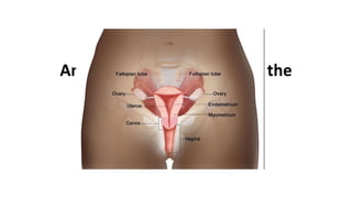
2. Gross Anatomy
Lower one third of uterus
3-4 cm in length and 2.5 cm in diameter
Lower end projects through anterior vaginal wall, dividing it into supra
vaginal and a lower vaginal portion
4. Supra vaginal part of the Cervix
Relations:
Anteriorly- base of bladder
Posteriorly- rectouterine pouch with intestinal coils and rectum
On each side-
• Ureter
• Uterine artery
• Attachment of Mackenrodt’s ligament
• Lower attached margin of the broad ligament
5. Vaginal Part of the Cervix
Conical in shape.
• Projects into the anterior wall of vagina forming the vaginal fornices
(anterior, posterior and 2 lateral).
• Cervical canal (Endocervical canal) opens into the vagina by an
opening called the external os.
• External os is small and circular in nulliparous women.
• In multiparous women, the external os is bounded by the anterior
and posterior lips.
7. Microscopic Anatomy
• The cervix is composed of epithelium (surface lining) and underlying
stroma (deeper fibrous tissue) separated by a thin barrier called basement
membrane.
• The ectocervix is lined by non-keratinized stratified squamous epithelium
that has multiple (1520) layers of cells and appears pale pink in colour.
• The squamous epithelium is divided into basal, parabasal, intermediate
and superficial layers from below upwards
8.
9. Microscopic Anatomy
• The endocervix is lined by columnar epithelium composed of a single layer
of tall cells with dark-staining nuclei close to the basement membrane. On
visual examination, columnar epithelium appears red in colour with a
granular velvet like surface.
• The epithelium forms several invaginations into the substance of the
stroma, resulting in the formation of endocervical crypts (sometimes
referred to as endocervical glands). The crypts lined by columnar
epithelium may extend 5–8 mm into the stroma.
10.
11. Microscopic Anatomy
• The columnar epithelium at its lower limit meets the squamous epithelium.
The junction between the two epithelia is known as the squamocolumnar
junction
• The SCJ is usually visible as a sharp border located near the external os.
• The position of the SCJ in relation to the external os changes with age,
pregnancy and use of oral contraceptive pills.
12.
13.
14.
15. Physiological changes in cervical Epithelium
• The cervix enlarges under the influence of oestrogen at puberty
and during pregnancy.
• As a result, columnar epithelium becomes visible on the
ectocervix and the SCJ moves out on the ectocervix. This
condition is known as ectropion or ectopy.
16. Physiological changes in cervical Epithelium
• The columnar epithelium on the ectocervix becomes exposed to
the acidic environment of the vagina. This causes destruction of
the columnar epithelium and its gradual replacement by the
newly formed squamous epithelium. This process through
which the columnar epithelium on the ectocervix is gradually
replaced with squamous epithelium is called squamous
metaplasia.
17. Physiological changes in cervical Epithelium
• Squamous metaplasia usually begins at the SCJ at the distal limit of the
ectopy (original SCJ) and gradually moves inwards (centripetally) towards
the external os
• The SCJ formed between the metaplastic squamous epithelium and the
columnar epithelium is known as the new SCJ.
• The area between the original SCJ and the newly formed SCJ as a result
of metaplasia is the transformation zone (TZ).
19. Transformation Zone
• The proximal extent of the TZ is the new SCJ and is easy to identify.
Tongue like projections of the thin newly formed metaplastic squamous
epithelium is a feature of the normal TZ.
• Patent crypts appear as small openings on the TZ. Some of the crypts are
blocked by the metaplastic epithelium, which leads to formation of
retention cysts known as nabothian follicles or cysts.
• The crypt openings and nabothian cysts are features of normal TZ. The
position of the crypt opening or the nabothian cyst farthest from the SCJ
helps to identify the outer limit of the TZ
20.
21. Changes in Transformation Zone
• During pregnancy the cervix enlarges, becomes congested and the
columnar epithelium extends to the ectocervix (ectropion). The SCJ is
easily visible on the ectocervix
• During the peri-menopausal period and after menopause, the cervix
shrinks due to the lack of oestrogen and SCJ moves inside the
endocervical canal from the external os. In post-menopausal women, the
SCJ is often invisible on visual examination.