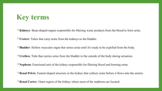
Key terms
Kidneys: Bean-shapedorgans responsible for filtering waste products from the blood to form urine.
Ureters: Tubes that carry urine from the kidneys to the bladder.
Bladder: Hollow muscular organ that stores urine until it's ready to be expelled from the body.
Urethra: Tube that carries urine from the bladder to the outside of the body during urination.
Nephron: Functional unit of the kidney responsible for filtering blood and forming urine.
Renal Pelvis: Funnel-shaped structure in the kidney that collects urine before it flows into the ureters.
Renal Cortex: Outer region of the kidney where most of the nephrons are located.
3.
Renal Medulla: Innerregion of the kidney containing structures like renal pyramids and renal
columns.
Glomerulus: A cluster of capillaries within the nephron where blood filtration takes place.
Filtration: The process by which blood is filtered in the kidneys, removing waste products and excess
substances to form urine.
Reabsorption: Process by which useful substances such as water, electrolytes, and nutrients are
reabsorbed from the filtrate back into the bloodstream.
Secretion: Process by which substances are actively transported from the bloodstream into the renal
tubules to be excreted in urine.
Micturition: Another term for urination, the process of expelling urine from the bladder.
4.
Renin: Enzyme secretedby the kidneys that plays a role in regulating blood pressure and electrolyte
balance.
Aldosterone: Hormone produced by the adrenal glands that helps regulate sodium and potassium
levels in the body by acting on the kidneys.
5.
Introduction
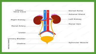
• The urinarysystem, also known as the renal system, comprises organs responsible for the production,
storage, and elimination of urine, which is the body's waste product formed from filtering blood. Its
primary components include the kidneys, ureters, urinary bladder, and urethra.
• The kidneys filter waste and excess substances from the blood to produce urine, which then flows
through the ureters to the urinary bladder for storage. When the bladder is full, urine is expelled from
the body through the urethra during urination.
• The urinary system plays a crucial role in maintaining the body's fluid balance, regulating electrolytes,
and removing waste products from metabolism.
7.
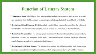
Function of UrinarySystem
Filtration of Blood: The kidneys filter waste products and excess substances, such as urea, uric acid,
and creatinine, from the bloodstream to maintain proper balance of electrolytes and fluids in the body.
Regulation of Blood Pressure: The kidneys help regulate blood pressure by controlling the volume of
blood and the concentration of electrolytes, such as sodium and potassium, in the bloodstream.
Regulation of Electrolytes: The urinary system maintains the balance of electrolytes, such as sodium,
potassium, calcium, and phosphate, in the body. These electrolytes are essential for proper nerve and
muscle function, as well as for maintaining fluid balance.
Regulation of Acid-Base Balance: The kidneys help regulate the pH balance of the body by excreting
hydrogen ions and reabsorbing bicarbonate ions, which helps maintain the body's acid-base balance.
8.
Production of Hormones:The kidneys produce hormones such as erythropoietin, which stimulates
the production of red blood cells in the bone marrow, and renin, which helps regulate blood pressure.
Excretion of Metabolic Waste: The urinary system removes metabolic waste products, such as urea
and uric acid, from the body through the formation of urine.
Regulation of Blood Volume and Osmolarity: The kidneys regulate blood volume and osmolarity by
adjusting the amount of water and solutes excreted in urine.
Detoxification: The urinary system helps remove toxins and drugs from the body by filtering them out
of the bloodstream and excreting them in urine.
9.
Kidney
• The kidneyis a vital organ in vertebrates that serves several essential functions in the body.
It is responsible for filtering waste products and excess substances, such as toxins, urea,
and salts, from the blood to form urine.
• Additionally, the kidney regulates the body's fluid balance, electrolyte levels, and blood
pressure by adjusting the composition and volume of urine produced.
• Moreover, the kidney plays a crucial role in maintaining acid-base balance and producing
hormones that regulate red blood cell production, blood pressure, and calcium metabolism.
10.
Position & Structure
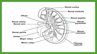
•The kidneys are bean-shaped organs located in the back of the abdominal cavity, one on each side of
the spine. They are situated below the rib cage, with the left kidney slightly higher than the right to
accommodate the liver. Each kidney is about the size of a fist and weighs around 120 to 150 grams in
adults.
Structure :
Renal Cortex: The outer layer of the kidney, containing structures like the renal corpuscles and
convoluted tubules.
Renal Medulla: The inner part of the kidney, which consists of renal pyramids, renal columns, and the
renal pelvis.
12.
Renal Pelvis: Afunnel-shaped structure at the center of the kidney that collects urine produced by the
nephrons.
Nephrons: The functional units of the kidney responsible for filtering the blood and producing urine.
Each kidney contains millions of nephrons, each composed of a renal corpuscle (glomerulus and
Bowman's capsule) and renal tubules (proximal convoluted tubule, loop of Henle, distal convoluted
tubule, and collecting duct).
Renal Arteries and Veins: Blood vessels that supply the kidneys with oxygenated blood and carry
away filtered blood and waste products.
13.
Function
Filtration: The primaryfunction of the kidneys is to filter waste products, excess ions, and water from
the bloodstream to form urine, which is then excreted from the body.
Fluid and Electrolyte Balance: The kidneys regulate the balance of fluids and electrolytes in the
body, ensuring that levels of substances like sodium, potassium, and calcium remain within a narrow
range necessary for proper cellular function.
Blood Pressure Regulation: The kidneys help regulate blood pressure by controlling the volume of
blood and the concentration of sodium and other ions in the bloodstream. They produce hormones like
renin, which plays a role in blood pressure regulation.
14.
Red Blood CellProduction: The kidneys produce erythropoietin, a hormone that stimulates the bone
marrow to produce red blood cells. Red blood cells are essential for carrying oxygen to tissues
throughout the body.
Acid-Base Balance: The kidneys help maintain the body's acid-base balance by excreting hydrogen
ions and reabsorbing bicarbonate ions, helping to regulate pH levels in the blood and body fluids.
15.
Nephron
• The nephronis the structural and functional unit of the kidney, responsible for filtering blood and
producing urine.
• Each kidney contains thousands of nephrons, which work together to maintain fluid balance, remove
waste products from the blood, regulate electrolyte concentrations, and help control blood pressure.
• The nephron consists of a renal corpuscle, composed of the glomerulus and Bowman's capsule, and a
renal tubule, which includes the proximal convoluted tubule, loop of Henle, distal convoluted tubule,
and collecting duct.
• This intricate structure allows for the efficient filtration, reabsorption, and secretion of substances to
maintain the body's internal environment.
17.
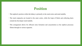
Position
• The nephron'sposition within the kidney is primarily in the renal cortex and renal medulla.
• The renal corpuscles are located in the outer cortex, while the loops of Henle and collecting ducts
extend into the deeper renal medulla.
• This arrangement allows for efficient urine formation and concentration as the nephron processes
filtrate through its various segments.
18.
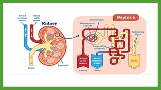
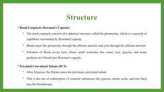
Structure
Renal Corpuscle (Bowman'sCapsule):
The renal corpuscle consists of a spherical structure called the glomerulus, which is a network of
capillaries surrounded by Bowman's capsule.
Blood enters the glomerulus through the afferent arteriole and exits through the efferent arteriole.
Filtration of blood occurs here, where small molecules like water, ions, glucose, and waste
products are filtered into Bowman's capsule.
Proximal Convoluted Tubule (PCT):
After filtration, the filtrate enters the proximal convoluted tubule.
This is the site of reabsorption of essential substances like glucose, amino acids, and ions back
into the bloodstream.
19.
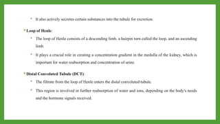
It alsoactively secretes certain substances into the tubule for excretion.
Loop of Henle:
The loop of Henle consists of a descending limb, a hairpin turn called the loop, and an ascending
limb.
It plays a crucial role in creating a concentration gradient in the medulla of the kidney, which is
important for water reabsorption and concentration of urine.
Distal Convoluted Tubule (DCT):
The filtrate from the loop of Henle enters the distal convoluted tubule.
This region is involved in further reabsorption of water and ions, depending on the body's needs
and the hormone signals received.
20.
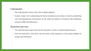
Collecting Duct:
Thecollecting duct receives urine from multiple nephrons.
It plays a major role in determining the final concentration and volume of urine by reabsorbing
water and adjusting the concentration of ions under the influence of hormones like antidiuretic
hormone (ADH) and aldosterone.
Renal Pelvis and Ureter:
The collecting ducts empty urine into the renal pelvis, which is a funnel-shaped structure.
From the renal pelvis, urine flows into the ureter, which transports it to the urinary bladder for
storage until elimination.
21.
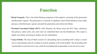
Function
Renal Corpuscle: Thisis the initial filtering component of the nephron, consisting of the glomerulus
and Bowman's capsule. The glomerulus is a network of capillaries where blood filtration occurs under
pressure, while Bowman's capsule surrounds the glomerulus and collects the filtrate.
Proximal Convoluted Tubule (PCT): After filtration, the filtrate enters the PCT. Here, substances
like glucose, amino acids, ions, and water are reabsorbed back into the bloodstream. This region is
highly convoluted, increasing its surface area for reabsorption.
Loop of Henle: The loop of Henle consists of a descending and an ascending limb. It plays a crucial
role in concentrating urine by creating an osmotic gradient in the renal medulla. The descending limb
is permeable to water but not to ions, while the ascending limb is permeable to ions but not to water.
22.
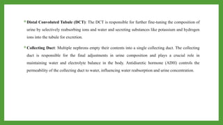
Distal Convoluted Tubule(DCT): The DCT is responsible for further fine-tuning the composition of
urine by selectively reabsorbing ions and water and secreting substances like potassium and hydrogen
ions into the tubule for excretion.
Collecting Duct: Multiple nephrons empty their contents into a single collecting duct. The collecting
duct is responsible for the final adjustments in urine composition and plays a crucial role in
maintaining water and electrolyte balance in the body. Antidiuretic hormone (ADH) controls the
permeability of the collecting duct to water, influencing water reabsorption and urine concentration.
23.
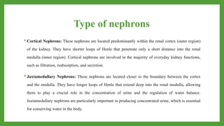
Type of nephrons
CorticalNephrons: These nephrons are located predominantly within the renal cortex (outer region)
of the kidney. They have shorter loops of Henle that penetrate only a short distance into the renal
medulla (inner region). Cortical nephrons are involved in the majority of everyday kidney functions,
such as filtration, reabsorption, and secretion.
Juxtamedullary Nephrons: These nephrons are located closer to the boundary between the cortex
and the medulla. They have longer loops of Henle that extend deep into the renal medulla, allowing
them to play a crucial role in the concentration of urine and the regulation of water balance.
Juxtamedullary nephrons are particularly important in producing concentrated urine, which is essential
for conserving water in the body.
24.
Ureters
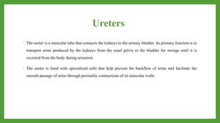
• The ureteris a muscular tube that connects the kidneys to the urinary bladder. Its primary function is to
transport urine produced by the kidneys from the renal pelvis to the bladder for storage until it is
excreted from the body during urination.
• The ureter is lined with specialized cells that help prevent the backflow of urine and facilitate the
smooth passage of urine through peristaltic contractions of its muscular walls.
25.
Position
• The uretersare paired tubes, with one ureter connected to each kidney.
• They extend from the renal pelvis of the kidney to the urinary bladder.
• In terms of anatomical position, the ureters travel down behind the abdominal cavity
and enter the pelvis.
• They run alongside the vertebral column and cross the pelvic brim to enter the
bladder.
26.
Structure
• The wallof the ureter is composed of three layers:
Inner Mucosa: This layer is made up of transitional epithelium, which can stretch as the ureter fills
with urine. It also has a layer of connective tissue underneath.
Middle Muscular Layer: This layer consists of smooth muscle fibers arranged in an inner
longitudinal and outer circular layer. These muscles create peristaltic contractions that help propel
urine downward toward the bladder.
Outer Adventitia: This is the outermost layer, made of connective tissue that anchors the ureter in
place and connects it to surrounding structures.
27.
Function
Transport of Urine:The primary function of the ureters is to transport urine from the kidneys to the
urinary bladder. Peristaltic contractions of the smooth muscle layers propel urine down the ureters.
Prevention of Reflux: The ureters have valves at their junction with the bladder known as
ureterovesical valves, which prevent urine from flowing back into the kidneys when the bladder
contracts during urination.
Role in Urinary Continence: While the ureters themselves do not directly contribute to urinary
continence (the ability to control urination), their proper function and connection with the bladder are
essential for maintaining normal urinary control.
28.
Urinary Bladder
The urinarybladder is a hollow, muscular organ located in the pelvic cavity of humans and some
animals. Its primary function is to store urine produced by the kidneys until it is expelled from the
body through the urethra.
The bladder can expand to accommodate varying volumes of urine and contracts to facilitate urination
when it's appropriate. It's lined with a specialized epithelium that allows it to stretch and maintain its
integrity during filling and emptying.
29.
Position
The urinary bladderis a hollow, muscular organ located in the pelvis just behind the pubic bone.
Its position can vary slightly depending on the individual's anatomy and bladder filling, but it typically
lies inferior to the peritoneum and anterior to the rectum in males, and anterior to the vagina and
uterus in females.
30.
Structure
Epithelial lining: Theinner lining of the bladder is made up of transitional epithelium, which allows
the bladder to stretch as it fills with urine without rupturing.
Muscular layers: The bladder wall consists of smooth muscle known as the detrusor muscle, which
contracts during urination to expel urine.
Sphincters: There are two sphincters that control the flow of urine into and out of the bladder. The
internal urethral sphincter is involuntary and made of smooth muscle, while the external urethral
sphincter is voluntary and made of skeletal muscle.
31.
Function
• The mainfunctions of the urinary bladder include:
Storage: The bladder stores urine produced by the kidneys until it is convenient to urinate. Its elastic
walls expand to accommodate increasing volumes of urine.
Voiding: When the bladder is sufficiently filled and there is a signal from the nervous system
indicating the need to urinate, the detrusor muscle contracts, and the sphincters relax to allow the
expulsion of urine through the urethra.
32.
Urethra
• The urethrais a tube-like structure in the body that serves as a passageway for urine to exit the bladder
and be expelled from the body. In males, the urethra also functions as a passage for semen during
ejaculation.
• It extends from the bladder to the external opening of the genitals. In females, the urethra is shorter
compared to males and is located between the clitoris and the vaginal opening. Its main function is to
transport urine from the bladder to the outside of the body.
33.
Position
Male Urethra:
Position: Inmales, the urethra runs through the penis. It extends from the internal
urethral orifice, where it connects to the urinary bladder, to the external urethral
orifice at the tip of the penis.
34.
Structure
• Structure: Themale urethra can be divided into three parts: the prostatic urethra, the membranous
urethra, and the spongy or penile urethra. The prostatic urethra passes through the prostate gland, the
membranous urethra passes through the urogenital diaphragm, and the spongy urethra runs through the
length of the penis.
• Functions: In males, the urethra serves both urinary and reproductive functions. It allows for the
passage of urine from the bladder during urination and also serves as the conduit for semen during
ejaculation.
35.
Position
• Female Urethra:
•Position: In females, the urethra is shorter compared to males and is located anterior to the vaginal
opening.
• Structure: The female urethra is relatively simple compared to the male urethra. It is a short tube that
extends from the bladder to the external urethral orifice.
• Functions: The primary function of the female urethra is to allow the passage of urine from the
bladder to the outside of the body during urination.
36.
Common Functions ofBoth Male and
Female Urethras
• Urinary Excretion: Both male and female urethras serve as the final pathway for urine excretion from
the body.
• Sphincter Control: Both genders have internal and external urethral sphincters that help control the
flow of urine and prevent leakage between urinations.
• Mucosal Lubrication: The urethra is lined with mucous membranes that help lubricate the passage of
urine and protect the lining from irritation.


![PERI-PROSTHETIC FRACTURE NAIL-PLATE CONSTRUCT [NPC].pptx](https://cdn.slidesharecdn.com/ss_thumbnails/drarunkumardrmohamedashrafperiprostheticfrasturenail-plateconstructnpc-260209164459-7e9d15a1-thumbnail.jpg?width=640&height=640&fit=bounds)


![CTEV [ clubfoot] DR ARUN LAL ,DR MOHAMED ASHRAF travancore medical college k...](https://cdn.slidesharecdn.com/ss_thumbnails/ctevclubfootdrarunlaldrmohamedashraftravancoremedicalcollegekollamkeralaindia-260208063247-18fc466c-thumbnail.jpg?width=640&height=640&fit=bounds)


