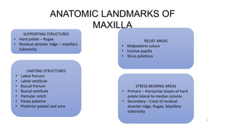
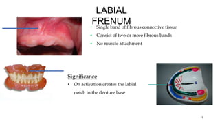
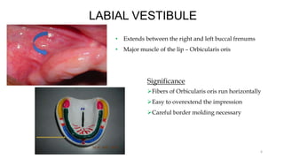
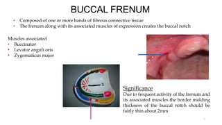
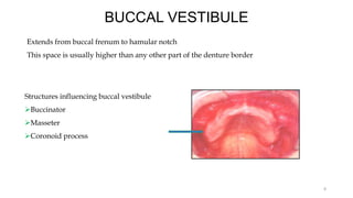
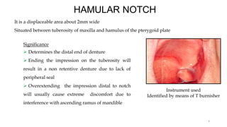
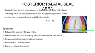
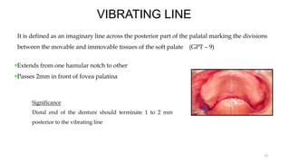
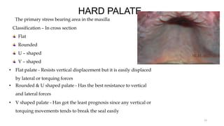
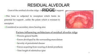
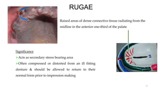
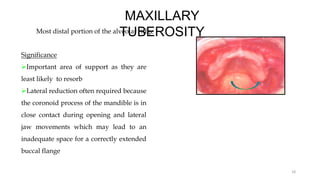
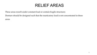
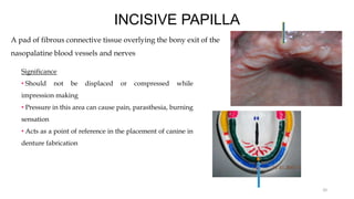
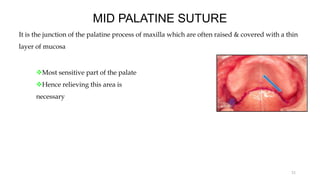
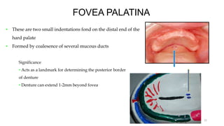
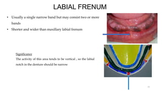
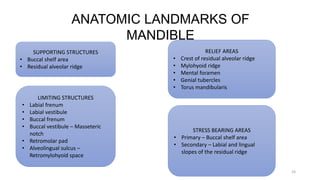
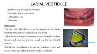
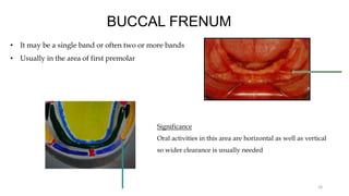
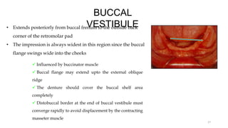
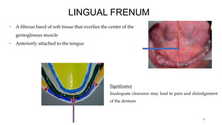
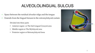
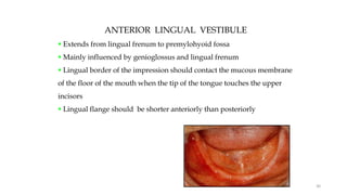
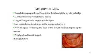
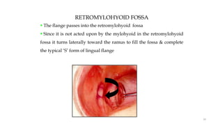
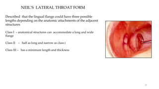
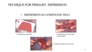
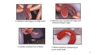
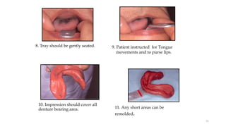
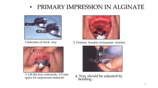
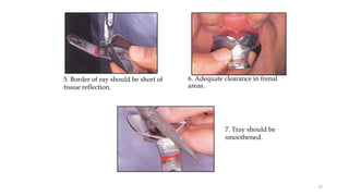
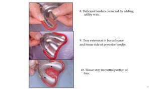
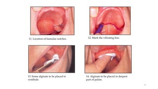
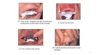
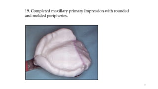
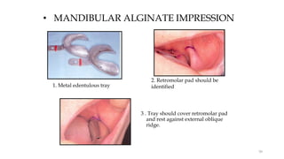
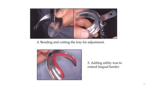
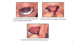
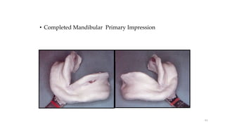
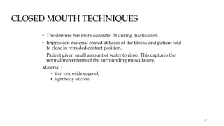
ANATOMICAL
LANDMARKS AND
PRIMARY IMPRESSION
DR ASWITHA G
1ST YEAR PG
DEPT OF PROSTHODONTICS
1
ANATOMICAL LANDMARK
• It is defined as a recognizable anatomic structure used as a point of reference
(GPT -9)
2
ANATOMIC LANDMARKS OF
MAXILLA
3
SUPPORTING STRUCTURES
• Hard palate – Rugae
• Residual alveolar ridge – maxillary
tuberosity
LIMITING STRUCTURES
• Labial frenum
• Labial vestibule
• Buccal frenum
• Buccal vestibule
• Hamular notch
• Fovea palatine
• Posterior palatal seal area
RELIEF AREAS
• Midpalatine suture
• Incisive papilla
• Torus palatinus
STRESS BEARING AREAS
• Primary – Horizontal slopes of hard
palate lateral to median sutures
• Secondary – Crest of residual
alveolar ridge, Rugae, Maxillary
tuberosity
LIMITING STRUCTURES
These are the structures which determine & confine the extent of
the denture
GPT-9
4
LABIAL
FRENUM
• Single band of fibrous connective tissue
• Consist of two or more fibrous bands
• No muscle attachment
5
Significance
• On activation creates the labial
notch in the denture base
LABIAL VESTIBULE
• Extends between the right and left buccal frenums
• Major muscle of the lip – Orbicularis oris
6
Significance
Fibers of Orbicularis oris run horizontally
Easy to overextend the impression
Careful border molding necessary
BUCCAL FRENUM
• Composed of one or more bands of fibrous connective tissue
• The frenum along with its associated muscles of expression creates the buccal notch
Muscles associated
• Buccinator
• Levator anguli oris
• Zygomaticus major
7
Significance
Due to frequent activity of the frenum and
its associated muscles the border molding
thickness of the buccal notch should be
fairly thin about 2mm
BUCCAL VESTIBULE
Extends from buccal frenum to hamular notch
This space is usually higher than any other part of the denture border
Structures influencing buccal vestibule
Buccinator
Masseter
Coronoid process
8
It is a displaceable area about 2mm wide
Situated between tuberosity of maxilla and hamulus of the pterygoid plate
HAMULAR NOTCH
Instrument used
Identified by means of T burnisher
9
Significance
 Determines the distal end of denture
 Ending the impression on the tuberosity will
result in a non retentive denture due to lack of
peripheral seal
 Overextending the impression distal to notch
will usually cause extreme discomfort due to
interference with ascending ramus of mandible
POSTERIOR PALATAL SEAL
AREA
It is defined as the soft tissue area at or beyond the junction of the hard
and soft palates on which pressure within the physiological limit can be
applied by a complete denture to aid in its retention
(GPT – 9)
10
Significance
Reduces the tendency for gag reflex
Aids in retention by maintaining constant contact with soft palate
 Compensates for polymerisation shrinkage
 Prevents food accumulation
 Reduces patient discomfort
M.M.HOUSE CLASSIFICATION
Describes the amount of posterior tissue that will accept the posterior palatal seal
Class I – more than 5mm of movable tissue available for post-damming; retention is usually good
Class II – 1-5mm of movable tissue available for post-damming good retention is usually possible
Class III – less than 1mm movable tissue available for post-damming; retention is usually poor
11
METHODS TO RECORD POSTERIOR PALATAL SEAL
• Conventional approach using ’T’ burnisher
• Fluid wax technique
• Arbitrary scrapping of the master cast
12
VIBRATING LINE
It is defined as an imaginary line across the posterior part of the palatal marking the divisions
between the movable and immovable tissues of the soft palate (GPT – 9)
Extends from one hamular notch to other
Passes 2mm in front of fovea palatina
13
Significance
Distal end of the denture should terminate 1 to 2 mm
posterior to the vibrating line
SUPPORTING
STRUCTURES
It is defined as the surfaces of oral structures that resists force, strains or pressures brought on them
during function
GPT – 9
14
HARD PALATE
The primary stress bearing area in the maxilla
Classification – In cross section
Flat
Rounded
U – shaped
V – shaped
15
• Flat palate - Resists vertical displacement but it is easily displaced
by lateral or torquing forces
• Rounded & U shaped palate - Has the best resistance to vertical
and lateral forces
• V shaped palate - Has got the least prognosis since any vertical or
torquing movements tends to break the seal easily
RESIDUAL ALVEOLAR
RIDGE
Crest of the residual alveolar ridge - important area of support
This bone is subjected to resorption which limits its
potential for support , unlike the palate which is resistant to
resorption
Considered as secondary stress bearing area
16
Factors influencing architecture of residual alveolar ridge
• Persons general health
• Forces developed by the surrounding musculature
• Severity of periodontal disease
• Forces acquiring from wearing of dental prosthesis
• Time length of edentulous span
RUGAE
Raised areas of dense connective tissue radiating from the
midline in the anterior one-third of the palate
17
Significance
Acts as secondary stress bearing area
Often compressed or distorted from an ill fitting
denture & should be allowed to return to their
normal form prior to impression making
MAXILLARY
TUBEROSITY
Most distal portion of the alveolar ridge
Significance
Important area of support as they are
least likely to resorb
Lateral reduction often required because
the coronoid process of the mandible is in
close contact during opening and lateral
jaw movements which may lead to an
inadequate space for a correctly extended
buccal flange
18
RELIEF AREAS
These areas resorb under constant load or contain fragile structures
Denture should be designed such that the masticatory load is not concentrated in these
areas
19
INCISIVE PAPILLA
A pad of fibrous connective tissue overlying the bony exit of the
nasopalatine blood vessels and nerves
20
Significance
• Should not be displaced or compressed while
impression making
• Pressure in this area can cause pain, parasthesia, burning
sensation
• Acts as a point of reference in the placement of canine in
denture fabrication
MID PALATINE SUTURE
It is the junction of the palatine process of maxilla which are often raised & covered with a thin
layer of mucosa
Most sensitive part of the palate
Hence relieving this area is
necessary
21
FOVEA PALATINA
• These are two small indentations fond on the distal end of the
hard palate
• Formed by coalesence of several mucous ducts
22
Significance
• Acts as a landmark for determining the posterior border
of denture
• Denture can extend 1-2mm beyond fovea
LABIAL FRENUM
• Usually a single narrow band but may consist two or more
bands
• Shorter and wider than maxillary labial frenum
23
Significance
The activity of this area tends to be vertical , so the labial
notch in the denture should be narrow
ANATOMIC LANDMARKS OF
MANDIBLE
24
SUPPORTING STRUCTURES
• Buccal shelf area
• Residual alveolar ridge
LIMITING STRUCTURES
• Labial frenum
• Labial vestibule
• Buccal frenum
• Buccal vestibule – Masseteric
notch
• Retromolar pad
• Alveolingual sulcus –
Retromylohyoid space
RELIEF AREAS
• Crest of residual alveolar ridge
• Mylohyoid ridge
• Mental foramen
• Genial tubercles
• Torus mandibularis
STRESS BEARING AREAS
• Primary – Buccal shelf area
• Secondary – Labial and lingual
slopes of the residual ridge
LABIAL VESTIBULE
It is the sulcus between the frenum's
The major muscle in this area
Orbicularis oris
Mentalis
25
Significance
The fibers of Orbicularis oris are horizontal, careful border
molding done it avoid overextension of denture
 Mentalis muscle due to its excessive activity results in short
flange which may not provide a real seal for the finished
denture
Labial and buccal borders are not as critical for border seal
because the drape of lips & cheeks create a facial seal
BUCCAL FRENUM
• It may be a single band or often two or more bands
• Usually in the area of first premolar
26
Significance
Oral activities in this area are horizontal as well as vertical
so wider clearance is usually needed
BUCCAL
VESTIBULE
• Extends posteriorly from buccal frenum to the outside back
corner of the retromolar pad
• The impression is always widest in this region since the buccal
flange swings wide into the cheeks
27
 Influenced by buccinator muscle
 Buccal flange may extend upto the external oblique
ridge
 The denture should cover the buccal shelf area
completely
 Distobuccal border at the end of buccal vestibule must
converge rapidly to avoid displacement by the contracting
masseter muscle
LINGUAL FRENUM
• A fibrous band of soft tissue that overlies the center of the
genioglossus muscle
• Anteriorly attached to the tongue
28
Significance
Inadequate clearance may lead to pain and dislodgement
of the denture
ALVEOLOLINGUAL SULCUS
• Space between the residual alveolar ridge and the tongue
• Extends from the lingual frenum to the retromylohyoid curtain
29
Divided into three parts
• Anterior region or The Sub Lingual Crescent area
• Middle region or The Mylohyoid area
• Posterior region or Retromylohyoid fossa
ANTERIOR LINGUAL VESTIBULE
 Extends from lingual frenum to premylohyoid fossa
 Mainly influenced by genioglossus and lingual frenum
 Lingual border of the impression should contact the mucous membrane
of the floor of the mouth when the tip of the tongue touches the upper
incisors
 Lingual flange should be shorter anteriorly than posteriorly
30
MYLOHYOID AREA
 Extends from premyohyoid fossa to the distal end of the mylohyoid ridge
 Mainly influenced by mylohyoid muscle
 Lingual flange should slope toward tongue
Aids in stabilizing the denture as the tongue rests over it
 Provides space for raising the floor of the mouth without displacing the
denture
 Peripheral seal is maintained
during function
31
RETROMYLOHYOID FOSSA
 The flange passes into the retromylohyoid fossa
 Since it is not acted upon by the mylohyoid in the retromylohyoid
fossa it turns laterally toward the ramus to fill the fossa & complete
the typical ‘S’ form of lingual flange
32
NEIL’S LATERAL THROAT FORM
Described that the lingual flange could have three possible
lengths depending on the anatomic attachments of the adjacent
structures
Class I – anatomical structures can accommodate a long and wide
flange
Class II – half as long and narrow as class i
Class III – has a minimum length and thickness
33
RETROMOLAR PAD
A triangular soft pad of tissue at the distal end of the lower ridge
Significance
Posterior seal of mandibular denture
34
DISTAL BORDRES
The distal border of the denture is limited by
• Ramus of the mandible
• Buccinator muscle
• Internal and external oblique ridge
Significance
Overextension at this border causes soreness & also limits of buccinator muscle
35
BUCCAL SHELF AREA
• Primary area of support of mandibular denture
• It is between the mandibular buccal frenum & anterior edge of
masseter muscle
It is bounded
Medially – crest of alveolar ridge
Laterally – external oblique ridge
Distally – retromolar pad
36
Significance
Bone of the buccal shelf is covered by a layer of
cortical bone & also it lies at right angles to the vertical
occlusal forces , makes it most suitable primary stress
bearing area
RESIDUAL ALVEOLAR
RIDGE
Anterior alveolar ridge tends to resorb & hence it is considered
as secondary stress bearing area
Posterior alveolar ridge can also be considered a primary stress
bearing area , however often the ridge is poor and the buccal shelf
must assume the major role
37
MYLOHYOID RIDGE
The distal end of the ridge is close to the crest of the ridge & anterior aspect close to the lower border
of mandible
Significance:
• A prominent sharp ridge interfere with the
development of correct lingual flange &
cause pain especially during mastication
•Proper relief should be given
38
MENTAL FORAMEN
 As resorption occurs mental foramen will come to lie closer
to the crest of residual ridge
 Unless relief is provided the nerves and blood vessels will
get compressed
39
GENIAL TUBERCLE
• They lie away from the crest of ridge
• Due to resorption they also become increasingly prominent
& hence relief is essential to avoid complications
40
IMPRESSIONS FOR COMPLETE
DENTURE
41
DEFINITIONS
IMPRESSION – A negative likeness or copy in reverse of the surface of an object, an imprint of the
teeth and adjacent structures for use in dentistry (GPT 9)
42
PRINCIPLES OF IMPRESSION MAKING
43
1. Tissues must be healthy, before impression making
2. Proper space must be provided for selected impression material
3. Tray and impression material should be dimensionally stable
4. For correct positioning of tray, a guiding mechanism should be provided
5. Impression should be adequately extended to include the entire basal seat area as dictated by
limiting and supporting structures
6. A border moulding must be performed in harmony with anatomical and physiological
limitations of the oral structures
7. Impression must be removed without damage to the oral structures
8. The tissue surface of impression and intaglio surface of the denture must be coincide
OBJECTIVES OF IMPRESSION MAKING
44
1. Retention
2. Stability
3. Support
4. Preservation of residual structures
5. Aesthetics
OPERATOR POSITION FOR MAXILLARY
IMPRESSION
CORRECT INCORRECT
45
OPERATOR POSITION FOR MANDIBULAR IMPRESSION
CORRECT INCORRECT
46
TECHNIQUES
• Open mouth technique
• Mucocompressive
• Mucostatic
• Selective pressure
• Closed mouth technique
47
PRIMARY IMPRESSION
• It records the useful anatomy of the edentulous mouth so that a model can be cast on
which an accurately fitting special tray can be made.
Materials used:
Alginate
Impression compound
48
• IMPRESSION IN COMPOUND TRAY
3. Compound placed in the tray.
1. Modelling compund. 2. Softenend in water bath
and kneaded.
TECHIQUE FOR PRIMARY IMPRESSION
49
5. Should cover mylohyoid ridge and
external oblique ridge.
4. Molded with fingers to ridge form.
6. Gently warmed over a flame. 7. Before insertion, tempering in
warm water bath.
50
9. Patient instructed for Tongue
movements and to purse lips.
8. Tray should be gently seated.
11. Any short areas can be
remolded.
10. Impression should cover all
denture bearing area.
51
• PRIMARY IMPRESSION IN ALGINATE
4. Tray should be adjusted by
bending .
1.Selection of stock tray. 2. Position borders at hamular notches.
3. Lift the tray anteriorly, 3-5 mm
space for impression material.
52
5. Border of ray should be short of
tissue reflection.
6. Adequate clearance in frenal
areas.
7. Tray should be
smoothened.
53
10. Tissue stop in central portion of
tray.
8. Deficient borders corrected by adding
utility wax.
9. Tray extension in buccal space
and tissue side of posterior border.
54
11. Location of hamular notches. 12. Mark the vibrating line.
13. Some alginate to be placed in
vestibule.
14. Alginate to be placed in deepest
part of palate.
55
15. Tray to be rotated into the mouth and
seated first at the back of the mouth. 16. Upper lip elevated.
17. Tray is held in the mouth.
18. Labial and buccal borders to be
molded.
56
19. Completed maxillary primary Impression with rounded
and molded peripheries.
57
• MANDIBULAR ALGINATE IMPRESSION
3 . Tray should cover retromolar pad
and rest against external oblique
ridge.
1. Metal edentulous tray
2. Retromolar pad should be
identified
58
4. Bending and cutting the tray for adjustment.
5. Adding utility was to
extend lingual border.
59
7. Patient told to do tongue movements.
6. Patient told to raise the tongue and
tray is rotated in the mouth.
8. Gently mold the labial and buccal areas.
60
• Completed Mandibular Primary Impression
61
CLOSED MOUTH TECHNIQUES
• The denture has more accurate fit during mastication.
• Impression material coated at bases of the blocks and patient told
to close in retruded contact position.
• Patient given small amount of water to rinse. This captures the
normal movements of the surrounding musculature.
Material :
• thin zinc oxide eugenol,
• light body silicone.
62
CLOSED MOUTH TECHNIQUE
Drawbacks
• Maxillary Disto buccal space is not recorded in function.
• Viscous impression material can lead to increase in vertical
dimension.
Advantages
• Discrepancies in the jaw relations, resulting from points of
premature contact of the rims are eliminated.
• Masseter muscle can be recorded in function.
63
MUCO-COMPRESSIVE TECHNIQUE
• The impression material must be capable of viscous flow as it is
extruded under pressure from between the tray and the tissue
surface
Materials used
• Impression compound
• High viscosity silicones
• Stiff zinc oxide eugenol
64
MUCOSTATIC TECHNIQUE
• Use a very fluid impression material, and use minimal pressure while it sets.
• Minimal pressure technique
• By Page
• Eg. Impression plaster, Alginate
65
SELECTIVE PRESSURE TECHNIQUE
• It is a combination of extension for maximum coverage within tissue
tolerance with light pressure or intimate contact with the movable,
loosely attached tissues in the vestibules.
• The impression is refined with a minimum of pressure.
• By Boucher
66
• Impressions For Complete Dentures , Bernard Levin
• Essentials Of Complete Denture Prosthodontics , Second Edition ,Winkler
• Textbook Of Complete Denture Prosthodontics, Boucher
• Significance Of Anatomical Landmarks In Complete Dentures, Jpd ,1964,vol :14
• Reliability Of The Fovea For Determining The Posterior Border Of The Maxillary Denture,
Jpd,1980 ,Vol :43
• Analysis Of The Posterior Palatal Forms Related To Complete Dentures, Jpd ,1982 ,Vol:47
• Location And Preparation Of Posterior Palatal Seal ,Jpd ,1983 ,Vol :49
• Relationship Of The Maxillary Canine To Incisive Papilla , Grove ,Jpd , 1989 ,Vol: 61.
• Frequency And Locations Of Traumatic Ulcerations Following Placement Of Complete Dentures,
Kivovics, Ijp ,Aug 2007
• Significance Of Fovea In Complete Dentures, Lye , JPD , 1975 ,VOL :33
• Variable Denture Limiting Structures Of The Edentulous Mouth ,Jpd , 1966 ,Vol : 16
• A Comparison of The Retention Of The Retention Of The Complete Denture Bases Having Different
Types Of Posterior Palatal Seal, Avant , JPD , 1973 ,VOL :29
67
REFERENCES

ANATOMICAL LANDMARKS AND PRIMARY IMPRESSION.pptx

  • 1.
    ANATOMICAL LANDMARKS AND PRIMARY IMPRESSION DRASWITHA G 1ST YEAR PG DEPT OF PROSTHODONTICS 1
  • 2.
    ANATOMICAL LANDMARK • Itis defined as a recognizable anatomic structure used as a point of reference (GPT -9) 2
  • 3.
    ANATOMIC LANDMARKS OF MAXILLA 3 SUPPORTINGSTRUCTURES • Hard palate – Rugae • Residual alveolar ridge – maxillary tuberosity LIMITING STRUCTURES • Labial frenum • Labial vestibule • Buccal frenum • Buccal vestibule • Hamular notch • Fovea palatine • Posterior palatal seal area RELIEF AREAS • Midpalatine suture • Incisive papilla • Torus palatinus STRESS BEARING AREAS • Primary – Horizontal slopes of hard palate lateral to median sutures • Secondary – Crest of residual alveolar ridge, Rugae, Maxillary tuberosity
  • 4.
    LIMITING STRUCTURES These arethe structures which determine & confine the extent of the denture GPT-9 4
  • 5.
    LABIAL FRENUM • Single bandof fibrous connective tissue • Consist of two or more fibrous bands • No muscle attachment 5 Significance • On activation creates the labial notch in the denture base
  • 6.
    LABIAL VESTIBULE • Extendsbetween the right and left buccal frenums • Major muscle of the lip – Orbicularis oris 6 Significance Fibers of Orbicularis oris run horizontally Easy to overextend the impression Careful border molding necessary
  • 7.
    BUCCAL FRENUM • Composedof one or more bands of fibrous connective tissue • The frenum along with its associated muscles of expression creates the buccal notch Muscles associated • Buccinator • Levator anguli oris • Zygomaticus major 7 Significance Due to frequent activity of the frenum and its associated muscles the border molding thickness of the buccal notch should be fairly thin about 2mm
  • 8.
    BUCCAL VESTIBULE Extends frombuccal frenum to hamular notch This space is usually higher than any other part of the denture border Structures influencing buccal vestibule Buccinator Masseter Coronoid process 8
  • 9.
    It is adisplaceable area about 2mm wide Situated between tuberosity of maxilla and hamulus of the pterygoid plate HAMULAR NOTCH Instrument used Identified by means of T burnisher 9 Significance  Determines the distal end of denture  Ending the impression on the tuberosity will result in a non retentive denture due to lack of peripheral seal  Overextending the impression distal to notch will usually cause extreme discomfort due to interference with ascending ramus of mandible
  • 10.
    POSTERIOR PALATAL SEAL AREA Itis defined as the soft tissue area at or beyond the junction of the hard and soft palates on which pressure within the physiological limit can be applied by a complete denture to aid in its retention (GPT – 9) 10 Significance Reduces the tendency for gag reflex Aids in retention by maintaining constant contact with soft palate  Compensates for polymerisation shrinkage  Prevents food accumulation  Reduces patient discomfort
  • 11.
    M.M.HOUSE CLASSIFICATION Describes theamount of posterior tissue that will accept the posterior palatal seal Class I – more than 5mm of movable tissue available for post-damming; retention is usually good Class II – 1-5mm of movable tissue available for post-damming good retention is usually possible Class III – less than 1mm movable tissue available for post-damming; retention is usually poor 11
  • 12.
    METHODS TO RECORDPOSTERIOR PALATAL SEAL • Conventional approach using ’T’ burnisher • Fluid wax technique • Arbitrary scrapping of the master cast 12
  • 13.
    VIBRATING LINE It isdefined as an imaginary line across the posterior part of the palatal marking the divisions between the movable and immovable tissues of the soft palate (GPT – 9) Extends from one hamular notch to other Passes 2mm in front of fovea palatina 13 Significance Distal end of the denture should terminate 1 to 2 mm posterior to the vibrating line
  • 14.
    SUPPORTING STRUCTURES It is definedas the surfaces of oral structures that resists force, strains or pressures brought on them during function GPT – 9 14
  • 15.
    HARD PALATE The primarystress bearing area in the maxilla Classification – In cross section Flat Rounded U – shaped V – shaped 15 • Flat palate - Resists vertical displacement but it is easily displaced by lateral or torquing forces • Rounded & U shaped palate - Has the best resistance to vertical and lateral forces • V shaped palate - Has got the least prognosis since any vertical or torquing movements tends to break the seal easily
  • 16.
    RESIDUAL ALVEOLAR RIDGE Crest ofthe residual alveolar ridge - important area of support This bone is subjected to resorption which limits its potential for support , unlike the palate which is resistant to resorption Considered as secondary stress bearing area 16 Factors influencing architecture of residual alveolar ridge • Persons general health • Forces developed by the surrounding musculature • Severity of periodontal disease • Forces acquiring from wearing of dental prosthesis • Time length of edentulous span
  • 17.
    RUGAE Raised areas ofdense connective tissue radiating from the midline in the anterior one-third of the palate 17 Significance Acts as secondary stress bearing area Often compressed or distorted from an ill fitting denture & should be allowed to return to their normal form prior to impression making
  • 18.
    MAXILLARY TUBEROSITY Most distal portionof the alveolar ridge Significance Important area of support as they are least likely to resorb Lateral reduction often required because the coronoid process of the mandible is in close contact during opening and lateral jaw movements which may lead to an inadequate space for a correctly extended buccal flange 18
  • 19.
    RELIEF AREAS These areasresorb under constant load or contain fragile structures Denture should be designed such that the masticatory load is not concentrated in these areas 19
  • 20.
    INCISIVE PAPILLA A padof fibrous connective tissue overlying the bony exit of the nasopalatine blood vessels and nerves 20 Significance • Should not be displaced or compressed while impression making • Pressure in this area can cause pain, parasthesia, burning sensation • Acts as a point of reference in the placement of canine in denture fabrication
  • 21.
    MID PALATINE SUTURE Itis the junction of the palatine process of maxilla which are often raised & covered with a thin layer of mucosa Most sensitive part of the palate Hence relieving this area is necessary 21
  • 22.
    FOVEA PALATINA • Theseare two small indentations fond on the distal end of the hard palate • Formed by coalesence of several mucous ducts 22 Significance • Acts as a landmark for determining the posterior border of denture • Denture can extend 1-2mm beyond fovea
  • 23.
    LABIAL FRENUM • Usuallya single narrow band but may consist two or more bands • Shorter and wider than maxillary labial frenum 23 Significance The activity of this area tends to be vertical , so the labial notch in the denture should be narrow
  • 24.
    ANATOMIC LANDMARKS OF MANDIBLE 24 SUPPORTINGSTRUCTURES • Buccal shelf area • Residual alveolar ridge LIMITING STRUCTURES • Labial frenum • Labial vestibule • Buccal frenum • Buccal vestibule – Masseteric notch • Retromolar pad • Alveolingual sulcus – Retromylohyoid space RELIEF AREAS • Crest of residual alveolar ridge • Mylohyoid ridge • Mental foramen • Genial tubercles • Torus mandibularis STRESS BEARING AREAS • Primary – Buccal shelf area • Secondary – Labial and lingual slopes of the residual ridge
  • 25.
    LABIAL VESTIBULE It isthe sulcus between the frenum's The major muscle in this area Orbicularis oris Mentalis 25 Significance The fibers of Orbicularis oris are horizontal, careful border molding done it avoid overextension of denture  Mentalis muscle due to its excessive activity results in short flange which may not provide a real seal for the finished denture Labial and buccal borders are not as critical for border seal because the drape of lips & cheeks create a facial seal
  • 26.
    BUCCAL FRENUM • Itmay be a single band or often two or more bands • Usually in the area of first premolar 26 Significance Oral activities in this area are horizontal as well as vertical so wider clearance is usually needed
  • 27.
    BUCCAL VESTIBULE • Extends posteriorlyfrom buccal frenum to the outside back corner of the retromolar pad • The impression is always widest in this region since the buccal flange swings wide into the cheeks 27  Influenced by buccinator muscle  Buccal flange may extend upto the external oblique ridge  The denture should cover the buccal shelf area completely  Distobuccal border at the end of buccal vestibule must converge rapidly to avoid displacement by the contracting masseter muscle
  • 28.
    LINGUAL FRENUM • Afibrous band of soft tissue that overlies the center of the genioglossus muscle • Anteriorly attached to the tongue 28 Significance Inadequate clearance may lead to pain and dislodgement of the denture
  • 29.
    ALVEOLOLINGUAL SULCUS • Spacebetween the residual alveolar ridge and the tongue • Extends from the lingual frenum to the retromylohyoid curtain 29 Divided into three parts • Anterior region or The Sub Lingual Crescent area • Middle region or The Mylohyoid area • Posterior region or Retromylohyoid fossa
  • 30.
    ANTERIOR LINGUAL VESTIBULE Extends from lingual frenum to premylohyoid fossa  Mainly influenced by genioglossus and lingual frenum  Lingual border of the impression should contact the mucous membrane of the floor of the mouth when the tip of the tongue touches the upper incisors  Lingual flange should be shorter anteriorly than posteriorly 30
  • 31.
    MYLOHYOID AREA  Extendsfrom premyohyoid fossa to the distal end of the mylohyoid ridge  Mainly influenced by mylohyoid muscle  Lingual flange should slope toward tongue Aids in stabilizing the denture as the tongue rests over it  Provides space for raising the floor of the mouth without displacing the denture  Peripheral seal is maintained during function 31
  • 32.
    RETROMYLOHYOID FOSSA  Theflange passes into the retromylohyoid fossa  Since it is not acted upon by the mylohyoid in the retromylohyoid fossa it turns laterally toward the ramus to fill the fossa & complete the typical ‘S’ form of lingual flange 32
  • 33.
    NEIL’S LATERAL THROATFORM Described that the lingual flange could have three possible lengths depending on the anatomic attachments of the adjacent structures Class I – anatomical structures can accommodate a long and wide flange Class II – half as long and narrow as class i Class III – has a minimum length and thickness 33
  • 34.
    RETROMOLAR PAD A triangularsoft pad of tissue at the distal end of the lower ridge Significance Posterior seal of mandibular denture 34
  • 35.
    DISTAL BORDRES The distalborder of the denture is limited by • Ramus of the mandible • Buccinator muscle • Internal and external oblique ridge Significance Overextension at this border causes soreness & also limits of buccinator muscle 35
  • 36.
    BUCCAL SHELF AREA •Primary area of support of mandibular denture • It is between the mandibular buccal frenum & anterior edge of masseter muscle It is bounded Medially – crest of alveolar ridge Laterally – external oblique ridge Distally – retromolar pad 36 Significance Bone of the buccal shelf is covered by a layer of cortical bone & also it lies at right angles to the vertical occlusal forces , makes it most suitable primary stress bearing area
  • 37.
    RESIDUAL ALVEOLAR RIDGE Anterior alveolarridge tends to resorb & hence it is considered as secondary stress bearing area Posterior alveolar ridge can also be considered a primary stress bearing area , however often the ridge is poor and the buccal shelf must assume the major role 37
  • 38.
    MYLOHYOID RIDGE The distalend of the ridge is close to the crest of the ridge & anterior aspect close to the lower border of mandible Significance: • A prominent sharp ridge interfere with the development of correct lingual flange & cause pain especially during mastication •Proper relief should be given 38
  • 39.
    MENTAL FORAMEN  Asresorption occurs mental foramen will come to lie closer to the crest of residual ridge  Unless relief is provided the nerves and blood vessels will get compressed 39
  • 40.
    GENIAL TUBERCLE • Theylie away from the crest of ridge • Due to resorption they also become increasingly prominent & hence relief is essential to avoid complications 40
  • 41.
  • 42.
    DEFINITIONS IMPRESSION – Anegative likeness or copy in reverse of the surface of an object, an imprint of the teeth and adjacent structures for use in dentistry (GPT 9) 42
  • 43.
    PRINCIPLES OF IMPRESSIONMAKING 43 1. Tissues must be healthy, before impression making 2. Proper space must be provided for selected impression material 3. Tray and impression material should be dimensionally stable 4. For correct positioning of tray, a guiding mechanism should be provided 5. Impression should be adequately extended to include the entire basal seat area as dictated by limiting and supporting structures 6. A border moulding must be performed in harmony with anatomical and physiological limitations of the oral structures 7. Impression must be removed without damage to the oral structures 8. The tissue surface of impression and intaglio surface of the denture must be coincide
  • 44.
    OBJECTIVES OF IMPRESSIONMAKING 44 1. Retention 2. Stability 3. Support 4. Preservation of residual structures 5. Aesthetics
  • 45.
    OPERATOR POSITION FORMAXILLARY IMPRESSION CORRECT INCORRECT 45
  • 46.
    OPERATOR POSITION FORMANDIBULAR IMPRESSION CORRECT INCORRECT 46
  • 47.
    TECHNIQUES • Open mouthtechnique • Mucocompressive • Mucostatic • Selective pressure • Closed mouth technique 47
  • 48.
    PRIMARY IMPRESSION • Itrecords the useful anatomy of the edentulous mouth so that a model can be cast on which an accurately fitting special tray can be made. Materials used: Alginate Impression compound 48
  • 49.
    • IMPRESSION INCOMPOUND TRAY 3. Compound placed in the tray. 1. Modelling compund. 2. Softenend in water bath and kneaded. TECHIQUE FOR PRIMARY IMPRESSION 49
  • 50.
    5. Should covermylohyoid ridge and external oblique ridge. 4. Molded with fingers to ridge form. 6. Gently warmed over a flame. 7. Before insertion, tempering in warm water bath. 50
  • 51.
    9. Patient instructedfor Tongue movements and to purse lips. 8. Tray should be gently seated. 11. Any short areas can be remolded. 10. Impression should cover all denture bearing area. 51
  • 52.
    • PRIMARY IMPRESSIONIN ALGINATE 4. Tray should be adjusted by bending . 1.Selection of stock tray. 2. Position borders at hamular notches. 3. Lift the tray anteriorly, 3-5 mm space for impression material. 52
  • 53.
    5. Border ofray should be short of tissue reflection. 6. Adequate clearance in frenal areas. 7. Tray should be smoothened. 53
  • 54.
    10. Tissue stopin central portion of tray. 8. Deficient borders corrected by adding utility wax. 9. Tray extension in buccal space and tissue side of posterior border. 54
  • 55.
    11. Location ofhamular notches. 12. Mark the vibrating line. 13. Some alginate to be placed in vestibule. 14. Alginate to be placed in deepest part of palate. 55
  • 56.
    15. Tray tobe rotated into the mouth and seated first at the back of the mouth. 16. Upper lip elevated. 17. Tray is held in the mouth. 18. Labial and buccal borders to be molded. 56
  • 57.
    19. Completed maxillaryprimary Impression with rounded and molded peripheries. 57
  • 58.
    • MANDIBULAR ALGINATEIMPRESSION 3 . Tray should cover retromolar pad and rest against external oblique ridge. 1. Metal edentulous tray 2. Retromolar pad should be identified 58
  • 59.
    4. Bending andcutting the tray for adjustment. 5. Adding utility was to extend lingual border. 59
  • 60.
    7. Patient toldto do tongue movements. 6. Patient told to raise the tongue and tray is rotated in the mouth. 8. Gently mold the labial and buccal areas. 60
  • 61.
    • Completed MandibularPrimary Impression 61
  • 62.
    CLOSED MOUTH TECHNIQUES •The denture has more accurate fit during mastication. • Impression material coated at bases of the blocks and patient told to close in retruded contact position. • Patient given small amount of water to rinse. This captures the normal movements of the surrounding musculature. Material : • thin zinc oxide eugenol, • light body silicone. 62
  • 63.
    CLOSED MOUTH TECHNIQUE Drawbacks •Maxillary Disto buccal space is not recorded in function. • Viscous impression material can lead to increase in vertical dimension. Advantages • Discrepancies in the jaw relations, resulting from points of premature contact of the rims are eliminated. • Masseter muscle can be recorded in function. 63
  • 64.
    MUCO-COMPRESSIVE TECHNIQUE • Theimpression material must be capable of viscous flow as it is extruded under pressure from between the tray and the tissue surface Materials used • Impression compound • High viscosity silicones • Stiff zinc oxide eugenol 64
  • 65.
    MUCOSTATIC TECHNIQUE • Usea very fluid impression material, and use minimal pressure while it sets. • Minimal pressure technique • By Page • Eg. Impression plaster, Alginate 65
  • 66.
    SELECTIVE PRESSURE TECHNIQUE •It is a combination of extension for maximum coverage within tissue tolerance with light pressure or intimate contact with the movable, loosely attached tissues in the vestibules. • The impression is refined with a minimum of pressure. • By Boucher 66
  • 67.
    • Impressions ForComplete Dentures , Bernard Levin • Essentials Of Complete Denture Prosthodontics , Second Edition ,Winkler • Textbook Of Complete Denture Prosthodontics, Boucher • Significance Of Anatomical Landmarks In Complete Dentures, Jpd ,1964,vol :14 • Reliability Of The Fovea For Determining The Posterior Border Of The Maxillary Denture, Jpd,1980 ,Vol :43 • Analysis Of The Posterior Palatal Forms Related To Complete Dentures, Jpd ,1982 ,Vol:47 • Location And Preparation Of Posterior Palatal Seal ,Jpd ,1983 ,Vol :49 • Relationship Of The Maxillary Canine To Incisive Papilla , Grove ,Jpd , 1989 ,Vol: 61. • Frequency And Locations Of Traumatic Ulcerations Following Placement Of Complete Dentures, Kivovics, Ijp ,Aug 2007 • Significance Of Fovea In Complete Dentures, Lye , JPD , 1975 ,VOL :33 • Variable Denture Limiting Structures Of The Edentulous Mouth ,Jpd , 1966 ,Vol : 16 • A Comparison of The Retention Of The Retention Of The Complete Denture Bases Having Different Types Of Posterior Palatal Seal, Avant , JPD , 1973 ,VOL :29 67 REFERENCES