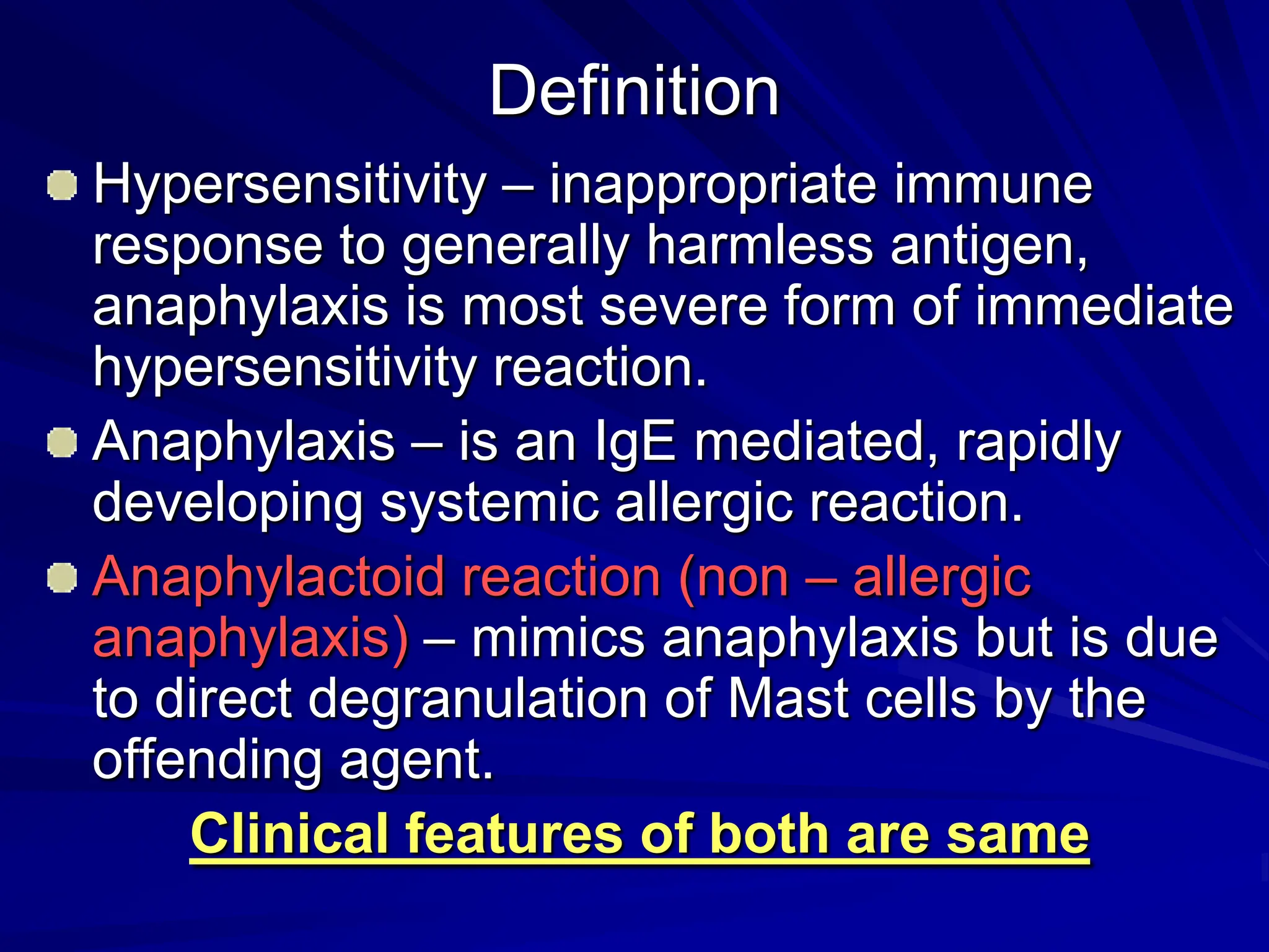
ANAPHYLAXIS
Definition
Hypersensitivity – inappropriate immune
response to generally harmless antigen,
anaphylaxis is most severe form of immediate
hypersensitivity reaction.
Anaphylaxis – is an IgE mediated, rapidly
developing systemic allergic reaction.
Anaphylactoid reaction (non – allergic
anaphylaxis) – mimics anaphylaxis but is due
to direct degranulation of Mast cells by the
offending agent.
Clinical features of both are same
Differences
Anaphylaxis Anaphylactoid
Exposure to Ag –
sensitization –
reexposure
No sensitization
IgE mediated Mast cell
degranulation
Non IgE mediated Mast
cell degranulation
Skin prick tests useful -
Serum tryptase level >
25ug/L
negative
Epidemiology
Risk factors
– Atopic individuals are at no greater risk
– Poorly controlled asthma – fatal outcome
– Previous anaphylaxis – recurrence risk 40 – 60% for
stings, 20 – 40 % for radio contrast & 10 – 20% for
penicillin.
Prevalence data limited – ranges from 5/1000 to
2/10000 ED visits. Less severe reactions never
reported.
Commonest causes
– Antibiotics
– Hymenoptera stings
– Food
Etiology – anaphylaxis/anaphylactoid reactions
1. Drugs
1. B lactam antibiotics - Penicillin and other antibiotics
2. ASA
3. Trimethoprim – sulphamethoxazole
4. Vancomycin
5. NSAID/ Opiates
6. IV anesthetics - suxamethonium, propofol
2. Food and additives
1. Nuts
2. Sea food - shellfish
3. Eggs/ milk
4. Soybeans
5. Seeds
6. Sulfites
3. Others
1. Hymenoptera stings
2. Ionic Radiocontrast
3. Physical – exercise/cold
4. Idiopathic – 30%
5. Latex
Pathophysiology
Basic event is Mast/basophil degranulation
due to
1. IgE cross linking
2. Complement activation
3. Direct activation
4. Modulation of arachidonic acid metabolism
5. Exercise or temperature dependent effects
6. Idiopathic
Classic anaphylaxis
Requires two separate exposures to Ag/ hapten
protein complex (penicillin)
First exposure Ag/ hapten protein complex – APC –
cell surface bound to MHC 2 complex – T helper
cell – IL 4 – Th 2 response- IgE synthesis.
IgE constant region binds to IgE receptors on Mast
cells & basophils, COVERS the cells with Ag
specific IgE.
After a latent period reexposure IgE bridging –
deregulation occurs- Antibiotics/foods/stings
Complement mediated
Blood products/mismatch – immune
complexes involving IgG/IgM antibodies
leading to complement pathway activation
and formation of anaphylatoxins C3a/ C5a
which directly lead to degranulation.
Non immunologic mechanisms
By
– Direct stimulation
– Unknown mechanisms
Agents which cause such reactions
– Radio contrast – high osmolarity ionic contrasts
– activates complement system
– Muscular depolarizing drugs - direct
– Opiates - direct
– Dextrans
Modulation of Cyclooxygenase pathway
Aspirin /NSAID cause anaphylaxis by non
Mast cell process.
Precise mechanism unknown. Alteration in
PG & LT synthesis (COX 1 )
5 – 10% of asthmatics – rarely full syndrome
Pathophysiology
Anaphylactic Reaction
antigen
Mast cell
IgE
Drug
Anaphylactoid reaction
Complement activation
Kinin system
Fibrinolytic system
Coagulation system
Histamine
SRSA
Leukotrines
Kinins
Prostaglandins
Target organs
Respiratory
CVS
Skin
Effects of mediators
Histamine has all the following effects
1. Decrease in systemic vascular
resistance
2. Leakage of fluid from capillaries and
post capillary venules - Kinins
3. Bronchoconstriction – PAF, SRS-A(LT
C4/D4/E4), prostaglandins
4. Cutaneous and mucosal inflammation
– Leukotrines, prostaglandins
Classification of severity
1. Mild (skin and subcutaneous tissue only)
erythema, urticaria, periorbital oedema or
angioedema
2. Moderate (features of Respiratory, CVS or
GI involvement) – dyspnea, stridor, wheeze,
nausea, vomiting, dizziness, diaphoresis, chest
tightness or abdominal pain.
3. Severe (hypoxia, hypotension, CNS
compromise) cyanosis, BP < 90 mm Hg,
confusion, unconsciousness
MILD FORMS CAN CHANGE TO SEVERE ANY
TIME.
Clinical features
Most serious reactions occur within minutes
of exposure
May be delayed for hours – 2.5 hours after
parenteral exposure.
Oral exposure manifestations unpredictable
Some have Biphasic reaction – recurrence
after 4 to 8 hrs ( 3- 20% patient) - SRSA
Some have protracted course – require
treatment for several hours
Classic presentation
Begins with pruritus, cutaneous flushing, urticaria
Sense of fullness in throat, anxiety, chest
tightness, SOB & lightheadedness
Level of consciousness, resp distress circulatory
collapse
Loss of consciousness & cardio respiratory arrest
‘LUMP in throat & hoarseness heralds life
threatening laryngeal oedema’.
Clinical features
1. Skin/mucosa
1. Pruritus
2. Urticaria
3. Angioedema
2. Respiratory distress (laryngeal oedema,
laryngospasm, bronchospasm)
3. CVS – hypotension
4. Abdomen
1. Abdominal cramping
2. Diarrhea
5. Feeling of impending doom before
CVS/respiratory symptoms
Diagnosis
History & examination
If onset delayed & if related to food diagnosis may
not be easy
D/D
– Vasovagal - MI
– Arrhythmia - asthma
– Seizure - H.angiooedema
– Epiglottitis - F body
Lab
– Histamine levels elevated for 5- 30 min post reaction
– Tryptase – neutral protease found only in mast cell
granules. Elevated for several hours.
Treatment
The most common cause of death
is airway obstruction followed by
shock
May vary from mild to severe but if
left untreated all reactions have the
potential to become severe rapidly
Previous mild reaction does not
predict the severity on reexposure
Primary Treatment
1. Discontinue suspected allergen – prevents
further mast cell recruitment
2. Maintain airway and give 100% oxygen
3. Discontinue all sedatives/vasodepressors
4. Epinephrine
1. 0.1 mg IV bolus (1 ml of 1: 10,000) repeat 1 to 5
minutes. (3 ml via central line/ 15 minutes)
2. 0.3 to 0.5 mg (1:1,000) IM every 15 minutes
3. 3- 5 ml 1;10,000 in 10 ml endotracheally
4. Repeated doses may be required
5. Volume expansion – 0.5 to 1 litre bolus and
than titrate with BP and urinary output
Secondary Treatment
1. Antihistaminics
1. Relieve skin symptoms. No effect on
cardiopulmonary responses once Histamine has
been released.
2. Both H1 and H2 blockers
3. Diphenhydramine 1mg/kg, Ranitidine
2. Inhaled Salbutamol 2.5 to 5 mg
3. Steroids
1. Severe reactions. No immediate effect
2. May prevent relapse of severe reactions
3. Methyl Prednisolone 125 mg/hydrocortisone 250 –
500 mg
Prevention
Skin testing – for IgE mediated reactions
– Prick puncture/intradermal small dose
– Negative Control on other hand
– 20 minutes
– Wheal 3 mm larger than control
– No case of penicillin induced anaphylaxis has
been reported if skin test was negative
History of allergic reactions
I Card
Drug reactions
Penicillins
1. Reactive bicyclic lactam ring covalently
binds to tissues – major determinant –
responsible for anaphylaxis
2. If skin test is negative a delayed non IgE
mediated reaction may still occur
3. 75% patient who report penicillin allergy
have negative skin test and are not at risk
for anaphylaxis
4. 4% have positive skin test and are at risk
for anaphylaxis
Cephalosporins
1. Cross reactivity to penicillin – 4
fold risk of hypersensitivity
reactions if patient is allergic to
penicillin
2. Most common with first
generation cephalosporins
3. Can use third and fourth
generation with occasional mild
reactions
Thank you

ANAPHYLAXIS: DRUG REACTIONS, SHOCK, HYPERSENSITIVITY.pdf

  • 1.
  • 2.
    Definition Hypersensitivity – inappropriateimmune response to generally harmless antigen, anaphylaxis is most severe form of immediate hypersensitivity reaction. Anaphylaxis – is an IgE mediated, rapidly developing systemic allergic reaction. Anaphylactoid reaction (non – allergic anaphylaxis) – mimics anaphylaxis but is due to direct degranulation of Mast cells by the offending agent. Clinical features of both are same
  • 3.
    Differences Anaphylaxis Anaphylactoid Exposure toAg – sensitization – reexposure No sensitization IgE mediated Mast cell degranulation Non IgE mediated Mast cell degranulation Skin prick tests useful - Serum tryptase level > 25ug/L negative
  • 4.
    Epidemiology Risk factors – Atopicindividuals are at no greater risk – Poorly controlled asthma – fatal outcome – Previous anaphylaxis – recurrence risk 40 – 60% for stings, 20 – 40 % for radio contrast & 10 – 20% for penicillin. Prevalence data limited – ranges from 5/1000 to 2/10000 ED visits. Less severe reactions never reported. Commonest causes – Antibiotics – Hymenoptera stings – Food
  • 5.
    Etiology – anaphylaxis/anaphylactoidreactions 1. Drugs 1. B lactam antibiotics - Penicillin and other antibiotics 2. ASA 3. Trimethoprim – sulphamethoxazole 4. Vancomycin 5. NSAID/ Opiates 6. IV anesthetics - suxamethonium, propofol 2. Food and additives 1. Nuts 2. Sea food - shellfish 3. Eggs/ milk 4. Soybeans 5. Seeds 6. Sulfites 3. Others 1. Hymenoptera stings 2. Ionic Radiocontrast 3. Physical – exercise/cold 4. Idiopathic – 30% 5. Latex
  • 6.
    Pathophysiology Basic event isMast/basophil degranulation due to 1. IgE cross linking 2. Complement activation 3. Direct activation 4. Modulation of arachidonic acid metabolism 5. Exercise or temperature dependent effects 6. Idiopathic
  • 7.
    Classic anaphylaxis Requires twoseparate exposures to Ag/ hapten protein complex (penicillin) First exposure Ag/ hapten protein complex – APC – cell surface bound to MHC 2 complex – T helper cell – IL 4 – Th 2 response- IgE synthesis. IgE constant region binds to IgE receptors on Mast cells & basophils, COVERS the cells with Ag specific IgE. After a latent period reexposure IgE bridging – deregulation occurs- Antibiotics/foods/stings
  • 8.
    Complement mediated Blood products/mismatch– immune complexes involving IgG/IgM antibodies leading to complement pathway activation and formation of anaphylatoxins C3a/ C5a which directly lead to degranulation.
  • 9.
    Non immunologic mechanisms By –Direct stimulation – Unknown mechanisms Agents which cause such reactions – Radio contrast – high osmolarity ionic contrasts – activates complement system – Muscular depolarizing drugs - direct – Opiates - direct – Dextrans
  • 10.
    Modulation of Cyclooxygenasepathway Aspirin /NSAID cause anaphylaxis by non Mast cell process. Precise mechanism unknown. Alteration in PG & LT synthesis (COX 1 ) 5 – 10% of asthmatics – rarely full syndrome
  • 11.
    Pathophysiology Anaphylactic Reaction antigen Mast cell IgE Drug Anaphylactoidreaction Complement activation Kinin system Fibrinolytic system Coagulation system Histamine SRSA Leukotrines Kinins Prostaglandins Target organs Respiratory CVS Skin
  • 12.
    Effects of mediators Histaminehas all the following effects 1. Decrease in systemic vascular resistance 2. Leakage of fluid from capillaries and post capillary venules - Kinins 3. Bronchoconstriction – PAF, SRS-A(LT C4/D4/E4), prostaglandins 4. Cutaneous and mucosal inflammation – Leukotrines, prostaglandins
  • 13.
    Classification of severity 1.Mild (skin and subcutaneous tissue only) erythema, urticaria, periorbital oedema or angioedema 2. Moderate (features of Respiratory, CVS or GI involvement) – dyspnea, stridor, wheeze, nausea, vomiting, dizziness, diaphoresis, chest tightness or abdominal pain. 3. Severe (hypoxia, hypotension, CNS compromise) cyanosis, BP < 90 mm Hg, confusion, unconsciousness MILD FORMS CAN CHANGE TO SEVERE ANY TIME.
  • 14.
    Clinical features Most seriousreactions occur within minutes of exposure May be delayed for hours – 2.5 hours after parenteral exposure. Oral exposure manifestations unpredictable Some have Biphasic reaction – recurrence after 4 to 8 hrs ( 3- 20% patient) - SRSA Some have protracted course – require treatment for several hours
  • 15.
    Classic presentation Begins withpruritus, cutaneous flushing, urticaria Sense of fullness in throat, anxiety, chest tightness, SOB & lightheadedness Level of consciousness, resp distress circulatory collapse Loss of consciousness & cardio respiratory arrest ‘LUMP in throat & hoarseness heralds life threatening laryngeal oedema’.
  • 16.
    Clinical features 1. Skin/mucosa 1.Pruritus 2. Urticaria 3. Angioedema 2. Respiratory distress (laryngeal oedema, laryngospasm, bronchospasm) 3. CVS – hypotension 4. Abdomen 1. Abdominal cramping 2. Diarrhea 5. Feeling of impending doom before CVS/respiratory symptoms
  • 17.
    Diagnosis History & examination Ifonset delayed & if related to food diagnosis may not be easy D/D – Vasovagal - MI – Arrhythmia - asthma – Seizure - H.angiooedema – Epiglottitis - F body Lab – Histamine levels elevated for 5- 30 min post reaction – Tryptase – neutral protease found only in mast cell granules. Elevated for several hours.
  • 18.
    Treatment The most commoncause of death is airway obstruction followed by shock May vary from mild to severe but if left untreated all reactions have the potential to become severe rapidly Previous mild reaction does not predict the severity on reexposure
  • 19.
    Primary Treatment 1. Discontinuesuspected allergen – prevents further mast cell recruitment 2. Maintain airway and give 100% oxygen 3. Discontinue all sedatives/vasodepressors 4. Epinephrine 1. 0.1 mg IV bolus (1 ml of 1: 10,000) repeat 1 to 5 minutes. (3 ml via central line/ 15 minutes) 2. 0.3 to 0.5 mg (1:1,000) IM every 15 minutes 3. 3- 5 ml 1;10,000 in 10 ml endotracheally 4. Repeated doses may be required 5. Volume expansion – 0.5 to 1 litre bolus and than titrate with BP and urinary output
  • 20.
    Secondary Treatment 1. Antihistaminics 1.Relieve skin symptoms. No effect on cardiopulmonary responses once Histamine has been released. 2. Both H1 and H2 blockers 3. Diphenhydramine 1mg/kg, Ranitidine 2. Inhaled Salbutamol 2.5 to 5 mg 3. Steroids 1. Severe reactions. No immediate effect 2. May prevent relapse of severe reactions 3. Methyl Prednisolone 125 mg/hydrocortisone 250 – 500 mg
  • 21.
    Prevention Skin testing –for IgE mediated reactions – Prick puncture/intradermal small dose – Negative Control on other hand – 20 minutes – Wheal 3 mm larger than control – No case of penicillin induced anaphylaxis has been reported if skin test was negative History of allergic reactions I Card
  • 22.
    Drug reactions Penicillins 1. Reactivebicyclic lactam ring covalently binds to tissues – major determinant – responsible for anaphylaxis 2. If skin test is negative a delayed non IgE mediated reaction may still occur 3. 75% patient who report penicillin allergy have negative skin test and are not at risk for anaphylaxis 4. 4% have positive skin test and are at risk for anaphylaxis
  • 23.
    Cephalosporins 1. Cross reactivityto penicillin – 4 fold risk of hypersensitivity reactions if patient is allergic to penicillin 2. Most common with first generation cephalosporins 3. Can use third and fourth generation with occasional mild reactions
  • 24.