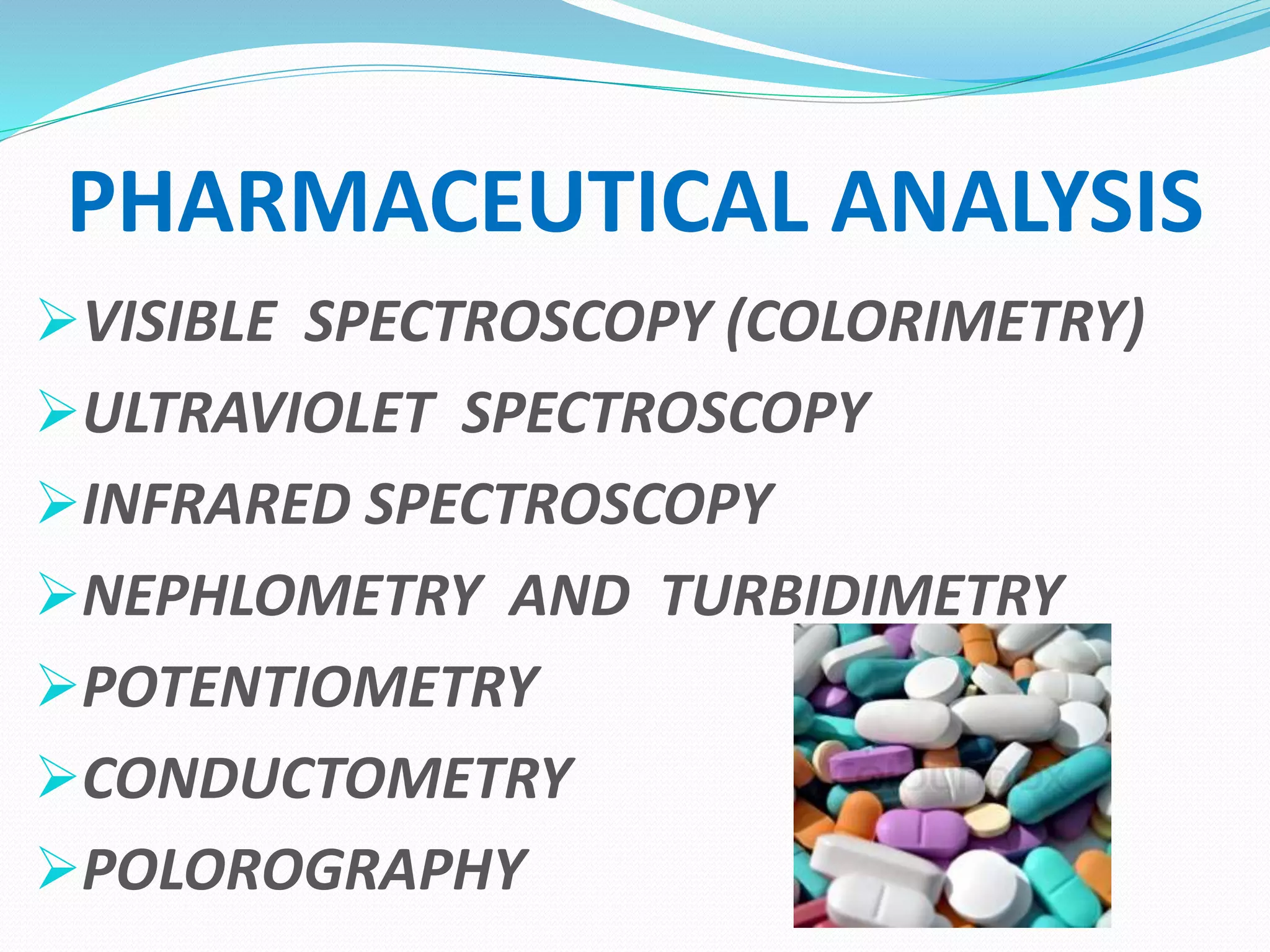
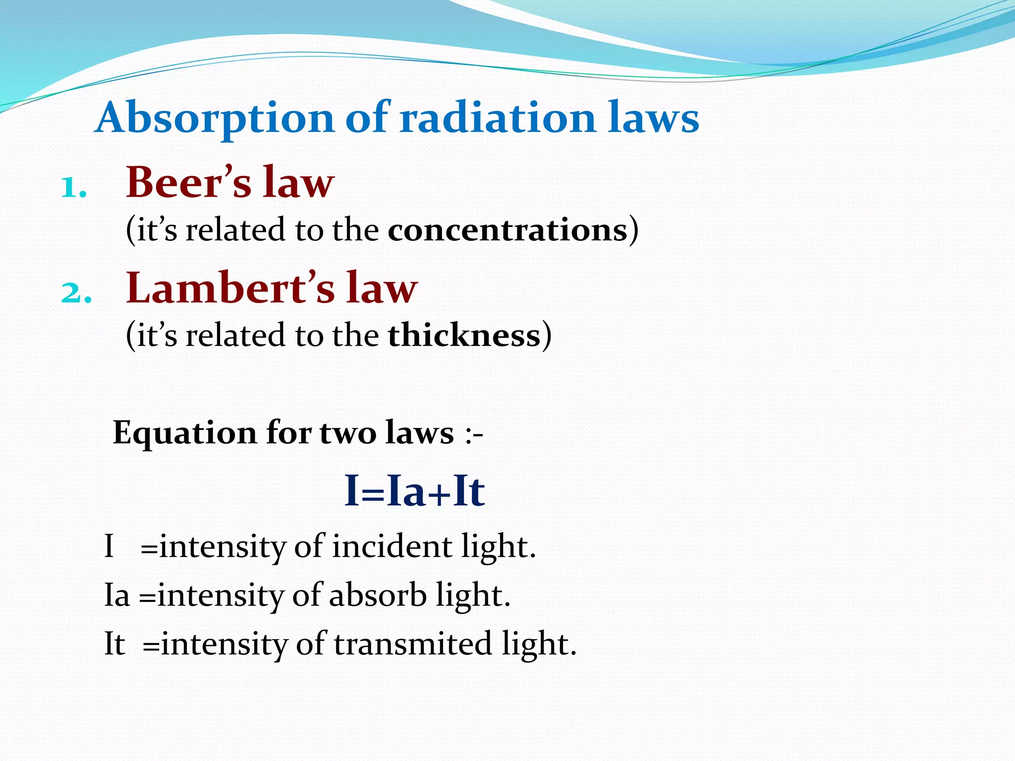
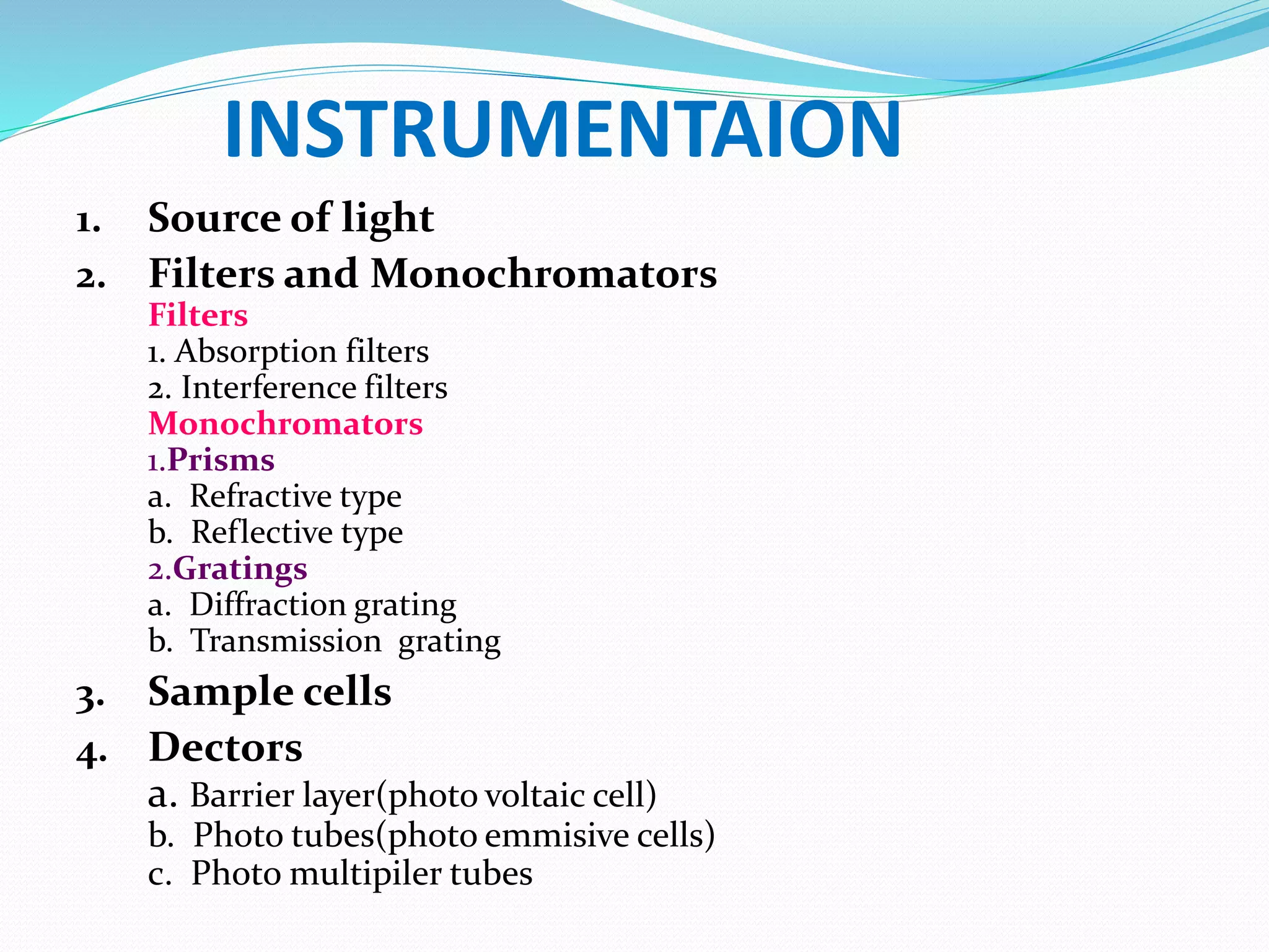
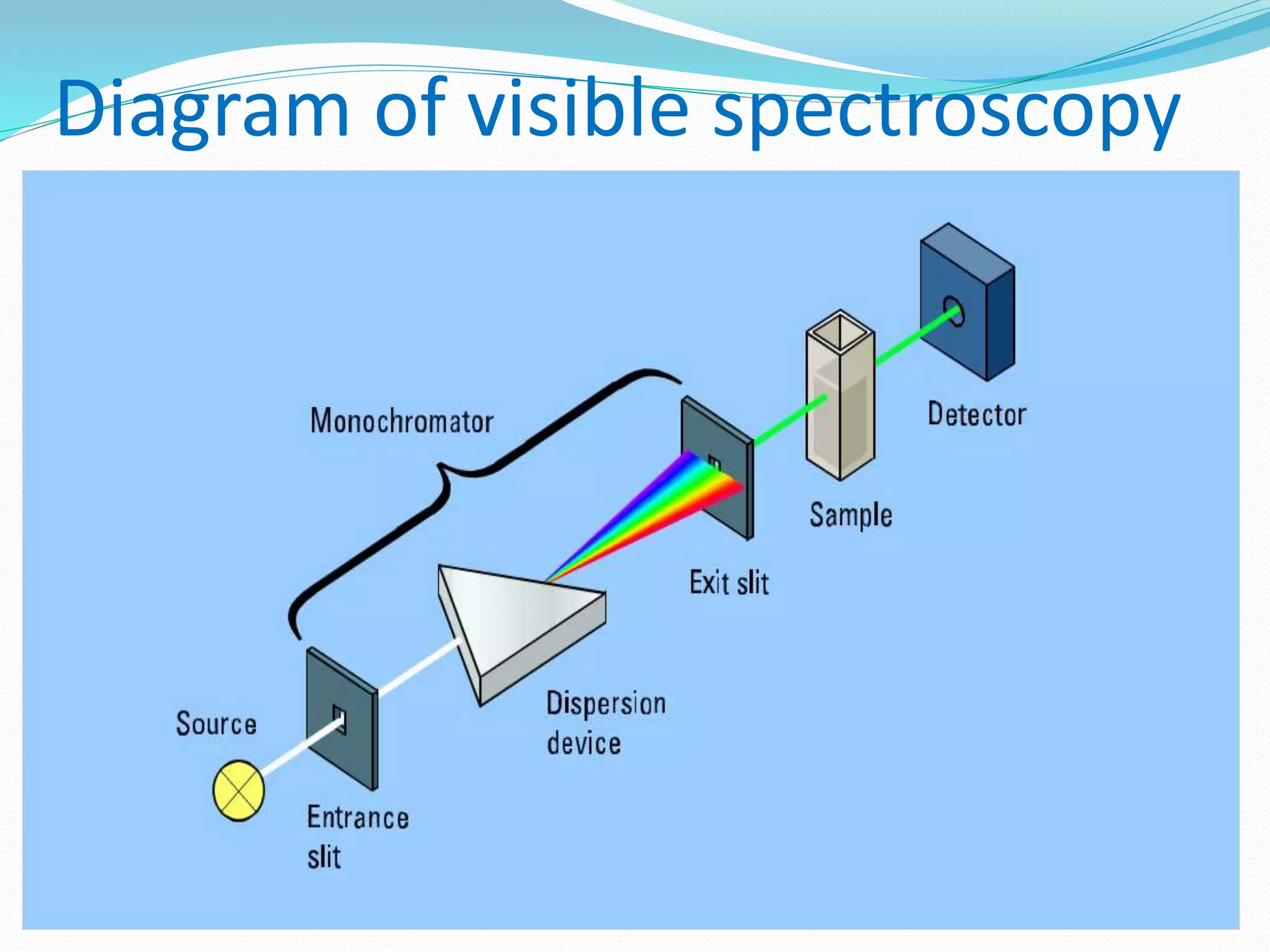
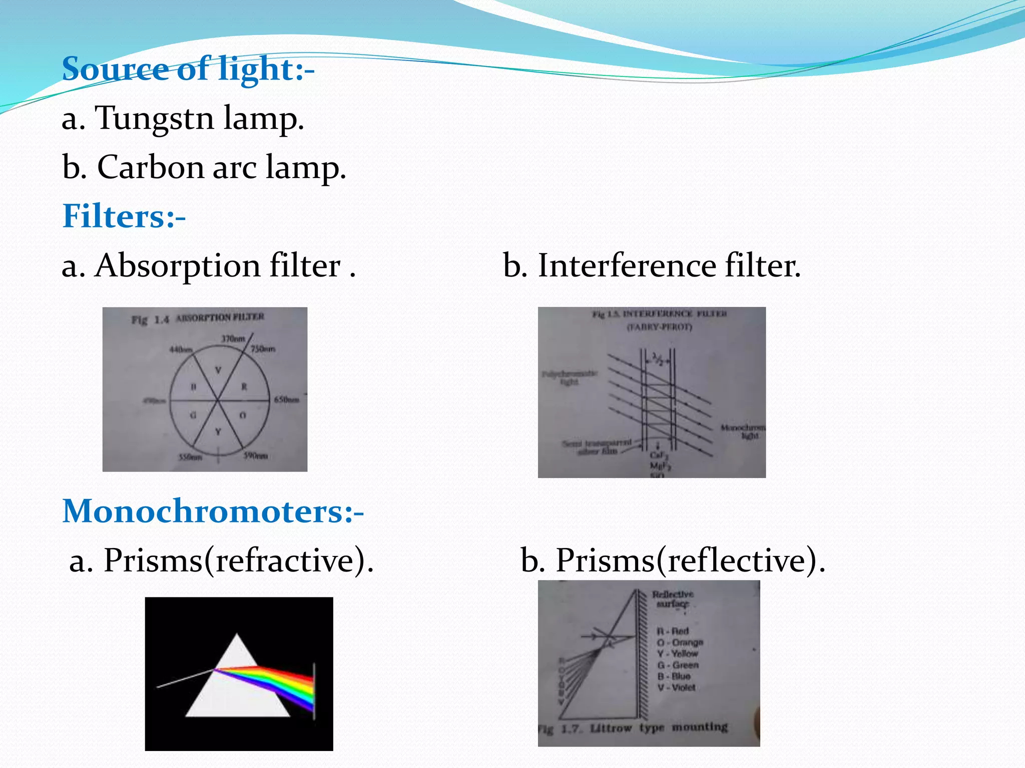
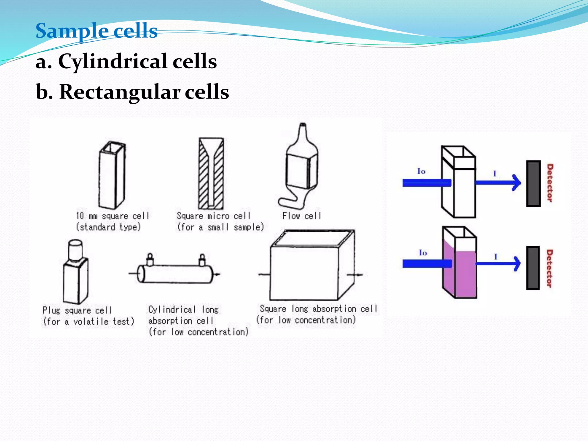
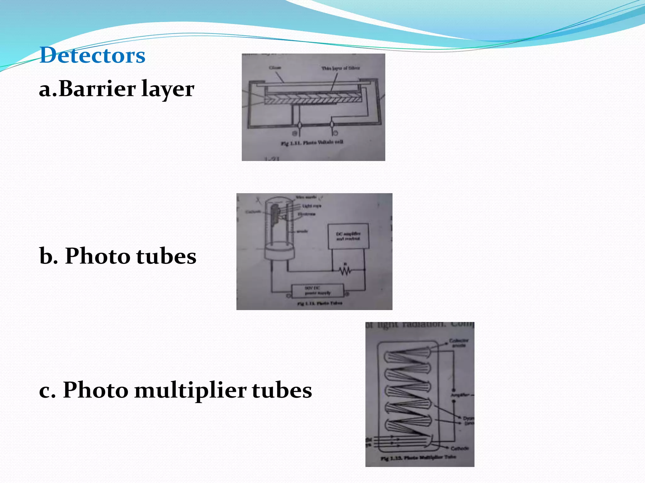
PHARMACEUTICAL ANALYSIS
VISIBLE SPECTROSCOPY (COLORIMETRY)
ULTRAVIOLET SPECTROSCOPY
INFRARED SPECTROSCOPY
NEPHLOMETRY AND TURBIDIMETRY
POTENTIOMETRY
CONDUCTOMETRY
POLOROGRAPHY
VISIBLE SPECTROSCOPY
 It is also called as colorimetry.
 It explain the absorption of visible radiation.
 The wavelenth range is 400-800nm.
 It is used to determinig concentration of drug present in
the given sample.
 In this plot a graph Absorbance Vs Wavelenght.
 At which maximum absorption of radiation takes place is
called as λmax.
Absorption of radiation laws
1. Beer’s law
(it’s related to the concentrations)
2. Lambert’s law
(it’s related to the thickness)
Equation for two laws :-
I=Ia+It
I =intensity of incident light.
Ia =intensity of absorb light.
It =intensity of transmited light.
INSTRUMENTAION
1. Source of light
2. Filters and Monochromators
Filters
1. Absorption filters
2. Interference filters
Monochromators
1.Prisms
a. Refractive type
b. Reflective type
2.Gratings
a. Diffraction grating
b. Transmission grating
3. Sample cells
4. Dectors
a. Barrier layer(photo voltaic cell)
b. Photo tubes(photo emmisive cells)
c. Photo multipiler tubes
Diagram of visible spectroscopy
Source of light:-
a. Tungstn lamp.
b. Carbon arc lamp.
Filters:-
a. Absorption filter . b. Interference filter.
Monochromoters:-
a. Prisms(refractive). b. Prisms(reflective).
Gratings:-
a. Diffraction gratings.
b. Transmission gratings.
Sample cells
a. Cylindrical cells
b. Rectangular cells
Detectors
a.Barrier layer
b. Photo tubes
c. Photo multiplier tubes
Applications:-
1. Quality control of purity.
2. Quantitative analysis.
3. Determination of ligands.
4. Structure elucidation organic compounds.
5. Determination of pKa value of indicators.
6. Determination of molecular weight of amines.
7. Determination of functional groups.
8. Determination of organic substances.
9. Spectrophtometric titrations.
Definations
Chromophore:-
It is a part of molecule responsible for characteristic absorption at a
wavelength.
Ex: =c=c=.
Chromophore are covalently unsaturated.
Auxochrome:-
They do not have any characteristic absorption but they can modify the
absorption of chromophore.
Ex: -OH,-NH2.
Isobestic point:-
The wavelength where the molar absoptivity is a same for two substances.
Bathochromic shift:- Shift in λmax towards longer wavelength.
It is also called as red shift.
Hypsochromic shift:- Shift in λmax towards shorter wavelength.
It also called as blue shift.
Hyperchromic effect:- Increase intensity of absorption.
Hypochromic effect:- Decrease intensity of absorption.
Chromogenic agent:-this are responsible for of colour.
Ex: ferric chloride.
PRESENTATIONBY:
V. MAHESH
Analysis intrumentation

Analysis intrumentation

  • 2.
    PHARMACEUTICAL ANALYSIS VISIBLE SPECTROSCOPY(COLORIMETRY) ULTRAVIOLET SPECTROSCOPY INFRARED SPECTROSCOPY NEPHLOMETRY AND TURBIDIMETRY POTENTIOMETRY CONDUCTOMETRY POLOROGRAPHY
  • 3.
    VISIBLE SPECTROSCOPY  Itis also called as colorimetry.  It explain the absorption of visible radiation.  The wavelenth range is 400-800nm.  It is used to determinig concentration of drug present in the given sample.  In this plot a graph Absorbance Vs Wavelenght.  At which maximum absorption of radiation takes place is called as λmax.
  • 4.
    Absorption of radiationlaws 1. Beer’s law (it’s related to the concentrations) 2. Lambert’s law (it’s related to the thickness) Equation for two laws :- I=Ia+It I =intensity of incident light. Ia =intensity of absorb light. It =intensity of transmited light.
  • 5.
    INSTRUMENTAION 1. Source oflight 2. Filters and Monochromators Filters 1. Absorption filters 2. Interference filters Monochromators 1.Prisms a. Refractive type b. Reflective type 2.Gratings a. Diffraction grating b. Transmission grating 3. Sample cells 4. Dectors a. Barrier layer(photo voltaic cell) b. Photo tubes(photo emmisive cells) c. Photo multipiler tubes
  • 6.
    Diagram of visiblespectroscopy
  • 7.
    Source of light:- a.Tungstn lamp. b. Carbon arc lamp. Filters:- a. Absorption filter . b. Interference filter. Monochromoters:- a. Prisms(refractive). b. Prisms(reflective).
  • 8.
  • 9.
    Sample cells a. Cylindricalcells b. Rectangular cells
  • 10.
    Detectors a.Barrier layer b. Phototubes c. Photo multiplier tubes
  • 11.
    Applications:- 1. Quality controlof purity. 2. Quantitative analysis. 3. Determination of ligands. 4. Structure elucidation organic compounds. 5. Determination of pKa value of indicators. 6. Determination of molecular weight of amines. 7. Determination of functional groups. 8. Determination of organic substances. 9. Spectrophtometric titrations.
  • 12.
    Definations Chromophore:- It is apart of molecule responsible for characteristic absorption at a wavelength. Ex: =c=c=. Chromophore are covalently unsaturated. Auxochrome:- They do not have any characteristic absorption but they can modify the absorption of chromophore. Ex: -OH,-NH2. Isobestic point:- The wavelength where the molar absoptivity is a same for two substances. Bathochromic shift:- Shift in λmax towards longer wavelength. It is also called as red shift. Hypsochromic shift:- Shift in λmax towards shorter wavelength. It also called as blue shift. Hyperchromic effect:- Increase intensity of absorption. Hypochromic effect:- Decrease intensity of absorption. Chromogenic agent:-this are responsible for of colour. Ex: ferric chloride.
  • 13.