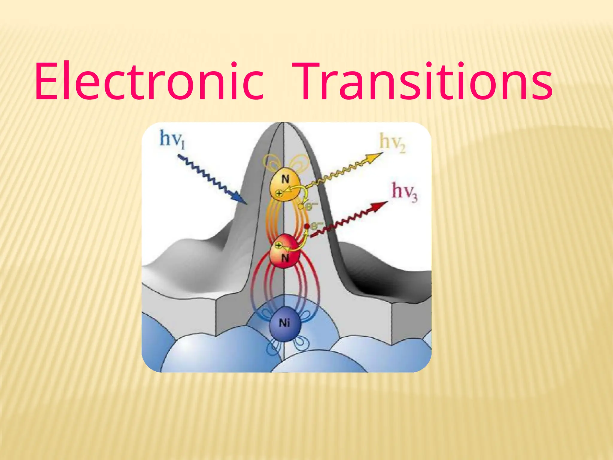
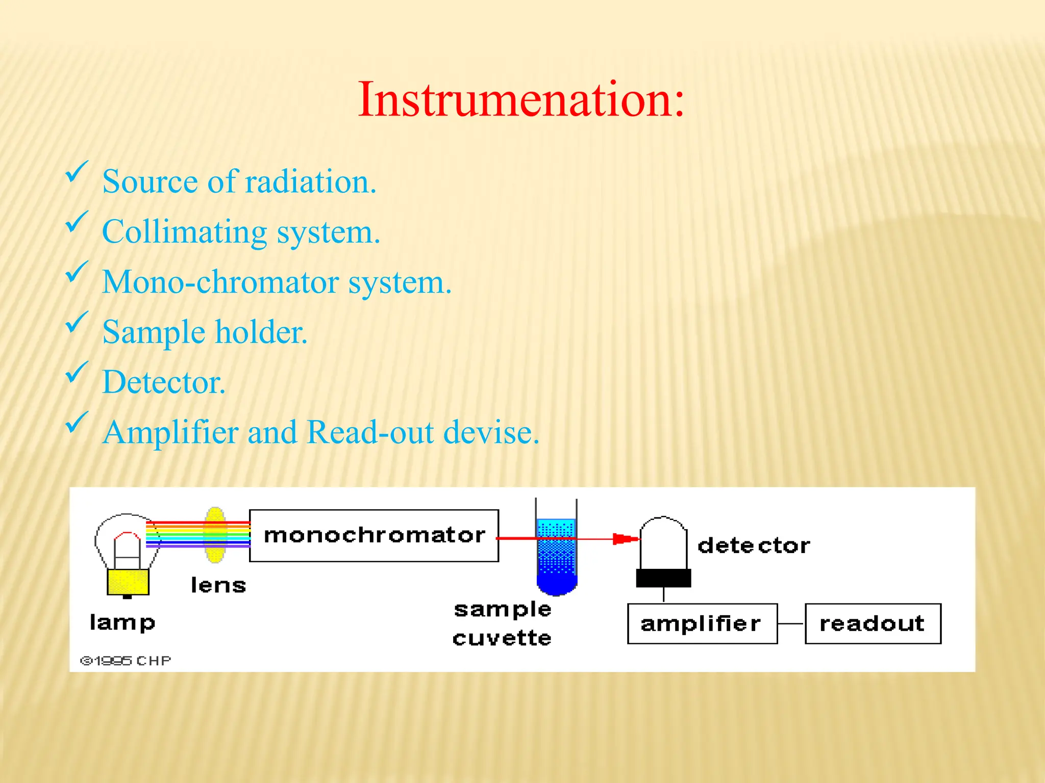
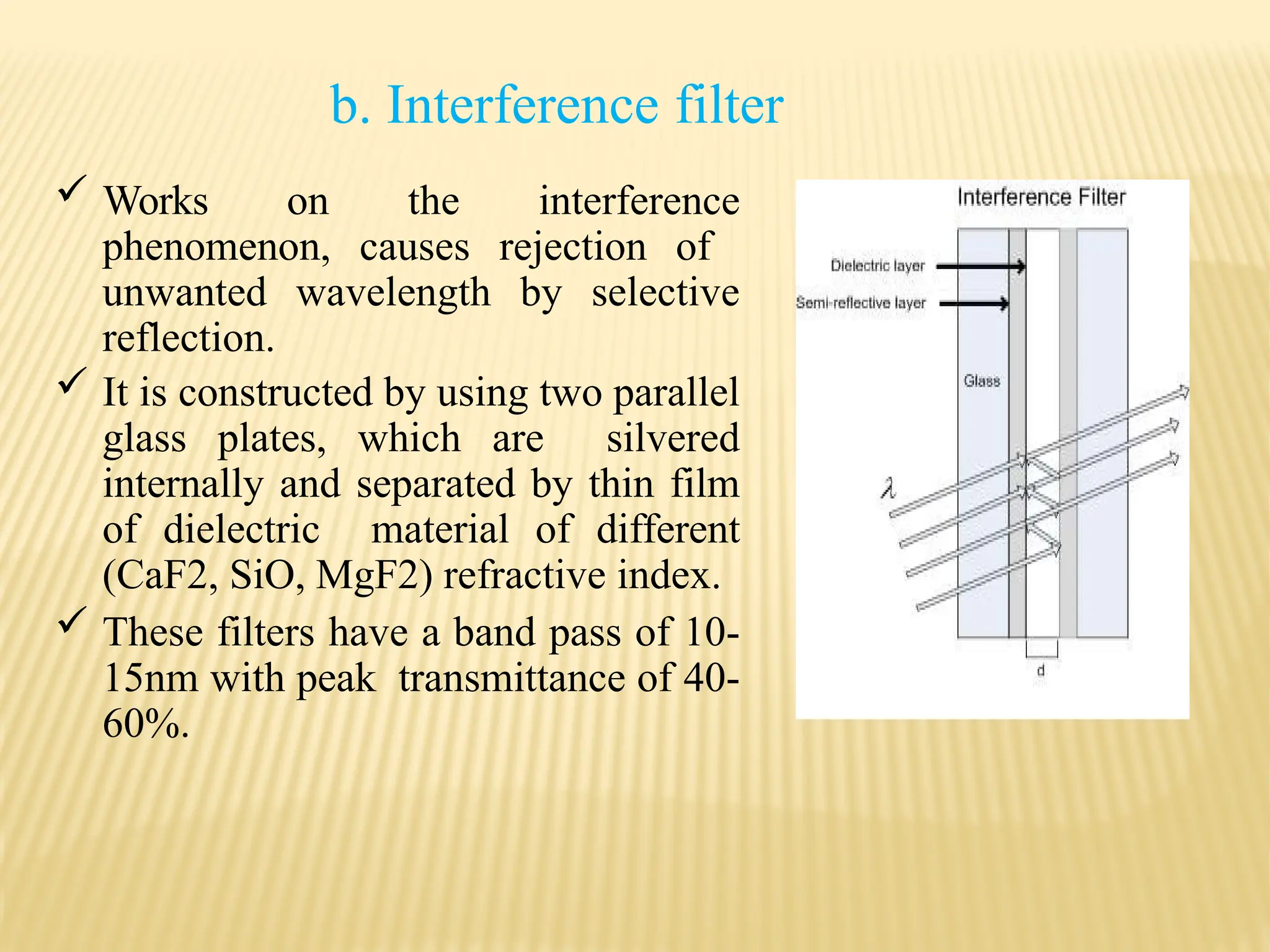
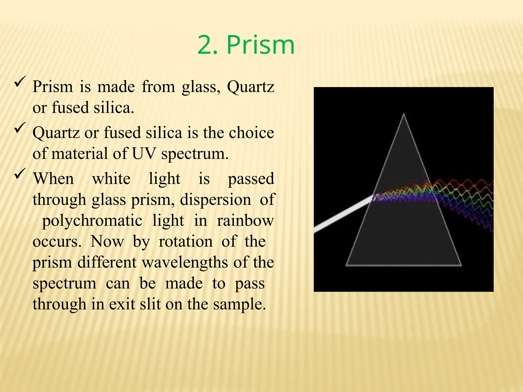
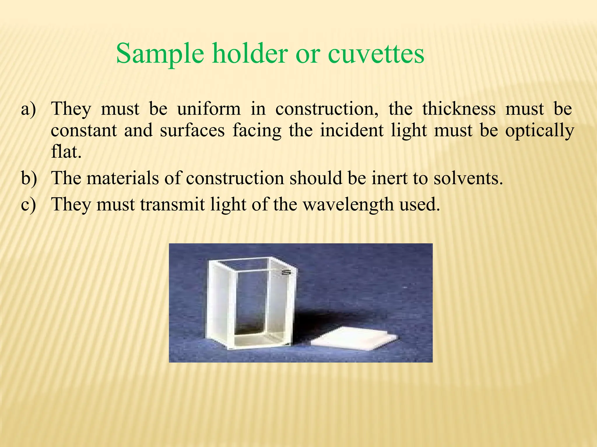
UV VISIBLE
SPECTROSCOPY……
by:- Sarika Singh.
Fy M-Pharmacy(QAT)
Spectroscopy
It is the branch of science that deals with the study of
interaction of matter with light.
OR
It is the branch of science that deals with the study of
interaction of electromagnetic radiation with matter.
Theory of UV
Visible
Spectroscopy…
Why we use UV spectroscopy ?
1. Detection of functional groups.
2. Detection of impurities.
3. Qualitative analysis.
4. Quantitative analysis.
5. Single compound without chromophore.
6. Drugs with chromophoric reagent.
Electromagnetic Radiation
 Electromagnetic radiation consist of discrete packages
 of energy which are called as photons.
 Frequency (ν):
◦ It is defined as the number of times electrical
field radiation oscillates in one second.
◦ The unit for frequency is Hertz (Hz).
1 Hz = 1 cycle per second
 Wavelength (λ):
◦ It is the distance between two nearest parts of the wave in the
same phase i.e. distance between two nearest crest or
troughs.
The relationship between wavelength & frequency can be written as:
c = ν λ
As photon is subjected to energy, so
E = h ν = h c / λ
ABSORBANCE LAWS
BEER’S LAW
“ The intensity of a beam of monochromatic light
decrease exponentially with the increase in
concentration of the absorbing substance” .
Arithmetically;
- dI/ dc ᾱ I
I= Io. eˉkc -------
eq (1)
LAMBERT’S LAW
“When a beam of light is allowed to pass through a
transparent medium, the rate of decrease of intensity
with the thickness of medium is directly
proportional to the intensity of the light”
mathematically;
-dI/ dt ᾱ I
-In . I = kt+b -------------- eq(2)
the combination of eq 1 & 2 we will get
A= Kct
A= ℇct
(K=ℇ)
Principle of UV Visible Spectroscopy
 The UV radiation region extends from 10 nm to 400 nm
and the visible radiation region extends from 400 nm to
800 nm.
Near UV Region: 200 nm to 400 nm
Far UV Region: below 200 nm
 Far UV spectroscopy is studied under vacuum
condition.
 The common solvent used for preparing sample to be
analyzed is either ethyl alcohol or hexane.
 Ultraviolet absorption spectra arise from transition of
electron with in a molecule from a lower level to a
higher level.
 A molecule absorb ultraviolet radiation of frequency
(𝜗), the electron in that molecule undergo transition
from lower to higher energy level.
The energy can be calculated by the equation,
E=h𝜗 erg
E₁-E =
ₒ h𝜗
Etotal = Eelectronic +
Evibrational +
Erotational
The energies decreases in the following
order: Electronic ⪢ Vibrational ⪢
Rotational
Electronic Transitions
The possible electronic transitions are
1- σ → σ* transition
2- π → π* transition
3- n → σ* transition
4- n → π* transition
Absorption & Intensity
Shifts
1- Bathochromic Shift (Red Shift)
2- Hypsochromic Shift (Blue Shift)
3- Hyperchromic Effect
4- Hypochromic Effect
Shifts and Effects
Hyperchromic shift
Absorbance
(
A
)
Blue
shift
Red
shift
Hypochromic shift
λmax Wavelength ( λ )
Instrumenation:
 Source of radiation.
 Collimating system.
 Mono-chromator system.
 Sample holder.
 Detector.
 Amplifier and Read-out devise.
Source of radiation
 Requirements of an ideal source
 It should be stable and should not allow fluctuations.
 It should emit light of continuous spectrum of high and uniform intensity
over the entire wavelength region in which it’s used.
 It should provide incident light of sufficient intensity for the transmitted
energy to be detected at the end of optic path.
 It should not show fatigue on continued use.
1.Tungsten Halogen Lamp
2. Hydrogen Discharge Lamp:
3. Xenon Discharge Lamp:
4. Mercury arc Lamp:
Momochromators
 It is a device used to isolate the radiation of the desired wavelength
from wavelength of the continuous spectra.
 The essential elements of monochromators are:
i. An entrance slit
ii. Dispersing element
iii. Exit slit
The entrance slit sharply define the incoming beam of heterochromatic
radiation. The dispersing element disperses the heterochromatic radiation
into its component wavelength. Exit slit allows the nominal wavelength
together with a bond of wavelength on either side of it.
Following types of monochromatic devices are
used.
1. Filters
2. Prisms
3. Gratings
1.
Filters
Two types of filters are used, they are:
a.Absorption filters- works by selective the
radiation which absorption of is unwanted
radiation andtransmits required.
Examples- Glass and Gelatin filters
b. Interference filter
 Works on the interference
phenomenon, causes rejection of
unwanted wavelength by selective
reflection.
 It is constructed by using two parallel
glass plates, which are silvered
internally and separated by thin film
of dielectric material of different
(CaF2, SiO, MgF2) refractive index.
 These filters have a band pass of 10-
15nm with peak transmittance of 40-
60%.
2. Prism
 Prism is made from glass, Quartz
or fused silica.
 Quartz or fused silica is the choice
of material of UV spectrum.
 When white light is passed
through glass prism, dispersion of
polychromatic light in rainbow
occurs. Now by rotation of the
prism different wavelengths of the
spectrum can be made to pass
through in exit slit on the sample.
3.
Gratings
o They are most efficient in converting a polychromatic light to
monochromatic light. As a resolution of +/- 0.1nm could be
achieved by using gratings. As the gratings are expensive, they are
commonly used in spectrophotometers.
o Gratings are of two types.
1. Diffraction grating.
2. Transmission gratings.
Sample holder or cuvettes
a) They must be uniform in construction, the thickness must be
constant and surfaces facing the incident light must be optically
flat.
b) The materials of construction should be inert to solvents.
c) They must transmit light of the wavelength used.
Detectors
 Device which converts light energy into electrical signals, that
are displayed on readout devices.
 The transmitted radiation falls on the detector which determines
the
intensity of radiation absorbed by sample.
 The following types of detectors are employed in instrumentation
of absorption spectrophotometer
1. Barrier layer cell/Photovoltaic cell
2. Phototubes/ Photo emissive tube
3. Photomultiplier tube
Requirements of an ideal detector:-
a. It should give quantitative response.
b. It should have high sensitivity and low noise level.
c. It should have a short response time.
d. It should provide signal or response quantitative to wide
spectrum of radiation received.
Single beam UV-Spectrophotometer
Double Beam UV-Spectrophotometer
Applications of
UV / Visible
Spectroscopy
1. Detection of Impurities.
2. Structure elucidation of organic
compounds.
3. Quantitative analysis
4. Qualitative analysis
5. Chemical kinetics
6. Detection of Groups
7. Quantitative analysis of pharmaceutical
substances
8. Molecular weight determination
9. As HPLC Detector
405737361-Ppt-3.pptx uv visible spectroscopy

405737361-Ppt-3.pptx uv visible spectroscopy

  • 1.
  • 2.
    Spectroscopy It is thebranch of science that deals with the study of interaction of matter with light. OR It is the branch of science that deals with the study of interaction of electromagnetic radiation with matter.
  • 3.
  • 4.
    Why we useUV spectroscopy ? 1. Detection of functional groups. 2. Detection of impurities. 3. Qualitative analysis. 4. Quantitative analysis. 5. Single compound without chromophore. 6. Drugs with chromophoric reagent.
  • 5.
    Electromagnetic Radiation  Electromagneticradiation consist of discrete packages  of energy which are called as photons.  Frequency (ν): ◦ It is defined as the number of times electrical field radiation oscillates in one second. ◦ The unit for frequency is Hertz (Hz). 1 Hz = 1 cycle per second  Wavelength (λ): ◦ It is the distance between two nearest parts of the wave in the same phase i.e. distance between two nearest crest or troughs.
  • 6.
    The relationship betweenwavelength & frequency can be written as: c = ν λ As photon is subjected to energy, so E = h ν = h c / λ
  • 7.
    ABSORBANCE LAWS BEER’S LAW “The intensity of a beam of monochromatic light decrease exponentially with the increase in concentration of the absorbing substance” . Arithmetically; - dI/ dc ᾱ I I= Io. eˉkc ------- eq (1)
  • 8.
    LAMBERT’S LAW “When abeam of light is allowed to pass through a transparent medium, the rate of decrease of intensity with the thickness of medium is directly proportional to the intensity of the light” mathematically; -dI/ dt ᾱ I -In . I = kt+b -------------- eq(2) the combination of eq 1 & 2 we will get A= Kct A= ℇct (K=ℇ)
  • 9.
    Principle of UVVisible Spectroscopy  The UV radiation region extends from 10 nm to 400 nm and the visible radiation region extends from 400 nm to 800 nm. Near UV Region: 200 nm to 400 nm Far UV Region: below 200 nm  Far UV spectroscopy is studied under vacuum condition.  The common solvent used for preparing sample to be analyzed is either ethyl alcohol or hexane.
  • 10.
     Ultraviolet absorptionspectra arise from transition of electron with in a molecule from a lower level to a higher level.  A molecule absorb ultraviolet radiation of frequency (𝜗), the electron in that molecule undergo transition from lower to higher energy level. The energy can be calculated by the equation, E=h𝜗 erg
  • 11.
    E₁-E = ₒ h𝜗 Etotal= Eelectronic + Evibrational + Erotational The energies decreases in the following order: Electronic ⪢ Vibrational ⪢ Rotational
  • 12.
  • 13.
    The possible electronictransitions are 1- σ → σ* transition 2- π → π* transition 3- n → σ* transition 4- n → π* transition
  • 14.
    Absorption & Intensity Shifts 1-Bathochromic Shift (Red Shift) 2- Hypsochromic Shift (Blue Shift) 3- Hyperchromic Effect 4- Hypochromic Effect
  • 15.
    Shifts and Effects Hyperchromicshift Absorbance ( A ) Blue shift Red shift Hypochromic shift λmax Wavelength ( λ )
  • 16.
    Instrumenation:  Source ofradiation.  Collimating system.  Mono-chromator system.  Sample holder.  Detector.  Amplifier and Read-out devise.
  • 17.
    Source of radiation Requirements of an ideal source  It should be stable and should not allow fluctuations.  It should emit light of continuous spectrum of high and uniform intensity over the entire wavelength region in which it’s used.  It should provide incident light of sufficient intensity for the transmitted energy to be detected at the end of optic path.  It should not show fatigue on continued use. 1.Tungsten Halogen Lamp 2. Hydrogen Discharge Lamp: 3. Xenon Discharge Lamp: 4. Mercury arc Lamp:
  • 18.
    Momochromators  It isa device used to isolate the radiation of the desired wavelength from wavelength of the continuous spectra.  The essential elements of monochromators are: i. An entrance slit ii. Dispersing element iii. Exit slit The entrance slit sharply define the incoming beam of heterochromatic radiation. The dispersing element disperses the heterochromatic radiation into its component wavelength. Exit slit allows the nominal wavelength together with a bond of wavelength on either side of it.
  • 19.
    Following types ofmonochromatic devices are used. 1. Filters 2. Prisms 3. Gratings
  • 20.
    1. Filters Two types offilters are used, they are: a.Absorption filters- works by selective the radiation which absorption of is unwanted radiation andtransmits required. Examples- Glass and Gelatin filters
  • 21.
    b. Interference filter Works on the interference phenomenon, causes rejection of unwanted wavelength by selective reflection.  It is constructed by using two parallel glass plates, which are silvered internally and separated by thin film of dielectric material of different (CaF2, SiO, MgF2) refractive index.  These filters have a band pass of 10- 15nm with peak transmittance of 40- 60%.
  • 22.
    2. Prism  Prismis made from glass, Quartz or fused silica.  Quartz or fused silica is the choice of material of UV spectrum.  When white light is passed through glass prism, dispersion of polychromatic light in rainbow occurs. Now by rotation of the prism different wavelengths of the spectrum can be made to pass through in exit slit on the sample.
  • 23.
    3. Gratings o They aremost efficient in converting a polychromatic light to monochromatic light. As a resolution of +/- 0.1nm could be achieved by using gratings. As the gratings are expensive, they are commonly used in spectrophotometers. o Gratings are of two types. 1. Diffraction grating. 2. Transmission gratings.
  • 24.
    Sample holder orcuvettes a) They must be uniform in construction, the thickness must be constant and surfaces facing the incident light must be optically flat. b) The materials of construction should be inert to solvents. c) They must transmit light of the wavelength used.
  • 25.
    Detectors  Device whichconverts light energy into electrical signals, that are displayed on readout devices.  The transmitted radiation falls on the detector which determines the intensity of radiation absorbed by sample.  The following types of detectors are employed in instrumentation of absorption spectrophotometer 1. Barrier layer cell/Photovoltaic cell 2. Phototubes/ Photo emissive tube 3. Photomultiplier tube
  • 26.
    Requirements of anideal detector:- a. It should give quantitative response. b. It should have high sensitivity and low noise level. c. It should have a short response time. d. It should provide signal or response quantitative to wide spectrum of radiation received.
  • 27.
  • 28.
  • 29.
    Applications of UV /Visible Spectroscopy
  • 30.
    1. Detection ofImpurities. 2. Structure elucidation of organic compounds. 3. Quantitative analysis 4. Qualitative analysis
  • 31.
    5. Chemical kinetics 6.Detection of Groups 7. Quantitative analysis of pharmaceutical substances 8. Molecular weight determination 9. As HPLC Detector