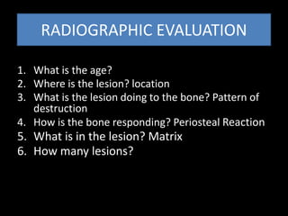
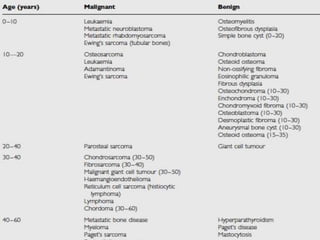
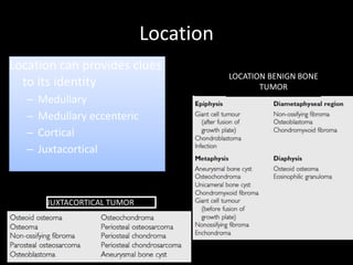
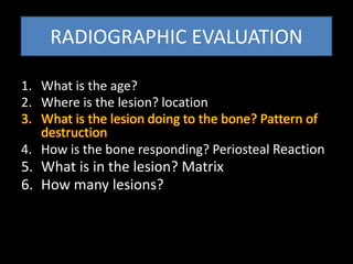
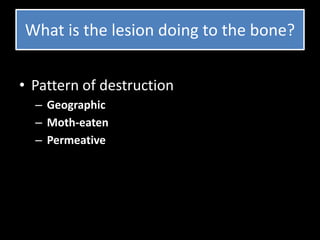
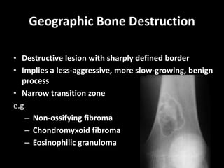
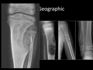
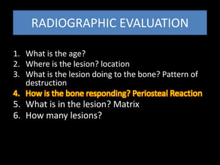
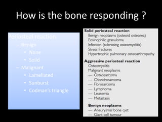
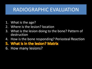
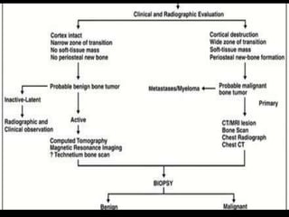
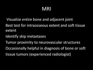
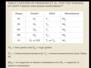
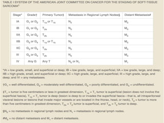
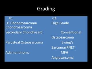
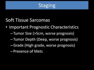
Radiological presentation and
staging of MSK tumours
Nor Azman MZ
Radiological presentation
• Plain radiograph very important for bone tumours
• For soft tissue tumour radiographs are useful to
identify osseous or mineralising lesions and what is
the tumour effect on bone
• MRI is superior in the assessment of a soft tissue
mass.
RADIOGRAPHIC EVALUATION
1. What is the age?
2. Where is the lesion? location
3. What is the lesion doing to the bone? Pattern of
destruction
4. How is the bone responding? Periosteal Reaction
5. What is in the lesion? Matrix
6. How many lesions?
What is the age?
Where is the lesion? location
Location
Location can provides clues
to its identity
– Medullary
– Medullary eccenteric
– Cortical
– Juxtacortical
LOCATION BENIGN BONE
TUMOR
JUXTACORTICAL TUMOR
Location
• Epiphyseal : PGCAT
– PVNS
– GCT
– Chondroblastoma
– ABC
– Tuberculous & other infections
• Metaphyseal
– NOF, UBC, OSC, CSC
• Diaphyseal
– ES, EG, Osteoid Osteoma
– metastasis
OSTEOID OSTEOMAOSTEOCHONDROMA
Location : Juxtacortical
RADIOGRAPHIC EVALUATION
1. What is the age?
2. Where is the lesion? location
3. What is the lesion doing to the bone? Pattern of
destruction
4. How is the bone responding? Periosteal Reaction
5. What is in the lesion? Matrix
6. How many lesions?
What is the lesion doing to the bone?
• Pattern of destruction
– Geographic
– Moth-eaten
– Permeative
Geographic Bone Destruction
• Destructive lesion with sharply defined border
• Implies a less-aggressive, more slow-growing, benign
process
• Narrow transition zone
e.g
– Non-ossifying fibroma
– Chondromyxoid fibroma
– Eosinophilic granuloma
Geographic
Moth-eaten Appearance
• Areas of destruction with ragged borders
• Implies more rapid growth
• Probably a malignancy
e.g
Myeloma
Metastasis
Lymphoma
Ewing sarcoma
Permeative Pattern
• Ill-defined lesion with multiple
“worm-holes”
• Spreads through marrow space
• Wide transition zone
• Implies an aggressive malignancy
– Round-cell lesions
Leukemia
Lymphoma, leukemia
Ewing’s Sarcoma
Myeloma
Osteomyelitis
Neuroblastoma
Less malignant More malignant
RADIOGRAPHIC EVALUATION
1. What is the age?
2. Where is the lesion? location
3. What is the lesion doing to the bone? Pattern of
destruction
4. How is the bone responding? Periosteal Reaction
5. What is in the lesion? Matrix
6. How many lesions?
How is the bone responding ?
Periosteal reaction
– Benign
• None
• Solid
– Malignant
• Lamellated
• Sunburst
• Codman’s triangle
SUNBURST
CODMANS
LAMELLATED
/ONION PEEL
Periosteal Reaction
SOLID
Less malignant More malignant
RADIOGRAPHIC EVALUATION
1. What is the age?
2. Where is the lesion? location
3. What is the lesion doing to the bone? Pattern of
destruction
4. How is the bone responding? Periosteal Reaction
5. What is in the lesion? Matrix
6. How many lesions?
WHAT IS THE LESION ?
MATRIX
• Osteoblastic
– Fluffy, cotton-like or cloud-
like densities
Osteosarcoma
• Cartilaginous
– Comma-shaped, punctate,
annular, popcorn-like
Enchondroma,
chondrosarcoma,
chondromyxoid fibroma
• Ground glass appearance
– Fibrous dysplasia
RADIOGRAPHIC EVALUATION
1. What is the age?
2. Where is the lesion? location
3. What is the lesion doing to the bone? Pattern of
destruction
4. How is the bone responding? Periosteal Reaction
5. What is in the lesion? Matrix
6. How many lesions?
How many lesions?
• Multiple bony lesion
DD for holes in the bone
• FOGMACHINE
– Fibrous dysplasia
– Osteoid osteoma, osteoblastoma, osteosarcoma
– Giant cell tumour
– Myeloma
– Aneurysmal Bone Cyst, adamantimoma
– Chondromyxoid fibroma, chondroblastoma,
chondrosarcoma
– Hystiocytosis
– Infection
– Nonossifying fibroma
– Enchondroma, Ewing sarcoma
Staging
Staging
Purpose
• Determine tumor type
• Determine prognosis
• Guide treatment
• Compare results between study groups
• Delineate extent of local and distant disease
Staging Studies
• Plain Radiograph
• MRI
• CT scan
• Chest CT
• Bone Scan
Plain Radiographs
Evaluate:
• Rate of tumor growth
• Tumor interaction with surrounding non-
neoplastic tissue
• Internal composition of tumor
MRI
Visualize entire bone and adjacent joint
Best test for intraosseous extent and soft tissue
extent
Identify skip metastases
Tumor proximity to neurovascular structures
Occasionally helpful in diagnosis of bone or soft
tissue tumors (experienced radiologist)
CT
• Good for evaluating cortical details and
destruction
• Subtle cortical erosions (endosteal;periosteal)
• not detectable on plain x-ray or MRI
• Subtle calcifications / ossification (Visible
tumor
matrix mineralization)
Chest CT
• Presence of metastatic disease
Bone Scan
• Whole body bone scan
• Sites of bony mets
• Active lesion??
Staging
• Benign Staging System
• Stage 1: Latent
– Grow slowly with growth of individual and
then stop; tendency to heal spontaneously
(ex. NOF; UBC)
• Stage 2: Active
Progressive growth
• Stage 3: Aggressive
Grading
G1 G2
LG Chondrosarcoma High Grade
Chondrosarcoma
Secondary Chondrosarc Conventional
Osteosarcoma
Parosteal Osteosarcoma Ewing’s
Sarcoma/PNET
Adamantinoma MFH
Angiosarcoma
Staging
Soft Tissue Sarcomas
• Important Prognostic Characteristics
–Tumor Size (>5cm, worse prognosis)
–Tumor Depth (Deep, worse prognosis)
–Grade (High grade, worse prognosis)
–Presence of Mets
Grading
Soft Tissue Sarcomas (Biological Behavior)
• Tumors that are definitionally high grade
– Ewing’s Sarcoma
– PNET
– Rhabdomyosarcoma
– Angiosarcoma
– Pleomorphic Liposarcoma
– Soft Tissue Osteosarcoma
– Mesenchymal Chondrosarcoma
Grading
Soft Tissue Sarcomas (Biological Behavior)
– Tumors that are definitionally low grade
• Well Differentiated Liposarcoma
• Dermatofibrosarcoma Protuberans
• Infantile Fibrosarcoma
• Angiomatoid MFH
Evaluating response to chemoRx
Lecture 1: Presentation and staging msk tumour

Lecture 1: Presentation and staging msk tumour

  • 1.
    Radiological presentation and stagingof MSK tumours Nor Azman MZ
  • 2.
    Radiological presentation • Plainradiograph very important for bone tumours • For soft tissue tumour radiographs are useful to identify osseous or mineralising lesions and what is the tumour effect on bone • MRI is superior in the assessment of a soft tissue mass.
  • 3.
    RADIOGRAPHIC EVALUATION 1. Whatis the age? 2. Where is the lesion? location 3. What is the lesion doing to the bone? Pattern of destruction 4. How is the bone responding? Periosteal Reaction 5. What is in the lesion? Matrix 6. How many lesions?
  • 4.
  • 6.
    Where is thelesion? location
  • 7.
    Location Location can providesclues to its identity – Medullary – Medullary eccenteric – Cortical – Juxtacortical LOCATION BENIGN BONE TUMOR JUXTACORTICAL TUMOR
  • 8.
    Location • Epiphyseal :PGCAT – PVNS – GCT – Chondroblastoma – ABC – Tuberculous & other infections • Metaphyseal – NOF, UBC, OSC, CSC • Diaphyseal – ES, EG, Osteoid Osteoma – metastasis
  • 9.
  • 10.
    RADIOGRAPHIC EVALUATION 1. Whatis the age? 2. Where is the lesion? location 3. What is the lesion doing to the bone? Pattern of destruction 4. How is the bone responding? Periosteal Reaction 5. What is in the lesion? Matrix 6. How many lesions?
  • 11.
    What is thelesion doing to the bone? • Pattern of destruction – Geographic – Moth-eaten – Permeative
  • 12.
    Geographic Bone Destruction •Destructive lesion with sharply defined border • Implies a less-aggressive, more slow-growing, benign process • Narrow transition zone e.g – Non-ossifying fibroma – Chondromyxoid fibroma – Eosinophilic granuloma
  • 13.
  • 14.
    Moth-eaten Appearance • Areasof destruction with ragged borders • Implies more rapid growth • Probably a malignancy e.g Myeloma Metastasis Lymphoma Ewing sarcoma
  • 15.
    Permeative Pattern • Ill-definedlesion with multiple “worm-holes” • Spreads through marrow space • Wide transition zone • Implies an aggressive malignancy – Round-cell lesions Leukemia Lymphoma, leukemia Ewing’s Sarcoma Myeloma Osteomyelitis Neuroblastoma
  • 16.
  • 17.
    RADIOGRAPHIC EVALUATION 1. Whatis the age? 2. Where is the lesion? location 3. What is the lesion doing to the bone? Pattern of destruction 4. How is the bone responding? Periosteal Reaction 5. What is in the lesion? Matrix 6. How many lesions?
  • 18.
    How is thebone responding ? Periosteal reaction – Benign • None • Solid – Malignant • Lamellated • Sunburst • Codman’s triangle
  • 19.
  • 20.
    RADIOGRAPHIC EVALUATION 1. Whatis the age? 2. Where is the lesion? location 3. What is the lesion doing to the bone? Pattern of destruction 4. How is the bone responding? Periosteal Reaction 5. What is in the lesion? Matrix 6. How many lesions?
  • 21.
    WHAT IS THELESION ? MATRIX • Osteoblastic – Fluffy, cotton-like or cloud- like densities Osteosarcoma • Cartilaginous – Comma-shaped, punctate, annular, popcorn-like Enchondroma, chondrosarcoma, chondromyxoid fibroma • Ground glass appearance – Fibrous dysplasia
  • 22.
    RADIOGRAPHIC EVALUATION 1. Whatis the age? 2. Where is the lesion? location 3. What is the lesion doing to the bone? Pattern of destruction 4. How is the bone responding? Periosteal Reaction 5. What is in the lesion? Matrix 6. How many lesions?
  • 23.
    How many lesions? •Multiple bony lesion
  • 24.
    DD for holesin the bone • FOGMACHINE – Fibrous dysplasia – Osteoid osteoma, osteoblastoma, osteosarcoma – Giant cell tumour – Myeloma – Aneurysmal Bone Cyst, adamantimoma – Chondromyxoid fibroma, chondroblastoma, chondrosarcoma – Hystiocytosis – Infection – Nonossifying fibroma – Enchondroma, Ewing sarcoma
  • 25.
  • 28.
    Staging Purpose • Determine tumortype • Determine prognosis • Guide treatment • Compare results between study groups • Delineate extent of local and distant disease
  • 29.
    Staging Studies • PlainRadiograph • MRI • CT scan • Chest CT • Bone Scan
  • 30.
    Plain Radiographs Evaluate: • Rateof tumor growth • Tumor interaction with surrounding non- neoplastic tissue • Internal composition of tumor
  • 31.
    MRI Visualize entire boneand adjacent joint Best test for intraosseous extent and soft tissue extent Identify skip metastases Tumor proximity to neurovascular structures Occasionally helpful in diagnosis of bone or soft tissue tumors (experienced radiologist)
  • 34.
    CT • Good forevaluating cortical details and destruction • Subtle cortical erosions (endosteal;periosteal) • not detectable on plain x-ray or MRI • Subtle calcifications / ossification (Visible tumor matrix mineralization)
  • 36.
    Chest CT • Presenceof metastatic disease
  • 37.
    Bone Scan • Wholebody bone scan • Sites of bony mets • Active lesion??
  • 40.
    Staging • Benign StagingSystem • Stage 1: Latent – Grow slowly with growth of individual and then stop; tendency to heal spontaneously (ex. NOF; UBC) • Stage 2: Active Progressive growth • Stage 3: Aggressive
  • 41.
    Grading G1 G2 LG ChondrosarcomaHigh Grade Chondrosarcoma Secondary Chondrosarc Conventional Osteosarcoma Parosteal Osteosarcoma Ewing’s Sarcoma/PNET Adamantinoma MFH Angiosarcoma
  • 42.
    Staging Soft Tissue Sarcomas •Important Prognostic Characteristics –Tumor Size (>5cm, worse prognosis) –Tumor Depth (Deep, worse prognosis) –Grade (High grade, worse prognosis) –Presence of Mets
  • 43.
    Grading Soft Tissue Sarcomas(Biological Behavior) • Tumors that are definitionally high grade – Ewing’s Sarcoma – PNET – Rhabdomyosarcoma – Angiosarcoma – Pleomorphic Liposarcoma – Soft Tissue Osteosarcoma – Mesenchymal Chondrosarcoma
  • 44.
    Grading Soft Tissue Sarcomas(Biological Behavior) – Tumors that are definitionally low grade • Well Differentiated Liposarcoma • Dermatofibrosarcoma Protuberans • Infantile Fibrosarcoma • Angiomatoid MFH
  • 45.