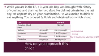
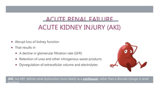
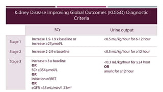
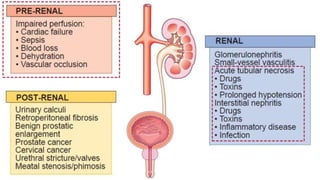
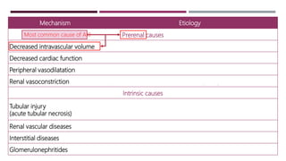
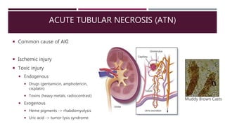
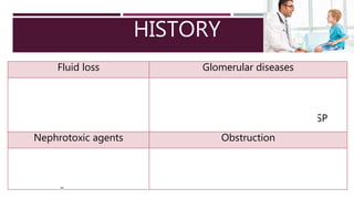
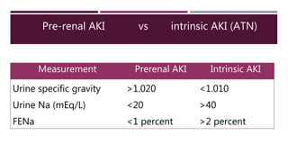
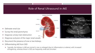
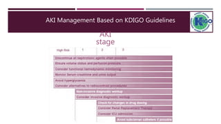
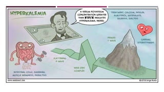
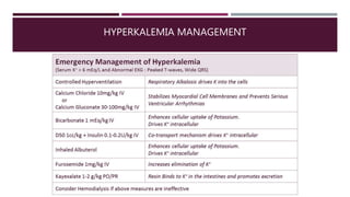
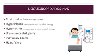
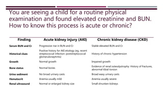
ACUTE RENAL FAILURE IN CHILDREN
IBRAHIM SANDOKJI, MD, FAAP
Pediatric Nephrologist, Assistant Professor
Board Certified by the American Board of Pediatrics
Taibah University
isandokji@taibahu.edu.sa
+966 50 632 5770
How do you approach this
child?
 While you are in the ER, a 3-year-old boy was brought with history
of vomiting and diarrhea for two days. He did not urinate for the last
day. He appears dry on your examination. He was unable to drink or
eat anything. You ordered IV fluids and obtained labs which show:
Lab Normal
Complete blood
count
Normal
Sodium 140 mmol/L 135–145 mmol/L
Potassium 6 mmol/L 3.5–5.0 mmol/L
Urea 12 mmol/L 1.8–6.4 mmol/L
Creatinine 150 μmol/L 44–88 μmol/L
Hyperkalemia
Uremia
Rise in creatinine (=decrease in GFR
ACUTE RENAL FAILURE
ACUTE KIDNEY INJURY (AKI)
 Abrupt loss of kidney function
 That results in
 A decline in glomerular filtration rate (GFR)
 Retention of urea and other nitrogenous waste products
 Dysregulation of extracellular volume and electrolytes
AKI, not ARF, defines renal dysfunction more clearly as a continuum, rather than a discrete change in renal
function
Kidney Disease Improving Global Outcomes (KDIGO) Diagnostic
Criteria
INTRINSIC RENAL
Vascular
Renal vein or artery
thrombosis
Hemolytic uremic syndrome
Tubular (ATN)
Ischemia, sepsis
Nephrotoxins:
Aminoglycosides, amphotericin,
chemotherapy, radiocontrast
Glomerular
PIGN, RPGN, HSP, SLE
Interstitium
Interstitial nephritis:
Antibiotics, PPIs, NSAIDs,
diuretics
Mechanism Etiology
Prerenal causes
Decreased intravascular volume Dehydration, hemorrhage, burns, shock, nephrotic syndrome
Decreased cardiac function Heart failure
Peripheral vasodilatation Sepsis, anaphylaxis, antihypertensive medication
Renal vasoconstriction Sepsis, nonsteroidal anti-inflammatory drugs, ACE inhibitor
Intrinsic causes
Tubular injury
(acute tubular necrosis)
Prolonged ischemia, nephrotoxins, hypotension, sepsis
Renal vascular diseases Hemolytic uremic syndrome, vasculitides, thrombosis
Interstitial diseases Interstitial nephritis, infections
Glomerulonephritides PIGN, RPGN, HSP
Most common cause of AKI
ACUTE TUBULAR NECROSIS (ATN)
Muddy Brown Casts
 Common cause of AKI
 Ischemic injury
 Toxic injury
 Endogenous
 Drugs (gentamicin, amphotericin,
cisplatin)
 Toxins (heavy metals, radiocontrast)
 Exogenous
 Heme pigments -> rhabdomyolysis
 Uric acid -> tumor lysis syndrome
HISTORY
Fluid loss Glomerular diseases
•Diarrhea, vomiting
•Burns
•Surgery
•Shock
•Streptococcal infection: PIGN
•Bloody diarrhea: HUS
•Joint symptoms, rash, or purpura: HSP
Nephrotoxic agents Obstruction
•NSAIDs
•Aminoglycosides
•Contrast agents
•Complete anuria
•Poor urinary stream
Intravascular volume depletion Palpably enlarged kidneys
•Tachycardia
•Delayed capillary refill
•Low blood pressure
•Weak peripheral pulses
•Dry mucous membranes
•Hydronephrosis
•Polycystic/multicystic kidney
disease
•Renal vein thrombosis
Fluid overload Obstruction
•Edema
•Hypertension
•Heart failure
•Pulmonary edema
•Poor urinary stream
•Palpably enlarged bladder
•Therapeutic catheterization
Systemic causes
•Rash: HSP, acute presentation of SLE, interstitial nephritis due to drug reaction
•Joint findings (tenderness or swelling): HSP, acute presentation of SLE
EXAMINATION
LABORATORY AND IMAGING EVALUATION
 Electrolytes
 Urinalysis
 Renal ultrasound
 Considered in some patients:
 CBC
 C3, C4
 Testing for streptococcal infection
 Fractional excretion of sodium (FeNa)
 Renal biopsy — rarely indicated in
AKI.
 Done if Suspected
 Acute GN
 Interstitial nephritis
 Lupus nephritis
PREVENTION
Fluid management Avoid nephrotoxins
MANAGEMENT STRATEGIES
 Treat the underlying cause
 Fluid management
 Hypovolemia –> fluid
 Euvolemia –> balanced intake/output
 Hypervolemia -> fluid removal and/or
restriction
 Management of AKI complications:
 Electrolyte disturbances
 Metabolic acidosis
 Hypertension
 Adequate nutrition
 Drug management
 Avoidance of nephrotoxic agents
 Dosing readjustment of renally
excreted drugs
 ? Dialysis
HOME READING
White Blood Cell Casts Red Blood Cell Casts
Hyaline Casts Muddy Brown Granular Casts
Waxy Casts
Fatty Casts
In which condition(s) do you see each one of these casts in the urine?
Pre-renal AKI vs intrinsic AKI (ATN)
Measurement Prerenal AKI Intrinsic AKI
Urine specific gravity >1.020 <1.010
Urine Na (mEq/L) <20 >40
FENa <1 percent >2 percent
Role of Renal Ultrasound in AKI
 Delineate renal size
 Survey the renal parenchyma
 Diagnose urinary tract obstruction
 Diagnose occlusion of the major renal vessels
 Document the presence of one or two kidneys
 Differentiating AKI from CKD
 Typically, the kidneys in AKI are normal in size or enlarged (due to inflammation or edema), with increased
echogenicity, whereas those in CKD are frequently small and shrunken
AKI Management Based on KDIGO Guidelines
HYPERKALEMIA MANAGEMENT
INDICATIONS OF DIALYSIS IN AKI
 Fluid overload unresponsive to diuretics
 Hyperkalemia unresponsive to non-dialytic therapy
 Hypertension unresponsive to pharmacologic therapy
 Uremic encephalopathy
 Pulmonary Edema
 Heart failure
You are seeing a child for a routine physical
examination and found elevated creatinine and BUN.
How to know this process is acute or chronic?
Finding Acute kidney injury (AKI) Chronic kidney disease (CKD)
Serum BUN and Cr Progressive rise in BUN and Cr Stable elevated BUN and Cr
Historical clues
Positive history for AKI etiology (eg, recent
streptococcal infection: poststreptococcal
glomerulonephritis)
History of chronic hypertension
Growth Normal growth Impaired growth
Bone status Normal bones
Evidence of renal osteodystrophy: History of fractures,
abnormal tibial torsion
Urine sediment No broad urinary casts Broad waxy urinary casts
Hematocrit Anemia usually mild Anemia usually severe
Renal ultrasound Normal or enlarged kidney size Small shrunken kidneys
OUTCOME
AFTER AKI

Pediatric Acute Kidney Injury.pptx

  • 1.
    ACUTE RENAL FAILUREIN CHILDREN IBRAHIM SANDOKJI, MD, FAAP Pediatric Nephrologist, Assistant Professor Board Certified by the American Board of Pediatrics Taibah University isandokji@taibahu.edu.sa +966 50 632 5770
  • 2.
    How do youapproach this child?  While you are in the ER, a 3-year-old boy was brought with history of vomiting and diarrhea for two days. He did not urinate for the last day. He appears dry on your examination. He was unable to drink or eat anything. You ordered IV fluids and obtained labs which show: Lab Normal Complete blood count Normal Sodium 140 mmol/L 135–145 mmol/L Potassium 6 mmol/L 3.5–5.0 mmol/L Urea 12 mmol/L 1.8–6.4 mmol/L Creatinine 150 μmol/L 44–88 μmol/L Hyperkalemia Uremia Rise in creatinine (=decrease in GFR
  • 3.
    ACUTE RENAL FAILURE ACUTEKIDNEY INJURY (AKI)  Abrupt loss of kidney function  That results in  A decline in glomerular filtration rate (GFR)  Retention of urea and other nitrogenous waste products  Dysregulation of extracellular volume and electrolytes AKI, not ARF, defines renal dysfunction more clearly as a continuum, rather than a discrete change in renal function
  • 4.
    Kidney Disease ImprovingGlobal Outcomes (KDIGO) Diagnostic Criteria
  • 6.
    INTRINSIC RENAL Vascular Renal veinor artery thrombosis Hemolytic uremic syndrome Tubular (ATN) Ischemia, sepsis Nephrotoxins: Aminoglycosides, amphotericin, chemotherapy, radiocontrast Glomerular PIGN, RPGN, HSP, SLE Interstitium Interstitial nephritis: Antibiotics, PPIs, NSAIDs, diuretics
  • 7.
    Mechanism Etiology Prerenal causes Decreasedintravascular volume Dehydration, hemorrhage, burns, shock, nephrotic syndrome Decreased cardiac function Heart failure Peripheral vasodilatation Sepsis, anaphylaxis, antihypertensive medication Renal vasoconstriction Sepsis, nonsteroidal anti-inflammatory drugs, ACE inhibitor Intrinsic causes Tubular injury (acute tubular necrosis) Prolonged ischemia, nephrotoxins, hypotension, sepsis Renal vascular diseases Hemolytic uremic syndrome, vasculitides, thrombosis Interstitial diseases Interstitial nephritis, infections Glomerulonephritides PIGN, RPGN, HSP Most common cause of AKI
  • 8.
    ACUTE TUBULAR NECROSIS(ATN) Muddy Brown Casts  Common cause of AKI  Ischemic injury  Toxic injury  Endogenous  Drugs (gentamicin, amphotericin, cisplatin)  Toxins (heavy metals, radiocontrast)  Exogenous  Heme pigments -> rhabdomyolysis  Uric acid -> tumor lysis syndrome
  • 9.
    HISTORY Fluid loss Glomerulardiseases •Diarrhea, vomiting •Burns •Surgery •Shock •Streptococcal infection: PIGN •Bloody diarrhea: HUS •Joint symptoms, rash, or purpura: HSP Nephrotoxic agents Obstruction •NSAIDs •Aminoglycosides •Contrast agents •Complete anuria •Poor urinary stream
  • 10.
    Intravascular volume depletionPalpably enlarged kidneys •Tachycardia •Delayed capillary refill •Low blood pressure •Weak peripheral pulses •Dry mucous membranes •Hydronephrosis •Polycystic/multicystic kidney disease •Renal vein thrombosis Fluid overload Obstruction •Edema •Hypertension •Heart failure •Pulmonary edema •Poor urinary stream •Palpably enlarged bladder •Therapeutic catheterization Systemic causes •Rash: HSP, acute presentation of SLE, interstitial nephritis due to drug reaction •Joint findings (tenderness or swelling): HSP, acute presentation of SLE EXAMINATION
  • 11.
    LABORATORY AND IMAGINGEVALUATION  Electrolytes  Urinalysis  Renal ultrasound  Considered in some patients:  CBC  C3, C4  Testing for streptococcal infection  Fractional excretion of sodium (FeNa)  Renal biopsy — rarely indicated in AKI.  Done if Suspected  Acute GN  Interstitial nephritis  Lupus nephritis
  • 12.
  • 13.
    MANAGEMENT STRATEGIES  Treatthe underlying cause  Fluid management  Hypovolemia –> fluid  Euvolemia –> balanced intake/output  Hypervolemia -> fluid removal and/or restriction  Management of AKI complications:  Electrolyte disturbances  Metabolic acidosis  Hypertension  Adequate nutrition  Drug management  Avoidance of nephrotoxic agents  Dosing readjustment of renally excreted drugs  ? Dialysis
  • 14.
  • 15.
    White Blood CellCasts Red Blood Cell Casts Hyaline Casts Muddy Brown Granular Casts Waxy Casts Fatty Casts In which condition(s) do you see each one of these casts in the urine?
  • 16.
    Pre-renal AKI vsintrinsic AKI (ATN) Measurement Prerenal AKI Intrinsic AKI Urine specific gravity >1.020 <1.010 Urine Na (mEq/L) <20 >40 FENa <1 percent >2 percent
  • 17.
    Role of RenalUltrasound in AKI  Delineate renal size  Survey the renal parenchyma  Diagnose urinary tract obstruction  Diagnose occlusion of the major renal vessels  Document the presence of one or two kidneys  Differentiating AKI from CKD  Typically, the kidneys in AKI are normal in size or enlarged (due to inflammation or edema), with increased echogenicity, whereas those in CKD are frequently small and shrunken
  • 18.
    AKI Management Basedon KDIGO Guidelines
  • 20.
  • 21.
    INDICATIONS OF DIALYSISIN AKI  Fluid overload unresponsive to diuretics  Hyperkalemia unresponsive to non-dialytic therapy  Hypertension unresponsive to pharmacologic therapy  Uremic encephalopathy  Pulmonary Edema  Heart failure
  • 23.
    You are seeinga child for a routine physical examination and found elevated creatinine and BUN. How to know this process is acute or chronic? Finding Acute kidney injury (AKI) Chronic kidney disease (CKD) Serum BUN and Cr Progressive rise in BUN and Cr Stable elevated BUN and Cr Historical clues Positive history for AKI etiology (eg, recent streptococcal infection: poststreptococcal glomerulonephritis) History of chronic hypertension Growth Normal growth Impaired growth Bone status Normal bones Evidence of renal osteodystrophy: History of fractures, abnormal tibial torsion Urine sediment No broad urinary casts Broad waxy urinary casts Hematocrit Anemia usually mild Anemia usually severe Renal ultrasound Normal or enlarged kidney size Small shrunken kidneys
  • 24.

Editor's Notes

  • #4 AKI, not ARF, defines renal dysfunction more clearly as a continuum, rather than a discrete change in renal function
  • #7 Vascular causes involve large vessels as well as microangiopathic processes Vascular – renal vein thrombosis, renal artery obstruction, hemolytic uremic syndrome Glomerular – acute glomerulonephritis (SLE nephritis, IgA nephropathy, MPGN) Acute interstitial nephritis – antibiotics, PPIs, NSAIDs, diuretics Nephrotoxins – aminoglycosides, amphotericin, chemotherapy (cisplastn, ifosfamide, methotrexate), radiocontrast
  • #14 Patients with oliguria or anuria should not receive potassium or phosphorus. Sodium intake should be restricted to 2 to 3 mEq/kg per day to prevent sodium and fluid retention with resultant hypertension.
  • #16 Hyaline Normal individuals Dehydration Heavy exercise Granular casts After strenuous exercise Chronic renal diseases Acute tubular necrosis Waxy casts (renal failure casts) Severe chronic renal disease renal amyloidosis Fatty casts Tubular degeneration Nephrotic syndrome Hypothyroidism RBC Casts Pyelonephritis Glomerulonephritis Acute interstitial nephritis Lupus nephritis WBC Casts Glomerulonephritis