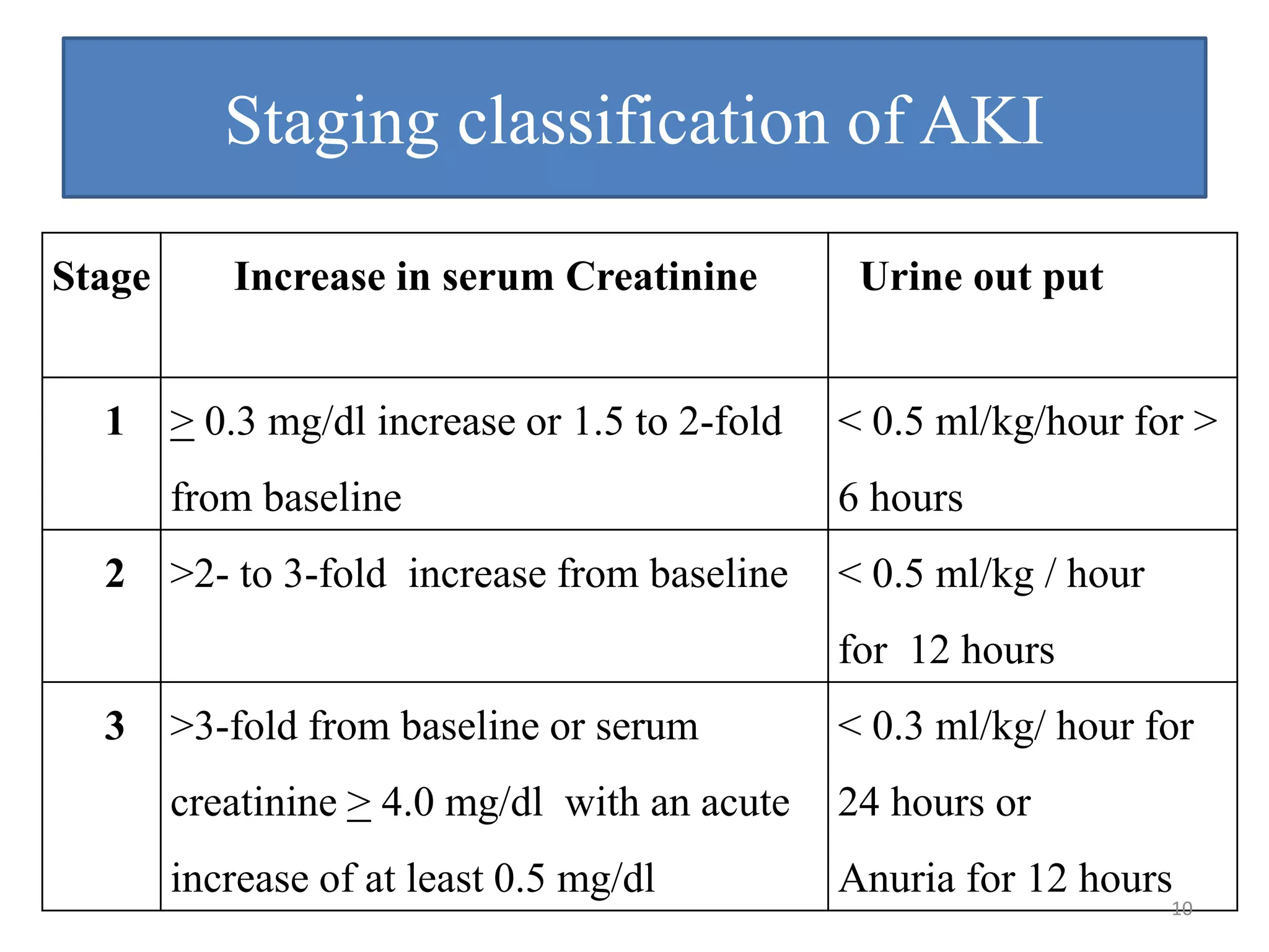
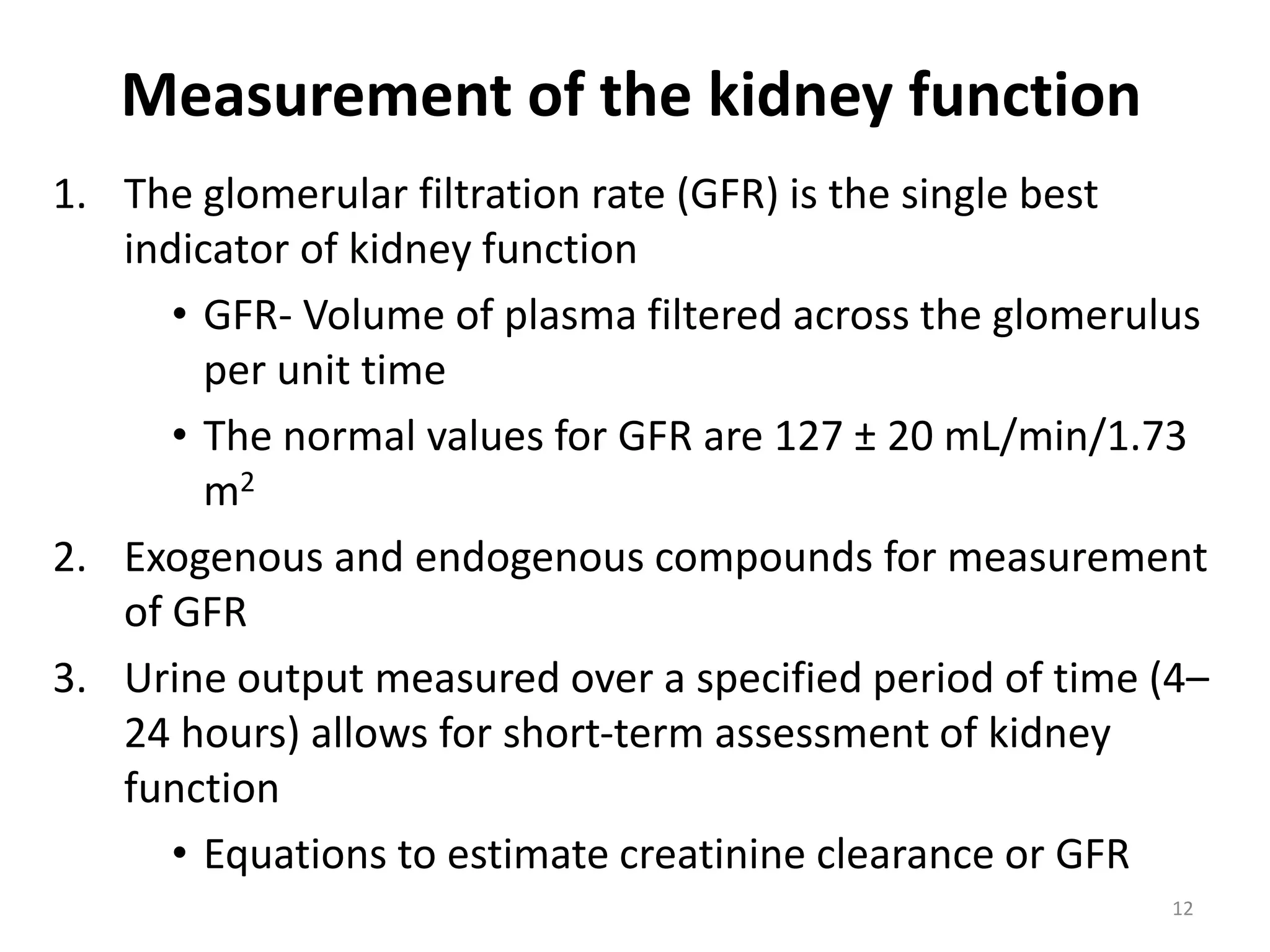
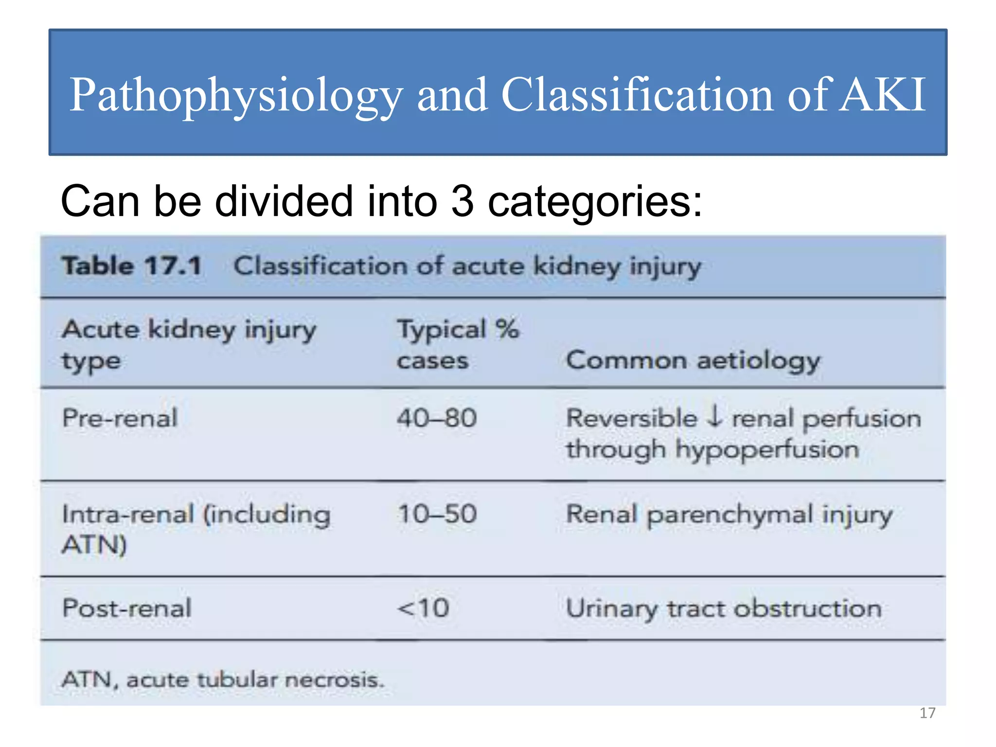
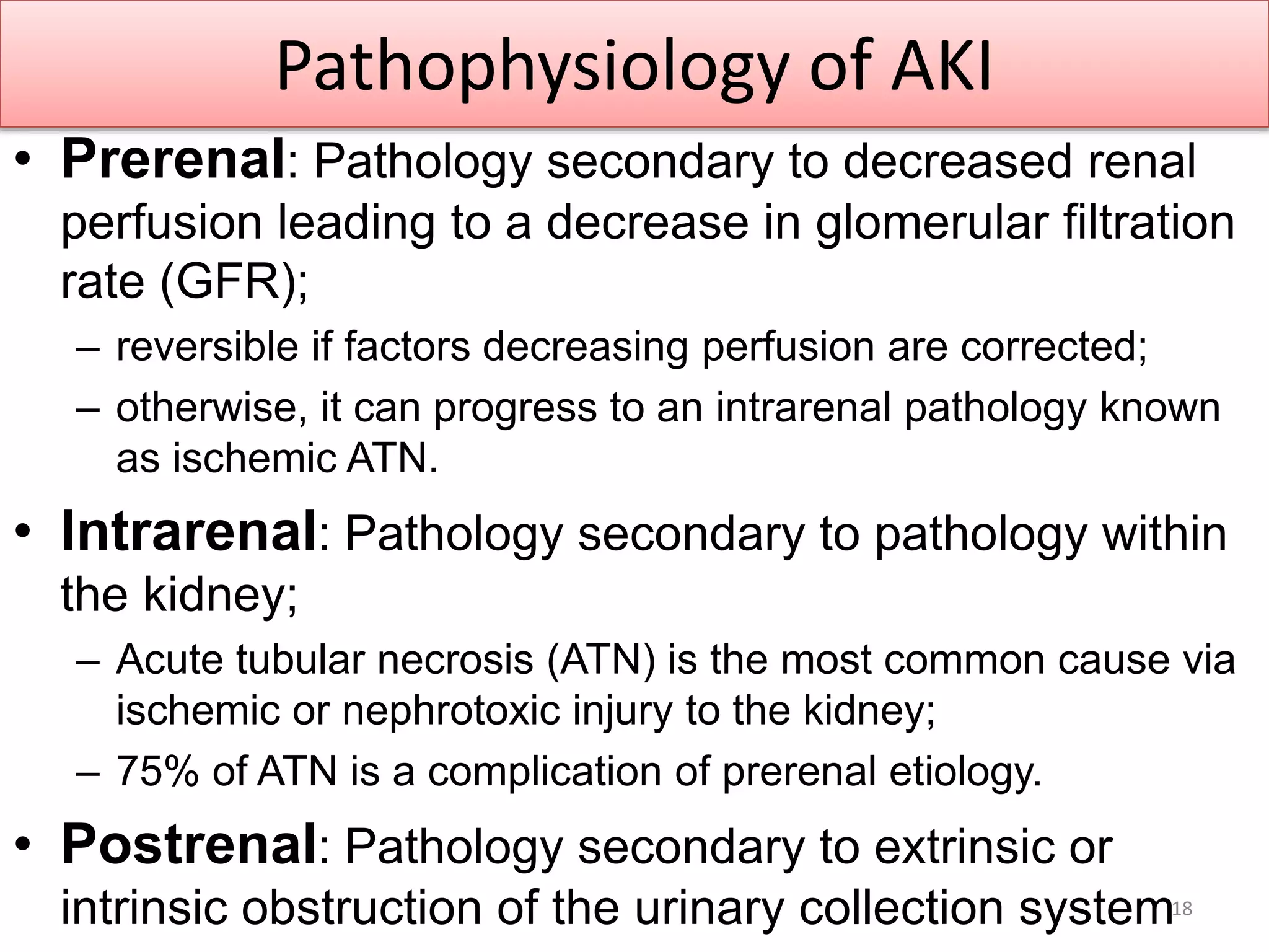
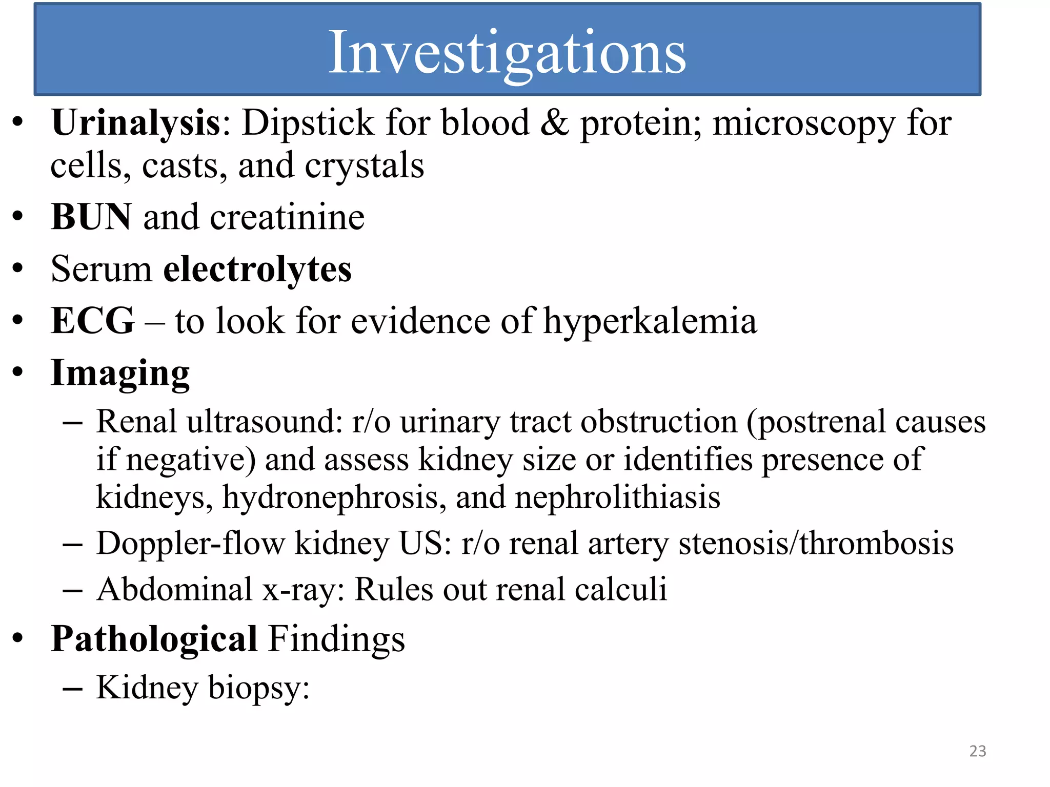
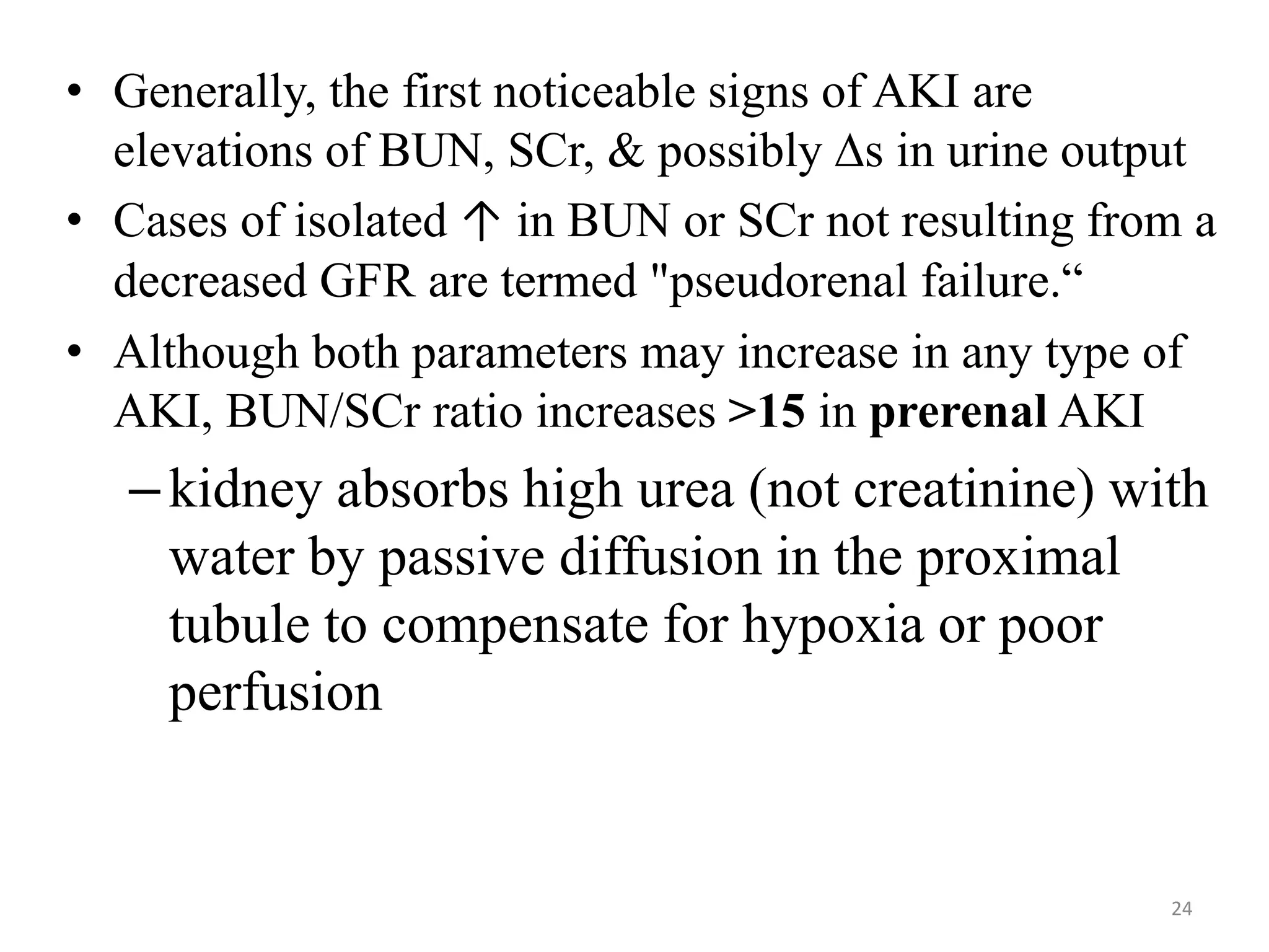
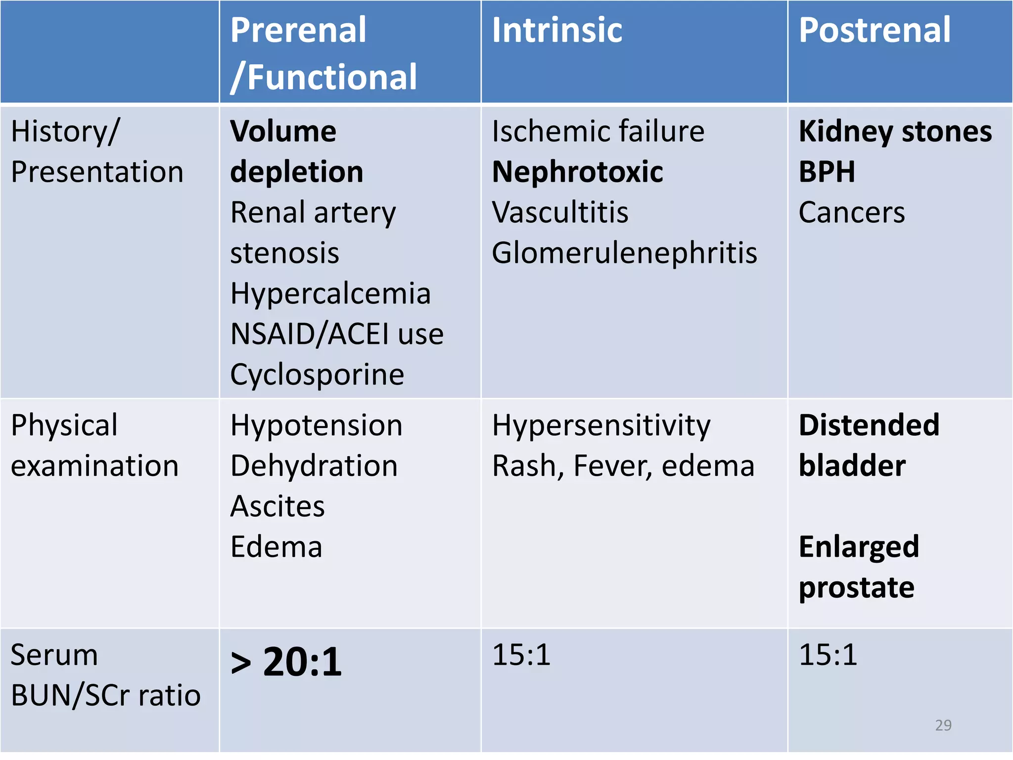
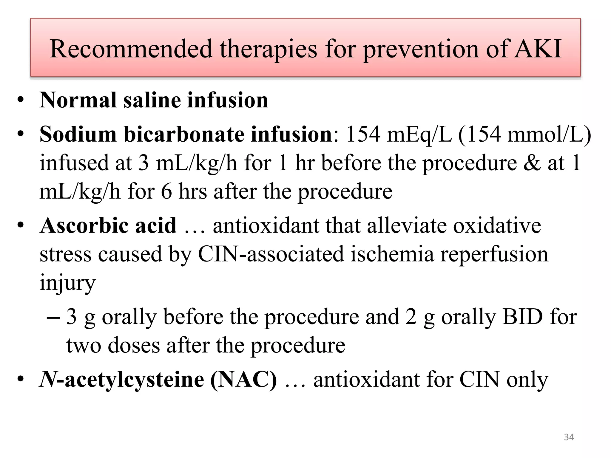
Pharmacotherapy of Renal Disorders
By Arega Gashaw
B.pharm, MSc in pharmacy practice
gemechu2002@gmail.com
1
Pharmacotherapy of renal disorders
 Acute Renal failure
 Chronic Renal Failure
 Drug induced Renal Disease
 Glomerulonephritis
 Nephrotic/nephritic syndromes
 Acid-base disorders
 Disorders of fluid and electrolyte
homeostasis
 Hemodialysis and peritoneal dialysis
 Case studies on Renal Disorders 2
Acute Renal Failure
3
• What do you know about ARF?
• What do you want to know about ARF?
4
I have following ……..
AS the Learning objectives
• Understand basic renal physiology and
the role of the kidney
• Identify patients at high risk of developing
acute kidney injury ARF
• Describe the pathophysiology and the
most common causes of prerenal,
intrinsic, and postrenal ARF
• Describe the prevention, management of
5
Introduction
–An organ attached to the posterior
wall of abdominal cavity bilaterally
–Is only 0.4% of body weight but
receives ~25% of CO
–Filters ~180L plasma/day
–Contains ~1 million nephrons each
6
– Glomerulus
– Bowman’s capsule
– Proximal tubule
– Loop of Henle
– Distal tubule
– Collecting duct
• Basic processes
– Filtration
– Reabsorption
– Secretion
7
What is the functions of kidney?
1. Regulation of Water & Electrolyte Balance
2. Excretion of Metabolic Waste Products
3. Excretion of Foreign Chemicals & Drugs
4. Regulation of Arterial Blood Pressure
5. Regulation of Erythropoiesis
6. Regulation of Vitamin D
7. Gluconeogenesis and metabolism of
endogenous compounds
8
Acute Kidney Injury (AKI)
• Sudden loss of kidney function resulting in retention of
nitrogenous waste as well as electrolyte and volume
homeostasis abnormalities with or without oliguria (urine
output <500 mL/d)
• It is a common and serious problem in clinical medicine.
• AKI is diagnosed when one of the following criteria is
met
– SrCr rises by ≥ 0.3mg/dl within 48 hours
– SrCr rises ≥ 1.5 fold from the baseline, which is
known or presumed to have occurred within one week
or
– urine output is < 0.5ml/kg/hr for >6 consecutive hours
No single serum creatinine value is a
threshold for ARF.(Ethiopian STG 2013
9
Staging classification of AKI
Stage Increase in serum Creatinine Urine out put
1 > 0.3 mg/dl increase or 1.5 to 2-fold
from baseline
< 0.5 ml/kg/hour for >
6 hours
2 >2- to 3-fold increase from baseline < 0.5 ml/kg / hour
for 12 hours
3 >3-fold from baseline or serum
creatinine > 4.0 mg/dl with an acute
increase of at least 0.5 mg/dl
< 0.3 ml/kg/ hour for
24 hours or
Anuria for 12 hours
10
11
Measurement of the kidney function
1. The glomerular filtration rate (GFR) is the single best
indicator of kidney function
• GFR- Volume of plasma filtered across the glomerulus
per unit time
• The normal values for GFR are 127 ± 20 mL/min/1.73
m2
2. Exogenous and endogenous compounds for measurement
of GFR
3. Urine output measured over a specified period of time (4–
24 hours) allows for short-term assessment of kidney
function
• Equations to estimate creatinine clearance or GFR
12
13
Laboratory Procedures to Detect the
Presence of Kidney problem
• Urine Analysis
–Urine pH, Glucose, Ketones, Nitrite, Heme,
Protein or Albumin, Specific Gravity, Blood
Urea Nitrogen, Serum Creatinine etc.
–Formed elements in the urine include
erythrocytes and leukocytes, casts, and
crystals
14
Classifications based on…….Urine output
– Anureic: < 50mL/24 hr (worse outcome)
– Oliguric: 50-500 mL/24 hrs
– Nonoliguric: >500 mL/24 hrs….better outcome
• Other classifications
– Community-acquired AKI
– Hospital acquired AKI
– ICU acquired AKI
15
Risk factors
• Volume depletion
– sepsis, hemorrhage, vomiting, diarrhea, poor fluid intake/
dehydration, diuretic use
– Poor renal perfusion
• Pre-existing CKD
• Nephrotic drugs:
– AG, Contrast Dye, NSAID, ACEI, ARBS, Cyclosporine and
Cimetidine, TMP: inhibit kidney tubular secretion of SCr
• Co-morbidities (e.g., liver failure, heart failure, diabetes)
• Advanced age
• Benign prostatic hypertrophy (BPH)
• Malignancy
16
Pathophysiology and Classification of AKI
Can be divided into 3 categories:
17
Pathophysiology of AKI
• Prerenal: Pathology secondary to decreased renal
perfusion leading to a decrease in glomerular filtration
rate (GFR);
– reversible if factors decreasing perfusion are corrected;
– otherwise, it can progress to an intrarenal pathology known
as ischemic ATN.
• Intrarenal: Pathology secondary to pathology within
the kidney;
– Acute tubular necrosis (ATN) is the most common cause via
ischemic or nephrotoxic injury to the kidney;
– 75% of ATN is a complication of prerenal etiology.
• Postrenal: Pathology secondary to extrinsic or
intrinsic obstruction of the urinary collection system18
Etiology
• Prerenal (~55%):
– Hypotension, volume contraction, severe heart failure, or liver failure
• Intrarenal (~40%):
– ATN (from prolonged prerenal failure, radiographic contrast material,
aminoglycosides, or nephrotoxic substances),
– Glomerulonephritis, acute interstitial nephritis (drug-induced), arteriolar
insults, vasculitis, accelerated hypertension, cholesterol embolization
(common after arterial procedures),
– Intrarenal deposition or sludging (seen in increased uric acid and
multiple myeloma—bence-jones proteins)
• Postrenal (~5%):
– Extrinsic compression (prostatic hypertrophy, carcinoma),
– Intrinsic obstruction (calculus, tumor, clot, stricture, sloughed papilli),
– Decreased function (neurogenic bladder)
19
Commonly Associated Conditions
 Hyperphosphatemia, hypercalcemia, hyperuricemia,
 Hydronephrosis, BPH, nephrolithiasis,
 Congestive heart failure (CHF), pericarditis,
cirrhosis, chronic renal insufficiency, malignant
hypertension, vasculitis,
 Drug reactions, sepsis, severe trauma, burns,
transfusion reactions, recent chemotherapy, muscle
injury, internal bleeding
20
Clinical Presentation of AKI
Dependent on the underlying etiology
– Oliguria/Anuria
– Urine discoloration (cola-colored urine is a blood in urine i.e.
commonly associated with acute glomerulonephritis)
– Fatigue, sudden weight gain, or severe abdominal or flank pain
– Peripheral edema, pulmonary edema, pleural effusion or ascites
– Pericardial effusion
– Decreased appetite, nausea and vomiting
– Hiccups
– Mucocutaneous bleeding
– Change in mental status/flapping tremor/seizure
21
Clinical presentation…
Physical Exam
• Prerenal signs: Tachycardia, orthostatic
hypotension, dry mucous membranes, decreased skin
turgor; look for stigmata of associated comorbidities
such as liver and heart failure, as well as sepsis.
• Intrinsic renal signs: Pruritic rash, livedo
reticularis, subcutaneous nodules, ischemic digits
despite good pulses
• Postrenal signs: Suprapubic distension, flank pain,
and enlarged prostate
22
Investigations
• Urinalysis: Dipstick for blood & protein; microscopy for
cells, casts, and crystals
• BUN and creatinine
• Serum electrolytes
• ECG – to look for evidence of hyperkalemia
• Imaging
– Renal ultrasound: r/o urinary tract obstruction (postrenal causes
if negative) and assess kidney size or identifies presence of
kidneys, hydronephrosis, and nephrolithiasis
– Doppler-flow kidney US: r/o renal artery stenosis/thrombosis
– Abdominal x-ray: Rules out renal calculi
• Pathological Findings
– Kidney biopsy:
23
• Generally, the first noticeable signs of AKI are
elevations of BUN, SCr, & possibly ∆s in urine output
• Cases of isolated ↑ in BUN or SCr not resulting from a
decreased GFR are termed "pseudorenal failure.“
• Although both parameters may increase in any type of
AKI, BUN/SCr ratio increases >15 in prerenal AKI
–kidney absorbs high urea (not creatinine) with
water by passive diffusion in the proximal
tubule to compensate for hypoxia or poor
perfusion
24
• AKI in CKD may develop when an abrupt rise in the
patient’s Scr occurs (increase by > 1mg/dl from baseline)
• In hospitalized patients, changes in urine output may be
helpful in characterizing the cause of the patient’s AKI
– Acute anuria is typically caused by either complete
urinary obstruction or a catastrophic event (e.g., shock
or acute cortical necrosis).
– Oliguria, which often develops over several days,
suggests prerenal azotemia, whereas nonoliguric renal
failure usually results from acute intrinsic renal failure
or incomplete urinary obstruction.
25
Fractional excretion of Na
• Evaluating the urine sodium concentration and percent
excreted (FeNa) is particularly helpful in differentiating
between a pre-renal AKI and ATN.
• In pre-renal, kidneys should excrete very little Na as a
compensatory mechanism to increase water reabsorption.
• Under normal conditions, the kidneys excrete
approximately 1 to 2% of the total sodium intake (normal
FeNa value).
• * FE Na= [(UNa × SCr)/(SNa × UCr)] × 100
Where U= urine and S=serum
* < 1 prerenal,
>1 renal
26
• Urinary Na < 20
• Urine Osms > 500
• Highly concentrated urine (>500 mOsm/kg [>500
mmol/kg]) suggests stimulation of antidiuretic and intact
tubular function.
• These findings are consistent with prerenal
azotemia.
• Common lab abnormalities in ARF:
– Increased: K+, phosphate, Mg, uric acid
– Decreased: Hematocrit (Hct), Na, Ca
27
Creatinine Clearance
• The first diagnostic tool…………
• (Very!) Rough estimate …….. 100/Cr
• GC (Cockroft-Gault)- To Calculate Creatine
Clearance
 (140 - age) x weight in kg (x 0.85 for women)
72 x serum creatinine
• Creatinine clearance of
– < 50 adjust medications
– < 25 refer to nephrology for pre-dialysis
– < 10 most people need dialysis
28
Prerenal
/Functional
Intrinsic Postrenal
History/
Presentation
Volume
depletion
Renal artery
stenosis
Hypercalcemia
NSAID/ACEI use
Cyclosporine
Ischemic failure
Nephrotoxic
Vascultitis
Glomerulenephritis
Kidney stones
BPH
Cancers
Physical
examination
Hypotension
Dehydration
Ascites
Edema
Hypersensitivity
Rash, Fever, edema
Distended
bladder
Enlarged
prostate
Serum
BUN/SCr ratio
> 20:1 15:1 15:1
29
Prerenal
/Functional
Intrinsic Postrenal
Urine
Concentrate
d?
Yes
Low urine Na
(<20 mEq/L)
Low FENa (<1)
High Uosm
No
Urine Na >40 mEq/L
FENa >2
Low Uosm
No
Urine Na>40
mEq/L
FENa >2
Low Uosm
Urine
Sediment
Normal
(Transparent
hyaline casts)
pigmented granular
/muddy brown casts—
ATN
Variable, may
be normal
Urinary WBC Negative 2-4+ casts (acute
interstitial nephritis)
Variable
Urinary RBC Negative 2-4+ casts
(glomerulonephritis)
1+
Proteinuria Negative Positive Negative 30
Management
• Desired outcome
–The primary goal of therapy is to prevent
ARF
–If ARF develops, the goals are
• To provide supportive measures until
kidney function returns
• To maintenance of blood pressure, fluid,
and electrolyte homeostasis
• To avoid or minimize further renal insults
that would delay recovery 31
How????
• Manage the underlying state appropriately like
– Acute infection
– Acute blood loss in trauma
– Dehydration
– Chronic kidney disease (CKD).
– Cardiovascular disease
• Avoid nephrotoxin administration (e.g.,
radiocontrast dye)
• Strict glycemic control with insulin in diabetics
has also reduced the development of ARF.
32
Non-pharmacologic Approaches
• Appropriate fluid replacement should be initiated.
• In outpatient, educate patient on preventive measures
– optimal daily fluid intake (~2 L/day) to avoid dehydration,
especially if they are to receive nephrotoxic medication
• In inpatient
– adequate hydration, standardized hemodynamic support in
critically ill, & avoidance of nephrotoxic medications
– Sometimes , the potential insult to the kidneys cannot be avoided but
may be preventable with aggressive hydration and removal of any
additional insults.
33
Recommended therapies for prevention of AKI
• Normal saline infusion
• Sodium bicarbonate infusion: 154 mEq/L (154 mmol/L)
infused at 3 mL/kg/h for 1 hr before the procedure & at 1
mL/kg/h for 6 hrs after the procedure
• Ascorbic acid … antioxidant that alleviate oxidative
stress caused by CIN-associated ischemia reperfusion
injury
– 3 g orally before the procedure and 2 g orally BID for
two doses after the procedure
• N-acetylcysteine (NAC) … antioxidant for CIN only
34
• Medications associated with diminished renal
blood flow should be stopped.
• Maintenance of adequate cardiac output and
blood pressure to optimize tissue perfusion
• Renal replacement therapy (RRT), such as
hemodialysis and peritoneal dialysis
35
Electrolyte management
• Hyperkalemia is the most common and serious
electrolyte abnormality in ARF.
• Typically, potassium must be restricted to less than 3
g/day and monitored daily.
• Hypernatremia and fluid retention commonly occur,
necessitating restricting daily sodium intake to no more
than 3 g.
• All sources of sodium, including antibiotics, need to be
considered when calculating daily sodium intake.
• Phosphorus and magnesium should be monitored;
neither is efficiently removed by dialysis.
36
1. Contrast-Induced Nephropathy (CIN)
• Common cause of ATN in inpatients (>0.5 mg/dl ↑ Scr)
• Onset …an ↑in Scr or Oliguria develops w/in 24 hrs
• Prevention:
– N-acetylcystine 600 mg po bid for four doses, with the
first two doses administered prior to contrast exposure
and then the last 2 doses after
– 600 mg PO b.i.d. on day prior to and day of contrast [A]
and
– isotonic NaHCO3 3 mL/kg/h × 1 h before administration
of contrast material and 1 mL/kg/h × 6 h after contrast
material [B].
37
– Give 0.45% NS IV …1 ml/kg/h for 12 hrs before &
after the procedure
• Hydration--dilutes the contrast media, prevents renal
vasoconstriction that contributes to hypoxia & ischemia,
and minimize tubular obstruction
…….CIN
38
2. Nephrolithiasis
• Kidney stones…….incidence……5-10%
• Strong genetic association
• Male : female ratio………..3:1
• Usually contains....Ca+ salts (account 70-80%)
• Drug-induced…..insoluble drugs in urine …..
– form crystals in distal tubules
– E.g acyclover, sufla, triamterene, MTX, indinavir
• Hydration is a key……………may add anti-pain
39
3. Diabetic Nephropathy
• Incidence………Type I (up to 50%) vs. Type II (few)
• The first sign………Proteinuria
• Recommended Test
– Microalbuminuria test…………..30-300 mg/day
• Type I…………have test yearly at 5 years from
diagnosis
• Type II ……….begin test at diagnosis
• Prevention:
– ACE inhibitors
– Control of hypertension
– Low protein diet………reduce excessive BUN
40
4. Penicillin-Induced AIN
• Acute interstitial necrosis
– Occurs 6-10 days
– Associated w/fever, rash, malaise…..?????
– Usually non-oligouric
– Treatment………..stop Penicillin & give supportive
therapy
– Prednisone 1 mg/kg for 7 days and then gradually
taper the dose over the next several weeks (total
therapy of can be up to 8-14 weeks)
– How??????????????
41
Treatment of AKI
• Current treatment is focused on primary prevention,
treating the underlying cause, and treating
associated complications.
• Diuretics if edema occurs……IV furosemide
• Prerenal azotemia: Correct primary hemodynamic
– Normal saline if volume depleted
– Pressure management if needed (↓BP)
– Blood products if needed (↓Hgb)
• Postrenal azotemia – relieve obstruction
– Consult Urology….BPH, Prostate Cancer
• The ultimate goal is to have the patient’s renal
function restored to pre-AKI baseline 42
Treatment
• Intrinsic: no universal therapy
– Avoid insult
– Consider fluid bolus…..perfusion/urine production
– Loop diuretics for oliguric/euvolemic or hypevolemic
• Furosemide ………..40-80 mg IV every 6-8 hrs or
• Furosemide infusion 40-80 mg IV bolus; then, 10-20 mg/hr iv
• Furosemide is a commonly used intervention.
N.B Studies show that it is ineffective in preventing and treating
ARF [A].
– Dopamine therapy …………1-2 mcg/kg/min….
• Not recommended anymore
– Natriuretic peptides, insulin-like growth factor, and
thyroxine also have no benefit in the treatment of ARF
– Acidosis……………. restrict dietary protein
• HCO3 to maintain arterial pH > 7.35
– Dialysis if needed
43
Additional Treatment
• General Measures
– Identify and correct all prerenal and postrenal causes.
– Review drug list: Stop nephrotoxic drugs and renally
adjust others.
– Always record ins and outs and daily weights.
– Watch for complications, including hyperkalemia,
pulmonary edema, and acidosis—all potential reasons
to start dialysis.
– Ensure good cardiac output and subsequent renal
blood flow.
– Follow nutrition suggestions and be aware of
infections; treat aggressively if they occur.
– Start patients on H2 inhibitors or proton pump
inhibitors, and avoid aspirin to avoid bleeding
44
• One of your family child was admitted to ACS
hospital. You are going to visit him. What are the
most likely and best thing you are planning?
A. Asking him with a 1 litter mango juice
B. Asking him with a kilo of banana
C. Asking him with a pack of biscuit
D. Asking with a bottled water
45
Diet for AKI patients
• Total caloric intake should be 35–50 kcal/kg/d to
avoid catabolism.
• Sodium should be restricted to 2 g/d [A].
• Potassium intake should be restricted to 40
mEq/d.
• Phosphorus should be restricted to 800 mg/d. If it
becomes high, treat with calcium carbonate or
other phosphate binder [A].
• Magnesium compounds should be avoided.
46
Patient Education
• Keep well hydrated.
• Avoid nephrotoxic drugs such as NSAIDs, ACE
inhibitors, and aminoglycosides.
47
Treatment of AKI complication
Hypertension
Hyperkalimia
Hypocalcemia
hyperphosphatemia
Acidocis
Anemia
48
1. Hypertension
• Cause:
– Volume overload; Intrinsic renal disease
• Volume overload: direct vasodilators
–calcium channel blockers, clonidine,
nicardipine drip, nitropruside, etc.
• Intrinsic renal disease:
–ACE Inhibitors
–Goal is to prevent stroke, CHF
49
2. Hyperkalemia
• Other risk factors:
–infection, hemolysis, acidosis
• How can you tell if it is “real”?
• It’s real. What’s the first thing to do?
• What’s next?
50
Hyperkalemia…
• Role of the following agents:
– insulin (+ glucose to prevent hypoglycemia)
– bicarbonate infusion
– albuterol (SQ/aerosol)
• What happens to ionized calcium level as you
correct the acidosis?
• What’s the third step?
51
3. Calcium and Phosphate
• Metastatic calcification: Ca+2 x PO4 > 60-70
• Often are reciprocal: PO4 Ca+
• Symptoms of hypocalcemia:
– irritability, tetany, Symptoms
• If hypoalbuminemic:
– check ionized Ca or
– correct (0.8 increase of Ca for each 1.0 of albumin
below 4)
52
Hypocalcemia and hyperphosphatemia…
• Reduce PO4 with calcium acetate if can
swallow pills, calcium carbonate if needs
liquid
• Diet restriction
• Avoid exogenous PO4: Fleet's, carafate, TPN
53
4. Acidosis
• Correct if bicarbonate is < 15
• Acidosis makes feel terrible
• Restict dietary protein (<0.5 g/kg/day)
• Sodium bicarbonate (arterial pH >7.2)
• Dialysis
• BUT...
– watch sodium and fluid overload
– watch lowering ionized calcium levels (by
increasing binding of calcium to albumin) 54

1 Acute Kidney Injury.pptx

  • 1.
    Pharmacotherapy of RenalDisorders By Arega Gashaw B.pharm, MSc in pharmacy practice gemechu2002@gmail.com 1
  • 2.
    Pharmacotherapy of renaldisorders  Acute Renal failure  Chronic Renal Failure  Drug induced Renal Disease  Glomerulonephritis  Nephrotic/nephritic syndromes  Acid-base disorders  Disorders of fluid and electrolyte homeostasis  Hemodialysis and peritoneal dialysis  Case studies on Renal Disorders 2
  • 3.
  • 4.
    • What doyou know about ARF? • What do you want to know about ARF? 4
  • 5.
    I have following…….. AS the Learning objectives • Understand basic renal physiology and the role of the kidney • Identify patients at high risk of developing acute kidney injury ARF • Describe the pathophysiology and the most common causes of prerenal, intrinsic, and postrenal ARF • Describe the prevention, management of 5
  • 6.
    Introduction –An organ attachedto the posterior wall of abdominal cavity bilaterally –Is only 0.4% of body weight but receives ~25% of CO –Filters ~180L plasma/day –Contains ~1 million nephrons each 6
  • 7.
    – Glomerulus – Bowman’scapsule – Proximal tubule – Loop of Henle – Distal tubule – Collecting duct • Basic processes – Filtration – Reabsorption – Secretion 7
  • 8.
    What is thefunctions of kidney? 1. Regulation of Water & Electrolyte Balance 2. Excretion of Metabolic Waste Products 3. Excretion of Foreign Chemicals & Drugs 4. Regulation of Arterial Blood Pressure 5. Regulation of Erythropoiesis 6. Regulation of Vitamin D 7. Gluconeogenesis and metabolism of endogenous compounds 8
  • 9.
    Acute Kidney Injury(AKI) • Sudden loss of kidney function resulting in retention of nitrogenous waste as well as electrolyte and volume homeostasis abnormalities with or without oliguria (urine output <500 mL/d) • It is a common and serious problem in clinical medicine. • AKI is diagnosed when one of the following criteria is met – SrCr rises by ≥ 0.3mg/dl within 48 hours – SrCr rises ≥ 1.5 fold from the baseline, which is known or presumed to have occurred within one week or – urine output is < 0.5ml/kg/hr for >6 consecutive hours No single serum creatinine value is a threshold for ARF.(Ethiopian STG 2013 9
  • 10.
    Staging classification ofAKI Stage Increase in serum Creatinine Urine out put 1 > 0.3 mg/dl increase or 1.5 to 2-fold from baseline < 0.5 ml/kg/hour for > 6 hours 2 >2- to 3-fold increase from baseline < 0.5 ml/kg / hour for 12 hours 3 >3-fold from baseline or serum creatinine > 4.0 mg/dl with an acute increase of at least 0.5 mg/dl < 0.3 ml/kg/ hour for 24 hours or Anuria for 12 hours 10
  • 11.
  • 12.
    Measurement of thekidney function 1. The glomerular filtration rate (GFR) is the single best indicator of kidney function • GFR- Volume of plasma filtered across the glomerulus per unit time • The normal values for GFR are 127 ± 20 mL/min/1.73 m2 2. Exogenous and endogenous compounds for measurement of GFR 3. Urine output measured over a specified period of time (4– 24 hours) allows for short-term assessment of kidney function • Equations to estimate creatinine clearance or GFR 12
  • 13.
  • 14.
    Laboratory Procedures toDetect the Presence of Kidney problem • Urine Analysis –Urine pH, Glucose, Ketones, Nitrite, Heme, Protein or Albumin, Specific Gravity, Blood Urea Nitrogen, Serum Creatinine etc. –Formed elements in the urine include erythrocytes and leukocytes, casts, and crystals 14
  • 15.
    Classifications based on…….Urineoutput – Anureic: < 50mL/24 hr (worse outcome) – Oliguric: 50-500 mL/24 hrs – Nonoliguric: >500 mL/24 hrs….better outcome • Other classifications – Community-acquired AKI – Hospital acquired AKI – ICU acquired AKI 15
  • 16.
    Risk factors • Volumedepletion – sepsis, hemorrhage, vomiting, diarrhea, poor fluid intake/ dehydration, diuretic use – Poor renal perfusion • Pre-existing CKD • Nephrotic drugs: – AG, Contrast Dye, NSAID, ACEI, ARBS, Cyclosporine and Cimetidine, TMP: inhibit kidney tubular secretion of SCr • Co-morbidities (e.g., liver failure, heart failure, diabetes) • Advanced age • Benign prostatic hypertrophy (BPH) • Malignancy 16
  • 17.
    Pathophysiology and Classificationof AKI Can be divided into 3 categories: 17
  • 18.
    Pathophysiology of AKI •Prerenal: Pathology secondary to decreased renal perfusion leading to a decrease in glomerular filtration rate (GFR); – reversible if factors decreasing perfusion are corrected; – otherwise, it can progress to an intrarenal pathology known as ischemic ATN. • Intrarenal: Pathology secondary to pathology within the kidney; – Acute tubular necrosis (ATN) is the most common cause via ischemic or nephrotoxic injury to the kidney; – 75% of ATN is a complication of prerenal etiology. • Postrenal: Pathology secondary to extrinsic or intrinsic obstruction of the urinary collection system18
  • 19.
    Etiology • Prerenal (~55%): –Hypotension, volume contraction, severe heart failure, or liver failure • Intrarenal (~40%): – ATN (from prolonged prerenal failure, radiographic contrast material, aminoglycosides, or nephrotoxic substances), – Glomerulonephritis, acute interstitial nephritis (drug-induced), arteriolar insults, vasculitis, accelerated hypertension, cholesterol embolization (common after arterial procedures), – Intrarenal deposition or sludging (seen in increased uric acid and multiple myeloma—bence-jones proteins) • Postrenal (~5%): – Extrinsic compression (prostatic hypertrophy, carcinoma), – Intrinsic obstruction (calculus, tumor, clot, stricture, sloughed papilli), – Decreased function (neurogenic bladder) 19
  • 20.
    Commonly Associated Conditions Hyperphosphatemia, hypercalcemia, hyperuricemia,  Hydronephrosis, BPH, nephrolithiasis,  Congestive heart failure (CHF), pericarditis, cirrhosis, chronic renal insufficiency, malignant hypertension, vasculitis,  Drug reactions, sepsis, severe trauma, burns, transfusion reactions, recent chemotherapy, muscle injury, internal bleeding 20
  • 21.
    Clinical Presentation ofAKI Dependent on the underlying etiology – Oliguria/Anuria – Urine discoloration (cola-colored urine is a blood in urine i.e. commonly associated with acute glomerulonephritis) – Fatigue, sudden weight gain, or severe abdominal or flank pain – Peripheral edema, pulmonary edema, pleural effusion or ascites – Pericardial effusion – Decreased appetite, nausea and vomiting – Hiccups – Mucocutaneous bleeding – Change in mental status/flapping tremor/seizure 21
  • 22.
    Clinical presentation… Physical Exam •Prerenal signs: Tachycardia, orthostatic hypotension, dry mucous membranes, decreased skin turgor; look for stigmata of associated comorbidities such as liver and heart failure, as well as sepsis. • Intrinsic renal signs: Pruritic rash, livedo reticularis, subcutaneous nodules, ischemic digits despite good pulses • Postrenal signs: Suprapubic distension, flank pain, and enlarged prostate 22
  • 23.
    Investigations • Urinalysis: Dipstickfor blood & protein; microscopy for cells, casts, and crystals • BUN and creatinine • Serum electrolytes • ECG – to look for evidence of hyperkalemia • Imaging – Renal ultrasound: r/o urinary tract obstruction (postrenal causes if negative) and assess kidney size or identifies presence of kidneys, hydronephrosis, and nephrolithiasis – Doppler-flow kidney US: r/o renal artery stenosis/thrombosis – Abdominal x-ray: Rules out renal calculi • Pathological Findings – Kidney biopsy: 23
  • 24.
    • Generally, thefirst noticeable signs of AKI are elevations of BUN, SCr, & possibly ∆s in urine output • Cases of isolated ↑ in BUN or SCr not resulting from a decreased GFR are termed "pseudorenal failure.“ • Although both parameters may increase in any type of AKI, BUN/SCr ratio increases >15 in prerenal AKI –kidney absorbs high urea (not creatinine) with water by passive diffusion in the proximal tubule to compensate for hypoxia or poor perfusion 24
  • 25.
    • AKI inCKD may develop when an abrupt rise in the patient’s Scr occurs (increase by > 1mg/dl from baseline) • In hospitalized patients, changes in urine output may be helpful in characterizing the cause of the patient’s AKI – Acute anuria is typically caused by either complete urinary obstruction or a catastrophic event (e.g., shock or acute cortical necrosis). – Oliguria, which often develops over several days, suggests prerenal azotemia, whereas nonoliguric renal failure usually results from acute intrinsic renal failure or incomplete urinary obstruction. 25
  • 26.
    Fractional excretion ofNa • Evaluating the urine sodium concentration and percent excreted (FeNa) is particularly helpful in differentiating between a pre-renal AKI and ATN. • In pre-renal, kidneys should excrete very little Na as a compensatory mechanism to increase water reabsorption. • Under normal conditions, the kidneys excrete approximately 1 to 2% of the total sodium intake (normal FeNa value). • * FE Na= [(UNa × SCr)/(SNa × UCr)] × 100 Where U= urine and S=serum * < 1 prerenal, >1 renal 26
  • 27.
    • Urinary Na< 20 • Urine Osms > 500 • Highly concentrated urine (>500 mOsm/kg [>500 mmol/kg]) suggests stimulation of antidiuretic and intact tubular function. • These findings are consistent with prerenal azotemia. • Common lab abnormalities in ARF: – Increased: K+, phosphate, Mg, uric acid – Decreased: Hematocrit (Hct), Na, Ca 27
  • 28.
    Creatinine Clearance • Thefirst diagnostic tool………… • (Very!) Rough estimate …….. 100/Cr • GC (Cockroft-Gault)- To Calculate Creatine Clearance  (140 - age) x weight in kg (x 0.85 for women) 72 x serum creatinine • Creatinine clearance of – < 50 adjust medications – < 25 refer to nephrology for pre-dialysis – < 10 most people need dialysis 28
  • 29.
    Prerenal /Functional Intrinsic Postrenal History/ Presentation Volume depletion Renal artery stenosis Hypercalcemia NSAID/ACEIuse Cyclosporine Ischemic failure Nephrotoxic Vascultitis Glomerulenephritis Kidney stones BPH Cancers Physical examination Hypotension Dehydration Ascites Edema Hypersensitivity Rash, Fever, edema Distended bladder Enlarged prostate Serum BUN/SCr ratio > 20:1 15:1 15:1 29
  • 30.
    Prerenal /Functional Intrinsic Postrenal Urine Concentrate d? Yes Low urineNa (<20 mEq/L) Low FENa (<1) High Uosm No Urine Na >40 mEq/L FENa >2 Low Uosm No Urine Na>40 mEq/L FENa >2 Low Uosm Urine Sediment Normal (Transparent hyaline casts) pigmented granular /muddy brown casts— ATN Variable, may be normal Urinary WBC Negative 2-4+ casts (acute interstitial nephritis) Variable Urinary RBC Negative 2-4+ casts (glomerulonephritis) 1+ Proteinuria Negative Positive Negative 30
  • 31.
    Management • Desired outcome –Theprimary goal of therapy is to prevent ARF –If ARF develops, the goals are • To provide supportive measures until kidney function returns • To maintenance of blood pressure, fluid, and electrolyte homeostasis • To avoid or minimize further renal insults that would delay recovery 31
  • 32.
    How???? • Manage theunderlying state appropriately like – Acute infection – Acute blood loss in trauma – Dehydration – Chronic kidney disease (CKD). – Cardiovascular disease • Avoid nephrotoxin administration (e.g., radiocontrast dye) • Strict glycemic control with insulin in diabetics has also reduced the development of ARF. 32
  • 33.
    Non-pharmacologic Approaches • Appropriatefluid replacement should be initiated. • In outpatient, educate patient on preventive measures – optimal daily fluid intake (~2 L/day) to avoid dehydration, especially if they are to receive nephrotoxic medication • In inpatient – adequate hydration, standardized hemodynamic support in critically ill, & avoidance of nephrotoxic medications – Sometimes , the potential insult to the kidneys cannot be avoided but may be preventable with aggressive hydration and removal of any additional insults. 33
  • 34.
    Recommended therapies forprevention of AKI • Normal saline infusion • Sodium bicarbonate infusion: 154 mEq/L (154 mmol/L) infused at 3 mL/kg/h for 1 hr before the procedure & at 1 mL/kg/h for 6 hrs after the procedure • Ascorbic acid … antioxidant that alleviate oxidative stress caused by CIN-associated ischemia reperfusion injury – 3 g orally before the procedure and 2 g orally BID for two doses after the procedure • N-acetylcysteine (NAC) … antioxidant for CIN only 34
  • 35.
    • Medications associatedwith diminished renal blood flow should be stopped. • Maintenance of adequate cardiac output and blood pressure to optimize tissue perfusion • Renal replacement therapy (RRT), such as hemodialysis and peritoneal dialysis 35
  • 36.
    Electrolyte management • Hyperkalemiais the most common and serious electrolyte abnormality in ARF. • Typically, potassium must be restricted to less than 3 g/day and monitored daily. • Hypernatremia and fluid retention commonly occur, necessitating restricting daily sodium intake to no more than 3 g. • All sources of sodium, including antibiotics, need to be considered when calculating daily sodium intake. • Phosphorus and magnesium should be monitored; neither is efficiently removed by dialysis. 36
  • 37.
    1. Contrast-Induced Nephropathy(CIN) • Common cause of ATN in inpatients (>0.5 mg/dl ↑ Scr) • Onset …an ↑in Scr or Oliguria develops w/in 24 hrs • Prevention: – N-acetylcystine 600 mg po bid for four doses, with the first two doses administered prior to contrast exposure and then the last 2 doses after – 600 mg PO b.i.d. on day prior to and day of contrast [A] and – isotonic NaHCO3 3 mL/kg/h × 1 h before administration of contrast material and 1 mL/kg/h × 6 h after contrast material [B]. 37
  • 38.
    – Give 0.45%NS IV …1 ml/kg/h for 12 hrs before & after the procedure • Hydration--dilutes the contrast media, prevents renal vasoconstriction that contributes to hypoxia & ischemia, and minimize tubular obstruction …….CIN 38
  • 39.
    2. Nephrolithiasis • Kidneystones…….incidence……5-10% • Strong genetic association • Male : female ratio………..3:1 • Usually contains....Ca+ salts (account 70-80%) • Drug-induced…..insoluble drugs in urine ….. – form crystals in distal tubules – E.g acyclover, sufla, triamterene, MTX, indinavir • Hydration is a key……………may add anti-pain 39
  • 40.
    3. Diabetic Nephropathy •Incidence………Type I (up to 50%) vs. Type II (few) • The first sign………Proteinuria • Recommended Test – Microalbuminuria test…………..30-300 mg/day • Type I…………have test yearly at 5 years from diagnosis • Type II ……….begin test at diagnosis • Prevention: – ACE inhibitors – Control of hypertension – Low protein diet………reduce excessive BUN 40
  • 41.
    4. Penicillin-Induced AIN •Acute interstitial necrosis – Occurs 6-10 days – Associated w/fever, rash, malaise…..????? – Usually non-oligouric – Treatment………..stop Penicillin & give supportive therapy – Prednisone 1 mg/kg for 7 days and then gradually taper the dose over the next several weeks (total therapy of can be up to 8-14 weeks) – How?????????????? 41
  • 42.
    Treatment of AKI •Current treatment is focused on primary prevention, treating the underlying cause, and treating associated complications. • Diuretics if edema occurs……IV furosemide • Prerenal azotemia: Correct primary hemodynamic – Normal saline if volume depleted – Pressure management if needed (↓BP) – Blood products if needed (↓Hgb) • Postrenal azotemia – relieve obstruction – Consult Urology….BPH, Prostate Cancer • The ultimate goal is to have the patient’s renal function restored to pre-AKI baseline 42
  • 43.
    Treatment • Intrinsic: nouniversal therapy – Avoid insult – Consider fluid bolus…..perfusion/urine production – Loop diuretics for oliguric/euvolemic or hypevolemic • Furosemide ………..40-80 mg IV every 6-8 hrs or • Furosemide infusion 40-80 mg IV bolus; then, 10-20 mg/hr iv • Furosemide is a commonly used intervention. N.B Studies show that it is ineffective in preventing and treating ARF [A]. – Dopamine therapy …………1-2 mcg/kg/min…. • Not recommended anymore – Natriuretic peptides, insulin-like growth factor, and thyroxine also have no benefit in the treatment of ARF – Acidosis……………. restrict dietary protein • HCO3 to maintain arterial pH > 7.35 – Dialysis if needed 43
  • 44.
    Additional Treatment • GeneralMeasures – Identify and correct all prerenal and postrenal causes. – Review drug list: Stop nephrotoxic drugs and renally adjust others. – Always record ins and outs and daily weights. – Watch for complications, including hyperkalemia, pulmonary edema, and acidosis—all potential reasons to start dialysis. – Ensure good cardiac output and subsequent renal blood flow. – Follow nutrition suggestions and be aware of infections; treat aggressively if they occur. – Start patients on H2 inhibitors or proton pump inhibitors, and avoid aspirin to avoid bleeding 44
  • 45.
    • One ofyour family child was admitted to ACS hospital. You are going to visit him. What are the most likely and best thing you are planning? A. Asking him with a 1 litter mango juice B. Asking him with a kilo of banana C. Asking him with a pack of biscuit D. Asking with a bottled water 45
  • 46.
    Diet for AKIpatients • Total caloric intake should be 35–50 kcal/kg/d to avoid catabolism. • Sodium should be restricted to 2 g/d [A]. • Potassium intake should be restricted to 40 mEq/d. • Phosphorus should be restricted to 800 mg/d. If it becomes high, treat with calcium carbonate or other phosphate binder [A]. • Magnesium compounds should be avoided. 46
  • 47.
    Patient Education • Keepwell hydrated. • Avoid nephrotoxic drugs such as NSAIDs, ACE inhibitors, and aminoglycosides. 47
  • 48.
    Treatment of AKIcomplication Hypertension Hyperkalimia Hypocalcemia hyperphosphatemia Acidocis Anemia 48
  • 49.
    1. Hypertension • Cause: –Volume overload; Intrinsic renal disease • Volume overload: direct vasodilators –calcium channel blockers, clonidine, nicardipine drip, nitropruside, etc. • Intrinsic renal disease: –ACE Inhibitors –Goal is to prevent stroke, CHF 49
  • 50.
    2. Hyperkalemia • Otherrisk factors: –infection, hemolysis, acidosis • How can you tell if it is “real”? • It’s real. What’s the first thing to do? • What’s next? 50
  • 51.
    Hyperkalemia… • Role ofthe following agents: – insulin (+ glucose to prevent hypoglycemia) – bicarbonate infusion – albuterol (SQ/aerosol) • What happens to ionized calcium level as you correct the acidosis? • What’s the third step? 51
  • 52.
    3. Calcium andPhosphate • Metastatic calcification: Ca+2 x PO4 > 60-70 • Often are reciprocal: PO4 Ca+ • Symptoms of hypocalcemia: – irritability, tetany, Symptoms • If hypoalbuminemic: – check ionized Ca or – correct (0.8 increase of Ca for each 1.0 of albumin below 4) 52
  • 53.
    Hypocalcemia and hyperphosphatemia… •Reduce PO4 with calcium acetate if can swallow pills, calcium carbonate if needs liquid • Diet restriction • Avoid exogenous PO4: Fleet's, carafate, TPN 53
  • 54.
    4. Acidosis • Correctif bicarbonate is < 15 • Acidosis makes feel terrible • Restict dietary protein (<0.5 g/kg/day) • Sodium bicarbonate (arterial pH >7.2) • Dialysis • BUT... – watch sodium and fluid overload – watch lowering ionized calcium levels (by increasing binding of calcium to albumin) 54

Editor's Notes

  • #10 Ethiopian STG 2013 No single serum creatinine value is a threshold for ARF.
  • #11 Ethiopian STG 2013 Acute renal failure is generally identified by oliguria (urine output <400mL/day).
  • #17 Sepsis-induced hypovolemia can be a result of vomiting, diarrhea, sweating, edema, peritonitis or other exogenous. Contrast agents are water-soluble, triiodinated, benzoic acid salts that cause an osmotic diuresis due to their osmolality, which exceeds that of plasma. Tacrolimus----immunosuppressant for Prevention of organ rejection.
  • #22 Edema (peripheral: feet or pretibial)---is pitting edema (not comeback) Hiccup..sikita--(usually plural) the state of having reflex spasms of the diaphragm accompanied by a rapid closure of the glottis producing an audible sound; sometimes a symptom of indigestion Flank----The side between ribs and hipbone. Pleural effusion---fluid collects in the pleural space. when this fluid is blood, it is known as a haemothorax; if it is due to pus it is known as an empyema. Effusion---Flow under pressure
  • #23 Decreased jugular venous pressure (JVP) Livedo reticularis=manifested by a reddish-cyanotic, reticular pattern of the skin, characterized by unbroken circles in the skin, is associated with arterial lesions and multiple thromboses, Turgor=the normal rigid state of fullness of a cell or blood vessel or capillary resulting from pressure of the contents against the wall or membrane. stigmata =marks
  • #24 Renal or Abdominal ultrasound Change in the apparent frequency of a wave as observer and source move toward or away from each other Kidney biopsy--Used only as a last resort when all other tests do not reveal a diagnosis; most useful for suspicion of rapidly progressive glomerulonephritis or kidney transplant patients Biopsy=Examination of tissues or liquids from a living body to determine the existence or cause of a disease. Hydronephrosis----Accumulation of urine in the kidney because of an obstruction in the ureter. Biopsy---Examination of tissues or liquids from a living body to determine the existence or cause of a disease.
  • #25 In response to hypoxia or poor perfusion, kidney compensates by reabsorbing water in the proximal tubule. An increased amount of urea (not creatinine) will be absorbed with water due to passive diffusion resulting in the disproportionate rise of BUN compared to SCr. Evaluation of the patient’s volume and hemodynamic status is critical as well, as it will guide management. For example, patients with prerenal AKI can present with either volume depletion or fluid overload. Volume depletion may be evidenced by the presence of postural hypotension, decreased jugular venous pressure (JVP), and dry mucous membranes. Fluid overload, on the other hand, is often reflected by elevated JVP, pitting edema, ascites, and pulmonary crackles.
  • #26 Increase by > 1mg/dl Scr……in pt w/CKD is a dx for. A past medical history for renal disease–related chronic conditions (e.g., poorly controlled hypertension and diabetes mellitus), previous laboratory data documenting the presence of proteinuria or an elevated Scr, and the finding of bilateral small kidneys on renal ultrasonography suggest the presence of CKD rather than AKI. However, it is important to note that patients with CKD may develop episodes of AKI as well. In that case, an abrupt rise in the patient’s baseline Scr is one of the most useful indicators of the presence of an acute insult to the kidneys.
  • #27 The FeNa is often calculated as a more accurate means of determining the kidneys' ability to reabsorb sodium, as it takes into account the decrease in urinary sodium excretion due to a decrease in creatinine excretion. Fractional excretion of Na (FeNa)
  • #28 In the urine.
  • #29 Cockroft-Gault
  • #30 Postrenal azotemia also results in a high B/C ratio because urea is reabsorbed to a much greater extent than creatinine.
  • #31 FENa, fractional excretion of sodium The ability of the kidneys to produce a concentrated urine urine osmolality may be above 500 mOsm/kg in prerenal azotemia, consistent with an intact medullary gradient and elevated serum vasopressin levels causing water reabsorption resulting in concentrated urine. Loss of concentrating ability is common in septic or ischemic AKI, resulting in urine osmolality below 350 mOsm/kg. WBC: An increase in leukocytes in the urine (pyuria) is a much reliable indicator of inflammation in the urinary tract. RBC: Increased erythrocytes in the urine may derive from renal disease, disease of the lower urinary tract. Casts: Transparent hyaline casts—prerenal etiology; pigmented granular/muddy brown casts—ATN; white blood cell (WBC) casts—acute interstitial nephritis; red blood cell (RBC) casts—glomerulonephritis Urine eosinophils: Interstitial nephritis
  • #36 In outpatient, educate the patient on preventive measures for AKI. Patients should receive counseling regarding their optimal daily fluid intake (~2 L/day) to avoid dehydration, especially if they are to receive a potentially nephrotoxic medication. In inpatient, adequate hydration, standardized hemodynamic support in critically ill, & avoidance of nephrotoxic medications are commonly recommended strategies for prevention of AKI. Coronary bypass surgery--Open-heart surgery in which the rib cage is opened and a section of a blood vessel is grafted from the aorta to the coronary artery to bypass the blocked section of the coronary artery and improve the blood supply to the heart
  • #38 Iodinated contrast agents used for cardiovascular and CT imaging are a leading cause of AKI. The risk of AKI, or "contrast nephropathy," is negligible in those with normal renal function but increases markedly in the setting of chronic kidney disease, particularly diabetic nephropathy. The most common clinical course of contrast nephropathy is characterized by a rise in SCr beginning 24–48 hours following exposure, peaking within 3–5 days, and resolving within 1 week. More severe, dialysis-requiring AKI is uncommon except in the setting of significant preexisting chronic kidney disease, often in association with congestive heart failure or other coexisting causes for ischemia-associated AKI. Patients with multiple myeloma and renal disease are particularly susceptible. Low fractional excretion of sodium and relatively benign urinary sediment without features of tubular necrosis (see below) are common findings. Contrast nephropathy is thought to occur from a combination of factors, including (1) hypoxia in the renal outer medulla due to perturbations in renal microcirculation and occlusion of small vessels; (2) cytotoxic damage to the tubules directly or via the generation of oxygen free radicals, especially since the concentration of the agent within the tubule is markedly increased; and (3) transient tubule obstruction with precipitated contrast material. Other diagnostic agents implicated as a cause of AKI are high-dose gadolinium used for MRI and oral sodium phosphate solutions used as bowel purgatives.
  • #40 Anti-pain---NSAIDS & opioids. percutaneous nephrolithotomy. patients with calcium stones who cannot be solely managed with dietary modifications can be treated with a thiazide diuretic and low sodium diet for hypercalciuria, allopurinol for hyperuricosuria, and potassium citrate for hypocitraturia. (See "Prevention of recurrent calcium stones in adults".) Patients with uric acid stones can be treated with potassium citrate to alkalinize the urine and occasionally allopurinol (for patients with severe hyperuricosuria). (See "Uric acid nephrolithiasis".)Patients with cystine (A crystalline amino acid found in proteins) stones can be treated with a high fluid intake, urinary alkalinization, and drugs such as tiopronin. (See "Cystine stones".) Struvite stones typically require complete stone removal with percutaneous nephrolithotomy and aggressive prevention
  • #41 Approximately 50% of Type 1 patients and somewhat fewer Type 2 patients develop progressive proteinuria; initially patients have increased GFRs Test for microalbuminuria (30-300 mg/day) yearly at 5 years from dx for Type 1, begin at dx for Type 2 ACE inhibitors, control of hypertension (and low protein diet) delay progression
  • #42 Penicillin induced AIN---predinsolone 1mg/kg/day for 8-14 weeks with tapering.
  • #43 Urology---The branch of medicine that deals with the diagnosis and treatment of disorders of the urinary tract or urogenital system
  • #50 Could be from volume overload or from intrinsic renal disease If has volume overload, need to directly vasodilate (calcium channel blockers, clonidine, nicardipine drip, nitropruside, etc.) If intrinsic renal disease, ACE may work also Goal is to prevent stroke, congestive heart failure
  • #51 With ARF, K+ will increase and will be worsened by infection, hemolysis, acidosis DON'T IGNORE A HIGH K+ just because the specimen is hemolyzed especially in a patient who could easily be hyperkalemic How can you tell if it is “real”? check EKG for peaked T waves, widened QRS It’s real. What’s the first thing to do? Emergently stabilize membranes with calcium to prevent arrhythmia What’s next? Shift K+ intracellularly with: insulin (+ glucose to prevent hypoglycemia) bicarbonate infusion albuterol (SQ/aerosol) Check IV fluids to ensure no intake
  • #52 Shift K+ intracellularly with: insulin (+ glucose to prevent hypoglycemia) bicarbonate infusion albuterol (SQ/aerosol) Check IV fluids to ensure no intake What happens to ionized calcium level as you correct the acidosis? Increases albumin binding so ionized calcium decreases What’s the third step? Remove from body with Lasix, Kayexalate, dialysis
  • #53 Hypocalcemia and hyperphosphatemia