Download to read offline
![Introduction
Acute kidney injury (AKI)—or acute renal failure (ARF), as it was previously
termed—is defined as an abrupt or rapid decline in renal filtration function. This
condition is usually marked by a rise in serum creatinine concentration or by
azotemia (a rise in blood urea nitrogen [BUN] concentration). [1] However,
immediately after a kidney injury, BUN or creatinine levels may be normal, and
the only sign of a kidney injury may be decreased urine production.](https://image.slidesharecdn.com/paediatricacuterenalfailure-210525164256/75/Paediatric-acute-renal-failure-2-2048.jpg)


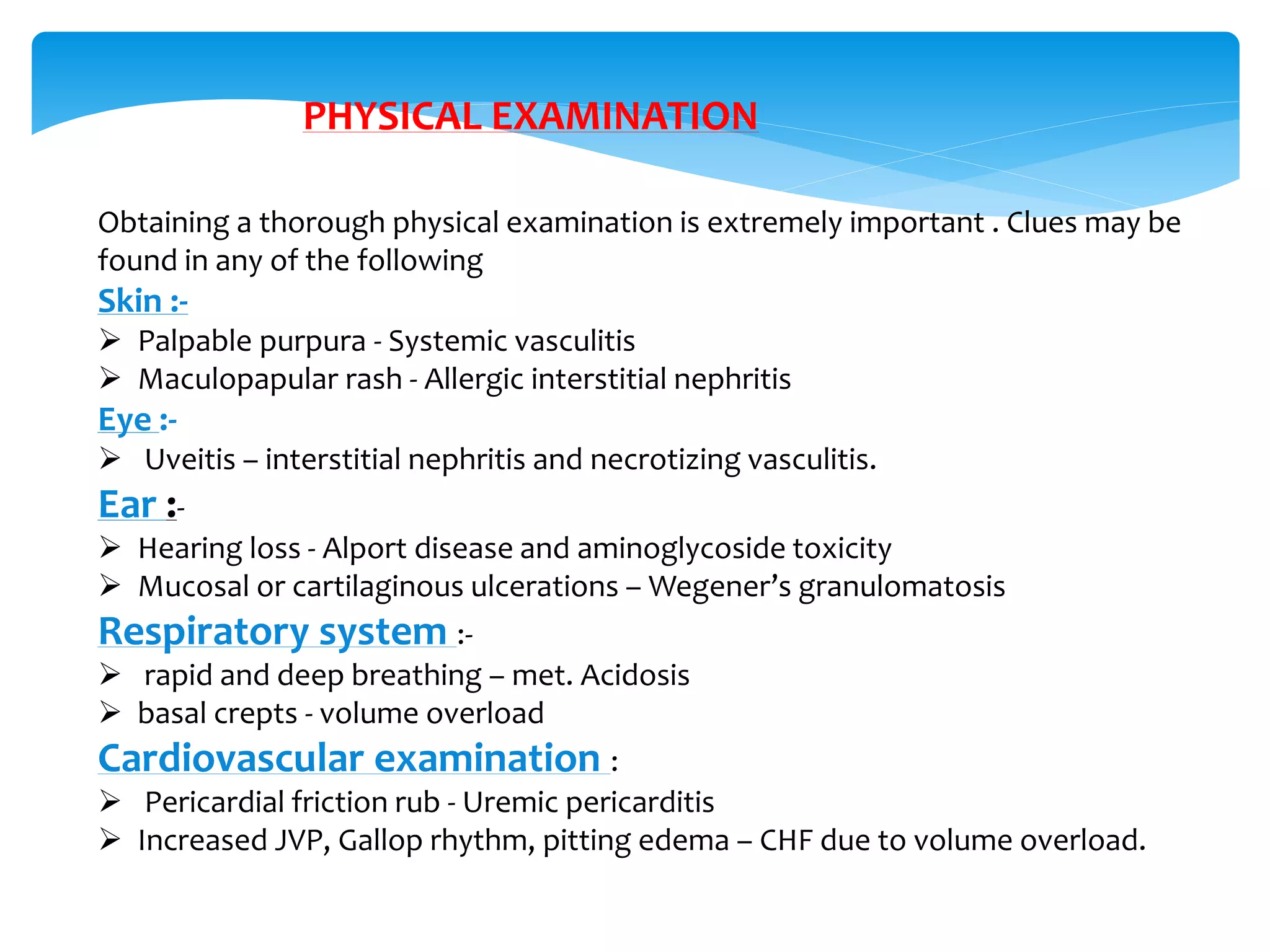
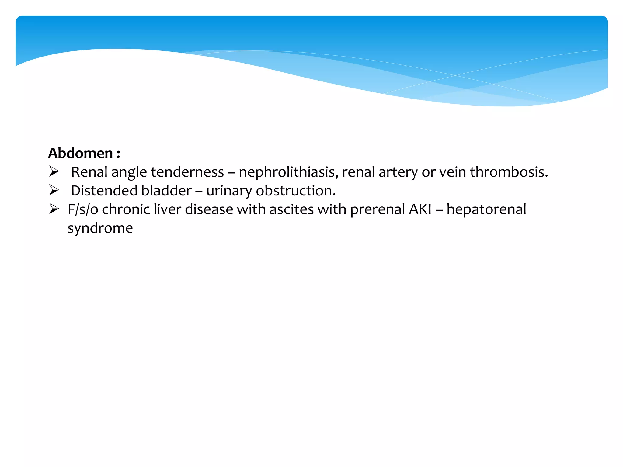
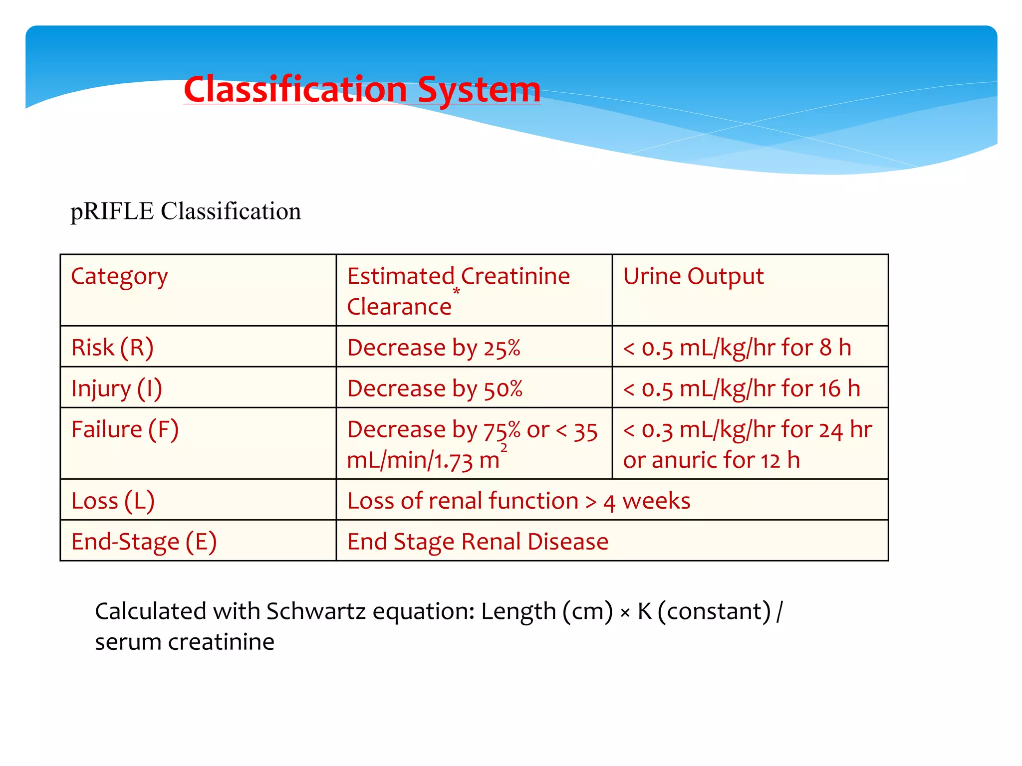
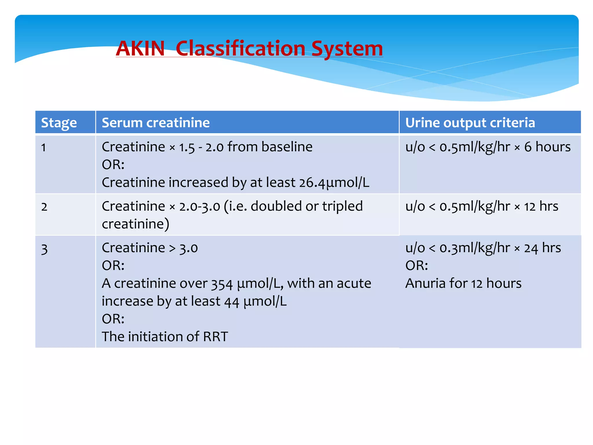

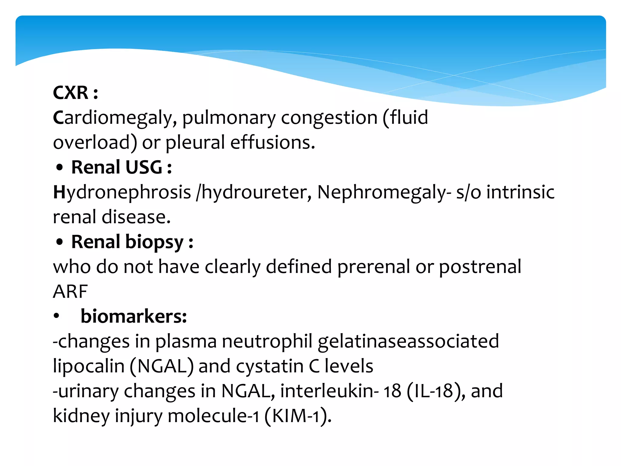

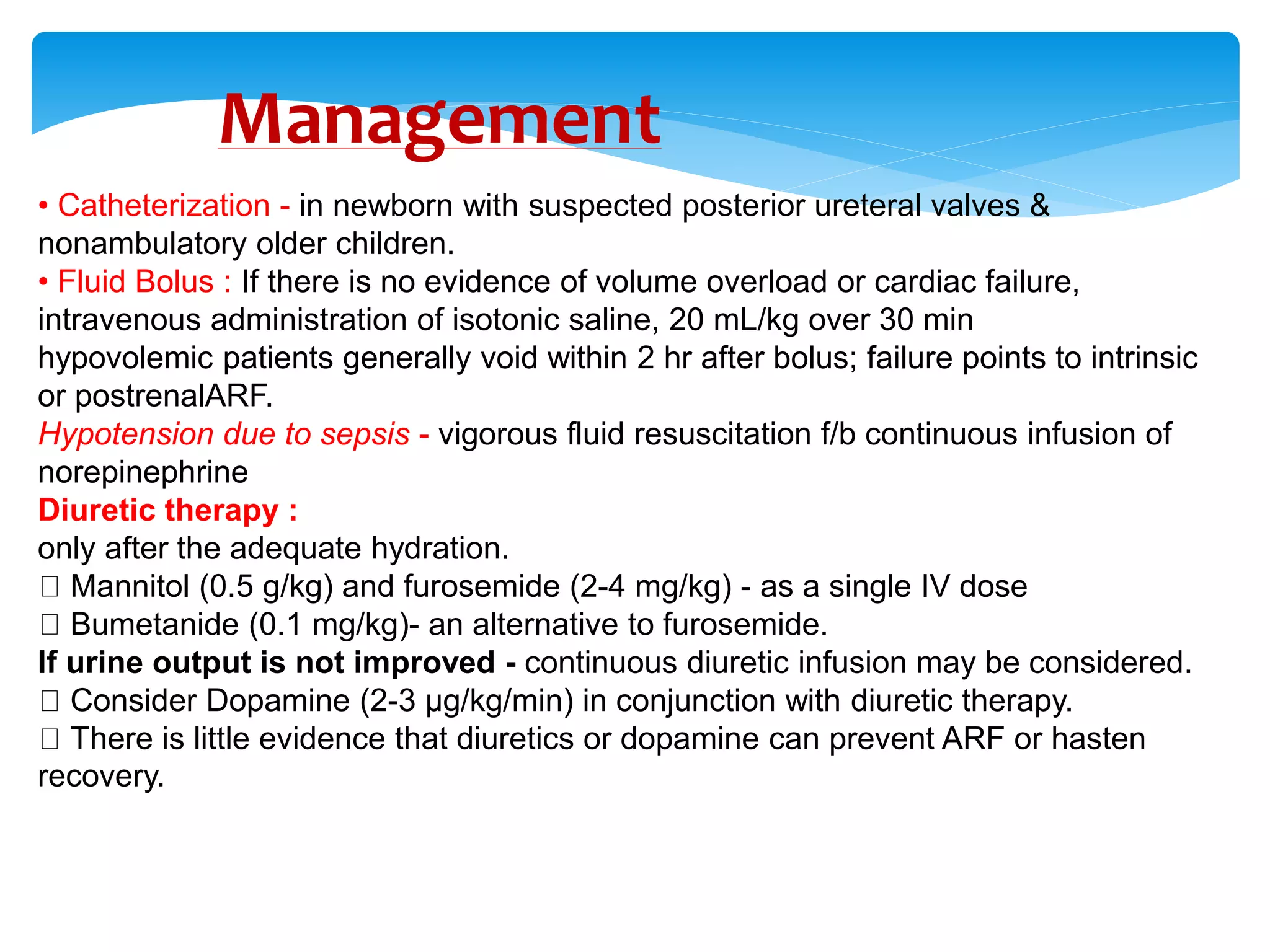


This document provides an overview of paediatric acute renal failure (ARF), including its definition, etiology, clinical evaluation, management, and classification systems. ARF is defined as an abrupt decline in renal function marked by rising creatinine or BUN. Causes of ARF include prerenal (decreased renal perfusion), intrinsic renal disease, and postrenal obstruction. In newborns and children, common etiologies include perinatal issues, gastroenteritis, heart disease, glomerulonephritis, and hemolytic uremic syndrome. ARF is clinically evaluated and managed based on fluid status, electrolyte abnormalities, and dialysis requirements. Staging systems include pRIFLE, AKIN


