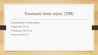
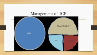
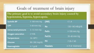
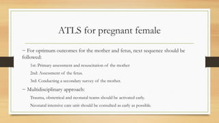
ATLS
PRESENTED BY :
DR.ABBAS RAJEH
CONSULTANT IN EMERGENCY MEDICINE AND
CRITICAL CARE
Advanced Trauma life support
Management consists of:
1. Rapid primary survey, resuscitation of vital functions
2. Secondary survey
3. Tertiary survey and initiation of definitive care.
Primary survey
Goal: Identification of life-threatening conditions by adhering to ABCDE
sequence:
− Airway maintenance with cervical spine protection
− Breathing and ventilation
− Circulation with hemorrhage control
− Disability: Neurologic status
− Exposure/Environmental control.
Airway maintenance with cervical spine
protection
1. Assessment:
− Check airway patency.
− Airway obstruction by debris, foreign bodies, teeth, and vomits.
2. Management:
− Establish a patent airway by:
▪ Perform a chin-lift or jaw-thrust maneuver.
▪ Clear the airway of foreign bodies.
▪ Insert an oropharyngeal airway.
Airway maintenance with cervical spine
protection
− Establish a definitive airway by:
▪ Intubation
▪ Surgical cricothyroidotomy
− Maintain the cervical spine in a neutral position:
▪ with manual immobilization during airway management.
▪ Appropriate devices after establishing airway.
Breathing: ventilation and oxygenation
Assessment
− Expose the neck and chest.
− Determine the rate and depth of respirations.
− Inspect and palpate the neck and chest for tracheal deviation, unilateral and bilateral chest
movement, use of accessory muscles, and any signs of injury.
− Percuss the chest for presence of dullness or hyperresonance.
− Auscultate the chest bilaterally.
− Exclude Pneumothorax.
Breathing: ventilation and oxygenation
Management:
− Administer high-concentration oxygen.
− Ventilate with a bag-mask device.
− Treat pneumothorax:
▪ Alleviate tension pneumothorax: cannula decompression in the 2nd intercostal space
midclavicular line (UPDATES: 4th – 5th in anterior to midaxillary line specially in obese patients
▪ For pediatrics: 2nd intercostal space midclavicular line.
− Attach pulse oximeter.
Circulation with hemorrhage control
Assessment:
− Signs of shock:
▪ Pulse: Quality, rate, regularity, and paradox.
▪ Skin colour.
▪ Blood pressure
▪ UOP
− Identify source bleeding: external, and internal hemorrhage.
Circulation with hemorrhage control
Management
− Apply direct pressure to external bleeding site(s).
− Consider presence of internal hemorrhage and potential need for operative
intervention and obtain surgical consult.
− Insert two large-caliber IV catheters.
− Initiate IV fluid therapy with warmed crystalloid solution and blood replacement.
− Prevent hypothermia.
Disability: brief neurologic examination
− Determine the level of consciousness using the GCS.
− Check pupils for size and reaction.
− Assess for lateralizing signs and spinal cord injury.
Exposure/environmental control
− Completely undress the patient.
− Prevent hypothermia.
▪ Ensure a warm environment.
▪ Use warm blankets.
▪ Warm fluids before administering.
Adjuncts to primary survey and resuscitation
− ABG.
− Monitor exhaled CO2.
− ECG.
− Blood sample for:
▪ Hematologic, cross match and blood grouping, coagulation profile.
▪ chemical analyses: electrolytes, toxicology, glucose, lactate.
▪ pregnancy test
Adjuncts to primary survey and resuscitation
− Insert urinary catheter and monitor UOP hourly.
− gastric catheters unless contraindicated.
− AP chest and AP pelvic x-rays.
− FAST or DPL.
Secondary Survey
− Does not begin until the primary survey (ABCDEs) is completed.
− Head-to-toe evaluation of the patient by:
▪ History
▪ Physical examination.
1. History and mechanism of injury
AMPLE Menominee for history:
− Allergies
− Medications currently used.
− Past illnesses/Pregnancy
− Last meal
− Events/Environment related to the injury to identify injury mechanisms.
2. Physical examination:
− Head and maxillofacial
Assessment:
▪ Lacerations, contusions, fractures, and thermal injury.
▪ Re-evaluate pupils and LOC.
▪ Eyes, mouth, cranial-nerve function
▪ CSF leakage.
2. Physical examination:
Management
▪ Maintain airway, and continue ventilation and oxygenation as indicated.
▪ Control hemorrhage.
▪ Prevent secondary brain injury.
▪ Remove contact lenses.
Cervical spine and neck
Assessment
▪ Palpate for tenderness, deformity, swelling, subcutaneous emphysema, tracheal
deviation, and symmetry of pulses.
▪ Accessory respiratory muscles.
▪ CT or a lateral x-ray of the cervical spine
Management:
▪ Maintain adequate in-line immobilization and protection of the cervical spine.
Chest
Assessment
▪ Inspection:
o chest wall for signs of blunt and penetrating injury
o use of accessory breathing muscles.
▪ Auscultation:
o Bilateral breath sounds and heart sounds.
Chest
▪ palpation
o For evidence of blunt and penetrating injury
o subcutaneous emphysema
o tenderness and crepitation.
▪ Percussion:
o Hyperresonance or dullness.
Chest
Management
▪ Chest tube if indicated.
▪ Dress an open chest wound.
▪ Pericardiocentesis, as indicated.
▪ Shift the patient to OR if required.
Abdomen
Assessment
▪ Inspection:
o For signs of blunt and penetrating injury.
▪ Auscultation:
o Presence of bowel sounds.
▪ Percussion:
o Rebound tenderness.
Abdomen
▪ palpation
o Tenderness
o Involuntary muscle guarding
o Gravid uterus.
▪ Pelvic x-ray film.
▪ abdominal ultrasound – DPL
▪ CT of the abdomen if the patient is hemodynamically normal.
Abdomen
Management
▪ Transfer the patient to the operating room, if indicated
▪ IF pelvic fracture: Wrap a sheet around the pelvis or apply a pelvic compression
binder as indicated to reduce pelvic volume and control of bleeding.
Perineum/rectum/vagina
▪ Perineal assessment.
o Contusions and hematomas, lacerations urethral bleeding
▪ Rectal assessment in selected patients.
o Rectal blood
o Anal sphincter tone
o Bowel wall integrity
Perineum/rectum/vagina
o Bony fragments
o Prostate position
▪ Vaginal assessment in selected patients.
o blood in vaginal vault, lacerations
Musculoskeletal
Assessment
▪ Upper and lower extremities for blunt and penetrating injury.
▪ complete neurovascular examination of the extremities.
▪ Thoracic and lumbar spines for tenderness, deformity, and sensation.
▪ x-ray films of suspected fracture sites as indicated.
▪ CT for spines.
Musculoskeletal
Management
▪ Splinting devices for extremity fractures.
▪ Maintain immobilization of the patient’s thoracic and lumbar spines.
▪ Administer tetanus immunization.
▪ Consider the possibility of compartment syndrome.
Adjuncts to secondary survey
▪ Contrast urography
▪ Angiography
▪ Transesophageal ultrasound
▪ Bronchoscopy
▪ Esophagoscopy
Tertiary survey
− An approach to address the issue of undiagnosed injuries in trauma patients.
− consists of re-evaluating patients 24 hours after admission by means of an
anamnesis protocol, physical examination, review of complementary tests and
request for new tests when necessary.
Airway management in trauma patient
Airway management algorithm in
(NON-airway) trauma patients
Airway management
Modified rapid sequence induction with inline manual stabilization using:
− Etomidate + Succ (avoid in sever crush injury and after 24 hour)
− Fentanyl + Ketamine + rocuronium (if sugammadex is available)
Airway management algorithm in (airway)
trauma patients
Management of surgical emphysema
− Stop PPV
− 100% oxygen: to replace air the subcutaneous tissue.
− Exclude pneumothorax.
− Advance ETT tip distal to the tear
− Fasciotomy in severe cases
Traumatic brain injury (TBI)
Classifications of Brain injury:
▪ Mild: GCS 13-15
▪ Moderate: GCS 9-12
▪ Severe: GCS 3-8
Management of trauma patient with TBI
1. primary survey, adhering to the ABCDE approach.
− If intubation is required:
▪ perform and document neurological examination before administering drugs for
intubation.
▪ Avoid increasing ICP during intubation.
Management of trauma patient with TBI
▪ Airway management:
o If airway is assured: Modified rapid sequence induction with manual inline
stabilization.
o If not: Drug assisted awake tracheal intubation.
▪ Assess the adequacy of breathing next, and monitor oxygen saturation.
− Treat hypotension, hypovolemia, and hypoxia
− Focused neurological examination.
− Type and crossmatch, coagulation studies
Management of trauma patient with TBI
2. Assessment of severity.
3. Secondary survey
− AMPLE history
− CT scan:
▪ To be done after patient stabilization (Systolic Bp >100 mmHg)
▪ If cannot achieve SBP target search for cause of hypotension and manage (fluids,
pressors or surgical intervention) then CT.
− Frequent serial neurological examinations with GCS.
Management of ICP
Management of ICP
− CPP = MAP – ICP.
− To maintain adequate CPP, both (MAP – ICP) are to be strictly optimized.
A. Keep SBP > 100 mmHg.
B. Decreases ICP:
Regarding Monro- kellie doctrine: the total volume of the intracranial contents
(brain, blood and CSF) must remain constant.
Goals of treatment of brain injury
The primary goal is to avoid secondary brain injury caused by
hypotension, hypoxia, hypercapnia.
The ICP can be controlled by controlling these
3 components:
1. Blood
2. Brain tissue
3. CSF drainage
Blood
▪ Elevate head of the bed 30 degree
▪ Loosen collars, ETT ties to promote venous drainage.
▪ Head midline
▪ Decrease intrathoracic pressures (change ventilator settings, decrease PEEP).
▪ Avoid internal jugular venous cannulation.
▪ Hyperventilation: Pco2 35-40 mmHg.
▪ Sedate
▪ Analgesia
Brain tissue
▪ Mannitol (0.5-1 g/kg)
▪ Furosemide (0.25-0.5 mg/kg)
▪ Hypertonic saline 6-8 ml/kg of 3% saline
▪ Correct sodium and osmolality
CSF drainage
If failed: ICP >20 mmHg.
▪ Paralysis
▪ Increase analgesia and sedation.
▪ Hyperventilation: Pco2 30-35 mmHg
▪ Barbiturate coma (1-5 mg/kg sodium thiopental then 1-3 mg/kg/hr)
▪ Hypothermia (or at least normothermia)
▪ Surgical decompression (head, abdomen)
CSF drainage
Consider:
▪ Seizure prophylaxis and management
▪ Shivering treatment
▪ Glycaemic control
Complications of TBI
− CNS: herniation, seizures
− CVS: myocardial dysfunction and ST changes, arrhythmias
− Pulmonary: neurogenic pulmonary edema
− DIC (disseminated intravascular coagulopathy)
− DI (diabetes insipidus), SIADH (syndrome of inappropriate ADH), CSW
(cerebral salt wasting)
ATLS for pregnant female
− For optimum outcomes for the mother and fetus, next sequence should be
followed:
1st: Primary assessment and resuscitation of the mother
2nd: Assessment of the fetus.
3rd: Conducting a secondary survey of the mother.
− Multidisciplinary approach:
Trauma, obstetrical and neonatal teams should be activated early.
Neonatal intensive care unit should be consulted as early as possible.
Primary Survey
Goal: Identification of mother life-threatening conditions by adhering to
ABCDE sequence:
− Check airway patency.
− Airway obstruction by debris, foreign bodies, teeth, and vomits.
− Maintain the cervical spine in a neutral position.
Airway
If Intubation is required, consider:
▪ preoxygenated adequately
▪ Airway management is further complicated due pregnancy-related changes.
▪ Aspiration prophylaxis.
▪ Modified rapid sequence induction with cricoid pressure and manual inline
stabilization.
▪ NGT after intubation.
Breathing
− Expose the neck and chest.
− Inspection, palpation, and percussion as non-pregnant.
− Exclude Pneumothorax.
− management of a pneumothorax proceeds with placement of a tube thoracostomy 1
to 2 intercostal spaces higher, anterior to the mid-axillary line.
Circulation
− Signs of shock developed late despite
loss of a large amount of blood due to
physiological changes of pregnancy.
− Identify source bleeding: external, and
internal hemorrhage.
Causes of shock
• Hemorrhage
• Direct cardiac injury
• Obstructive mechanisms
such as tamponade
• Tension pneumothorax
Circulation
− 2 large-bore IV cannulas placed supradiaphragmatic.
− Uterine displacement manually or by placing the patient in left lateral decubitus
position.
− Initiate IV fluid therapy with warmed crystalloid solution and blood
replacement.
− Vasopressors as a last resort to avoid reduction in uterine blood flow.
− Un-crossmatched type O, Rh (-) blood reduces the risk of alloimmunization
until type specific blood is available.
Disability
− GCS, pupillary examination, signs of lateralization.
trauma triad of death
− Hypothermia
− Coagulopathy
− Acidosis.
Exposure and environmental control
− As in the general population
− Prevent hypothermia.
Fetal assessment
− Ultrasound for:
▪ fetal heart rate
▪ fetal movement
▪ amniotic fluid quantity
▪ position of the placenta
▪ gestational age
Fetal assessment
− cardiotocographic (CTG): if 24 weeks for a minimum of 4 hours.
− The presence of prolonged, painful, and/or regular contractions necessitates
further monitoring and obstetric intervention.
Secondary Survey
History
− use the mnemonic AMPLE.
Physical exam
− As non-pregnant.
− Uterine and vaginal examination
Imaging Studies
− Focused assessment with sonography for trauma (FAST) to replace X-ray if possible.
− X-ray: if required, use abdominal shield
Secondary Survey
− CT scan:
▪ Selective CT rather than pan CT.
▪ Reduce radiation dose.
▪ Use abdominal and pelvic shield.
− CT scan:
▪ Selective CT rather than pan CT.
▪ Reduce radiation dose.
▪ Use abdominal and pelvic shield.
Secondary Survey
Laboratory Testing
− Rh status
− CBC
− The Kleihauer-Betke (KB) test: to detect the approximate the volume of
fetal-maternal hemorrhage (FMH)
− INR, PTT, fibrin degradation, Fibrinogen.
Medications
− Rh-negative: RhIG 1 ampule (300 g) IM
− Corticosteroids: between 24- and 34-weeks gestational age.
− Tocolytics
▪ magnesium sulfate
▪ corticosteroids.
▪ Avoid beta adrenergic activity (e.g. terbutaline), causes tachycardia and/or hypotension.
− Tranexamic Acid:1 g infused over 10 minutes, followed by an infusion of 1 g over eight
hours.
Complications Specific to Trauma in Pregnancy
− Placental abruption:
▪ The second highest cause of perinatal mortality from trauma.
▪ Diagnosis:
o Clinically with cardiotocographic fetal monitoring
o CT imaging is more sensitive for diagnosis than ultrasonography.
Complications Specific to Trauma in Pregnancy
Uterine rupture:
▪ Irregularly shaped uterus, palpable fetal parts, or abdominal tenderness.
▪ Treatment: laparotomy with fetal delivery and either hysterectomy or uterine
Complications Specific to Trauma in Pregnancy
Amniotic fluid embolism:
▪ sudden hypoxemia and cardiovascular collapse and can progress to DIC and
multi-organ failure.
▪ The mechanism:
▪ amniotic fluid entering the maternal circulation and leading to either vascular
obstruction or anaphylaxis.
▪ Treatment is supportive with resuscitation and potential delivery of the fetus.
Complications Specific to Trauma in Pregnancy
• − preterm labour,
• − preterm premature rupture of membranes (PPROM)
Cardiac arrest
Modifications of ACLS in maternal cardiac arrest:
1. Chest compression is higher and deeper.
2. Early intubation (suspected difficult intubation, most experienced should perform)
3. IV line to be supradiaphragmatic.
4. Relieving aortocaval compression.
5. If No ROSC in 4 min, Perimortem CS over one min.
Cardiac arrest
Other considerations:
▪ If on mgSo4, stop and start Ca chloride or gluconate.
▪ Remove fetal monitoring.
Common causes of maternal cardiac arrest
− Anesthesia complications
▪ High neuraxial block
▪ Aspiration
▪ Local anesthetic toxicity
▪ Hypotension
▪ Respiratory depression
− Accidents
▪ Trauma
▪ Suicide
Bleeding
▪ Uterine atony
▪ Placenta accreta
▪ Placental abruption and previa
▪ Uterine rupture
▪ Coagulopathy
▪ Transfusion reaction
Common causes of maternal cardiac arrest
− Cardiovascular
▪ Arrhythmia
▪ Myocardial infarction
▪ Congenital heart disease
▪ Aortic dissection
▪ Heart failure
-Drugs
▪ Oxytocin
▪ Magnesium
▪ Opioids
▪ Anaphylaxis
▪ Drug administration error
− Embolism
▪ Pulmonary embolus
▪ Amniotic fluid embolus
▪ Venous air embolism
▪ Cerebrovascular event
− Fever
▪ Sepsis
▪ Infection
Common causes of maternal cardiac arrest
− Hypertension
▪ Pre-eclampsia and
eclampsia
▪ HELLP syndrome
− General
▪ Hypoxia
▪ Hypovolemia
▪ Hypokalemia/hyperkalemia
▪ Tamponade
▪ Toxins
THANKS FOR YOUR ATTENTION

lectures ATLS الدعم المتقدم للصدمات.pptx

  • 1.
    ATLS PRESENTED BY : DR.ABBASRAJEH CONSULTANT IN EMERGENCY MEDICINE AND CRITICAL CARE
  • 2.
  • 3.
    Advanced Trauma lifesupport Management consists of: 1. Rapid primary survey, resuscitation of vital functions 2. Secondary survey 3. Tertiary survey and initiation of definitive care.
  • 4.
    Primary survey Goal: Identificationof life-threatening conditions by adhering to ABCDE sequence: − Airway maintenance with cervical spine protection − Breathing and ventilation − Circulation with hemorrhage control − Disability: Neurologic status − Exposure/Environmental control.
  • 5.
    Airway maintenance withcervical spine protection 1. Assessment: − Check airway patency. − Airway obstruction by debris, foreign bodies, teeth, and vomits. 2. Management: − Establish a patent airway by: ▪ Perform a chin-lift or jaw-thrust maneuver. ▪ Clear the airway of foreign bodies. ▪ Insert an oropharyngeal airway.
  • 6.
    Airway maintenance withcervical spine protection − Establish a definitive airway by: ▪ Intubation ▪ Surgical cricothyroidotomy − Maintain the cervical spine in a neutral position: ▪ with manual immobilization during airway management. ▪ Appropriate devices after establishing airway.
  • 7.
    Breathing: ventilation andoxygenation Assessment − Expose the neck and chest. − Determine the rate and depth of respirations. − Inspect and palpate the neck and chest for tracheal deviation, unilateral and bilateral chest movement, use of accessory muscles, and any signs of injury. − Percuss the chest for presence of dullness or hyperresonance. − Auscultate the chest bilaterally. − Exclude Pneumothorax.
  • 8.
    Breathing: ventilation andoxygenation Management: − Administer high-concentration oxygen. − Ventilate with a bag-mask device. − Treat pneumothorax: ▪ Alleviate tension pneumothorax: cannula decompression in the 2nd intercostal space midclavicular line (UPDATES: 4th – 5th in anterior to midaxillary line specially in obese patients ▪ For pediatrics: 2nd intercostal space midclavicular line. − Attach pulse oximeter.
  • 9.
    Circulation with hemorrhagecontrol Assessment: − Signs of shock: ▪ Pulse: Quality, rate, regularity, and paradox. ▪ Skin colour. ▪ Blood pressure ▪ UOP − Identify source bleeding: external, and internal hemorrhage.
  • 10.
    Circulation with hemorrhagecontrol Management − Apply direct pressure to external bleeding site(s). − Consider presence of internal hemorrhage and potential need for operative intervention and obtain surgical consult. − Insert two large-caliber IV catheters. − Initiate IV fluid therapy with warmed crystalloid solution and blood replacement. − Prevent hypothermia.
  • 11.
    Disability: brief neurologicexamination − Determine the level of consciousness using the GCS. − Check pupils for size and reaction. − Assess for lateralizing signs and spinal cord injury.
  • 12.
    Exposure/environmental control − Completelyundress the patient. − Prevent hypothermia. ▪ Ensure a warm environment. ▪ Use warm blankets. ▪ Warm fluids before administering.
  • 13.
    Adjuncts to primarysurvey and resuscitation − ABG. − Monitor exhaled CO2. − ECG. − Blood sample for: ▪ Hematologic, cross match and blood grouping, coagulation profile. ▪ chemical analyses: electrolytes, toxicology, glucose, lactate. ▪ pregnancy test
  • 14.
    Adjuncts to primarysurvey and resuscitation − Insert urinary catheter and monitor UOP hourly. − gastric catheters unless contraindicated. − AP chest and AP pelvic x-rays. − FAST or DPL.
  • 15.
    Secondary Survey − Doesnot begin until the primary survey (ABCDEs) is completed. − Head-to-toe evaluation of the patient by: ▪ History ▪ Physical examination.
  • 16.
    1. History andmechanism of injury AMPLE Menominee for history: − Allergies − Medications currently used. − Past illnesses/Pregnancy − Last meal − Events/Environment related to the injury to identify injury mechanisms.
  • 17.
    2. Physical examination: −Head and maxillofacial Assessment: ▪ Lacerations, contusions, fractures, and thermal injury. ▪ Re-evaluate pupils and LOC. ▪ Eyes, mouth, cranial-nerve function ▪ CSF leakage.
  • 18.
    2. Physical examination: Management ▪Maintain airway, and continue ventilation and oxygenation as indicated. ▪ Control hemorrhage. ▪ Prevent secondary brain injury. ▪ Remove contact lenses.
  • 19.
    Cervical spine andneck Assessment ▪ Palpate for tenderness, deformity, swelling, subcutaneous emphysema, tracheal deviation, and symmetry of pulses. ▪ Accessory respiratory muscles. ▪ CT or a lateral x-ray of the cervical spine Management: ▪ Maintain adequate in-line immobilization and protection of the cervical spine.
  • 20.
    Chest Assessment ▪ Inspection: o chestwall for signs of blunt and penetrating injury o use of accessory breathing muscles. ▪ Auscultation: o Bilateral breath sounds and heart sounds.
  • 21.
    Chest ▪ palpation o Forevidence of blunt and penetrating injury o subcutaneous emphysema o tenderness and crepitation. ▪ Percussion: o Hyperresonance or dullness.
  • 22.
    Chest Management ▪ Chest tubeif indicated. ▪ Dress an open chest wound. ▪ Pericardiocentesis, as indicated. ▪ Shift the patient to OR if required.
  • 23.
    Abdomen Assessment ▪ Inspection: o Forsigns of blunt and penetrating injury. ▪ Auscultation: o Presence of bowel sounds. ▪ Percussion: o Rebound tenderness.
  • 24.
    Abdomen ▪ palpation o Tenderness oInvoluntary muscle guarding o Gravid uterus. ▪ Pelvic x-ray film. ▪ abdominal ultrasound – DPL ▪ CT of the abdomen if the patient is hemodynamically normal.
  • 25.
    Abdomen Management ▪ Transfer thepatient to the operating room, if indicated ▪ IF pelvic fracture: Wrap a sheet around the pelvis or apply a pelvic compression binder as indicated to reduce pelvic volume and control of bleeding.
  • 26.
    Perineum/rectum/vagina ▪ Perineal assessment. oContusions and hematomas, lacerations urethral bleeding ▪ Rectal assessment in selected patients. o Rectal blood o Anal sphincter tone o Bowel wall integrity
  • 27.
    Perineum/rectum/vagina o Bony fragments oProstate position ▪ Vaginal assessment in selected patients. o blood in vaginal vault, lacerations
  • 28.
    Musculoskeletal Assessment ▪ Upper andlower extremities for blunt and penetrating injury. ▪ complete neurovascular examination of the extremities. ▪ Thoracic and lumbar spines for tenderness, deformity, and sensation. ▪ x-ray films of suspected fracture sites as indicated. ▪ CT for spines.
  • 29.
    Musculoskeletal Management ▪ Splinting devicesfor extremity fractures. ▪ Maintain immobilization of the patient’s thoracic and lumbar spines. ▪ Administer tetanus immunization. ▪ Consider the possibility of compartment syndrome.
  • 30.
    Adjuncts to secondarysurvey ▪ Contrast urography ▪ Angiography ▪ Transesophageal ultrasound ▪ Bronchoscopy ▪ Esophagoscopy
  • 31.
    Tertiary survey − Anapproach to address the issue of undiagnosed injuries in trauma patients. − consists of re-evaluating patients 24 hours after admission by means of an anamnesis protocol, physical examination, review of complementary tests and request for new tests when necessary.
  • 32.
    Airway management intrauma patient
  • 34.
    Airway management algorithmin (NON-airway) trauma patients
  • 36.
    Airway management Modified rapidsequence induction with inline manual stabilization using: − Etomidate + Succ (avoid in sever crush injury and after 24 hour) − Fentanyl + Ketamine + rocuronium (if sugammadex is available)
  • 37.
    Airway management algorithmin (airway) trauma patients
  • 39.
    Management of surgicalemphysema − Stop PPV − 100% oxygen: to replace air the subcutaneous tissue. − Exclude pneumothorax. − Advance ETT tip distal to the tear − Fasciotomy in severe cases
  • 40.
    Traumatic brain injury(TBI) Classifications of Brain injury: ▪ Mild: GCS 13-15 ▪ Moderate: GCS 9-12 ▪ Severe: GCS 3-8
  • 41.
    Management of traumapatient with TBI 1. primary survey, adhering to the ABCDE approach. − If intubation is required: ▪ perform and document neurological examination before administering drugs for intubation. ▪ Avoid increasing ICP during intubation.
  • 42.
    Management of traumapatient with TBI ▪ Airway management: o If airway is assured: Modified rapid sequence induction with manual inline stabilization. o If not: Drug assisted awake tracheal intubation. ▪ Assess the adequacy of breathing next, and monitor oxygen saturation. − Treat hypotension, hypovolemia, and hypoxia − Focused neurological examination. − Type and crossmatch, coagulation studies
  • 43.
    Management of traumapatient with TBI 2. Assessment of severity. 3. Secondary survey − AMPLE history − CT scan: ▪ To be done after patient stabilization (Systolic Bp >100 mmHg) ▪ If cannot achieve SBP target search for cause of hypotension and manage (fluids, pressors or surgical intervention) then CT. − Frequent serial neurological examinations with GCS.
  • 44.
  • 45.
    Management of ICP −CPP = MAP – ICP. − To maintain adequate CPP, both (MAP – ICP) are to be strictly optimized. A. Keep SBP > 100 mmHg. B. Decreases ICP: Regarding Monro- kellie doctrine: the total volume of the intracranial contents (brain, blood and CSF) must remain constant.
  • 46.
    Goals of treatmentof brain injury The primary goal is to avoid secondary brain injury caused by hypotension, hypoxia, hypercapnia.
  • 47.
    The ICP canbe controlled by controlling these 3 components: 1. Blood 2. Brain tissue 3. CSF drainage
  • 48.
    Blood ▪ Elevate headof the bed 30 degree ▪ Loosen collars, ETT ties to promote venous drainage. ▪ Head midline ▪ Decrease intrathoracic pressures (change ventilator settings, decrease PEEP). ▪ Avoid internal jugular venous cannulation. ▪ Hyperventilation: Pco2 35-40 mmHg. ▪ Sedate ▪ Analgesia
  • 49.
    Brain tissue ▪ Mannitol(0.5-1 g/kg) ▪ Furosemide (0.25-0.5 mg/kg) ▪ Hypertonic saline 6-8 ml/kg of 3% saline ▪ Correct sodium and osmolality
  • 50.
    CSF drainage If failed:ICP >20 mmHg. ▪ Paralysis ▪ Increase analgesia and sedation. ▪ Hyperventilation: Pco2 30-35 mmHg ▪ Barbiturate coma (1-5 mg/kg sodium thiopental then 1-3 mg/kg/hr) ▪ Hypothermia (or at least normothermia) ▪ Surgical decompression (head, abdomen)
  • 51.
    CSF drainage Consider: ▪ Seizureprophylaxis and management ▪ Shivering treatment ▪ Glycaemic control
  • 52.
    Complications of TBI −CNS: herniation, seizures − CVS: myocardial dysfunction and ST changes, arrhythmias − Pulmonary: neurogenic pulmonary edema − DIC (disseminated intravascular coagulopathy) − DI (diabetes insipidus), SIADH (syndrome of inappropriate ADH), CSW (cerebral salt wasting)
  • 53.
    ATLS for pregnantfemale − For optimum outcomes for the mother and fetus, next sequence should be followed: 1st: Primary assessment and resuscitation of the mother 2nd: Assessment of the fetus. 3rd: Conducting a secondary survey of the mother. − Multidisciplinary approach: Trauma, obstetrical and neonatal teams should be activated early. Neonatal intensive care unit should be consulted as early as possible.
  • 54.
    Primary Survey Goal: Identificationof mother life-threatening conditions by adhering to ABCDE sequence: − Check airway patency. − Airway obstruction by debris, foreign bodies, teeth, and vomits. − Maintain the cervical spine in a neutral position.
  • 55.
    Airway If Intubation isrequired, consider: ▪ preoxygenated adequately ▪ Airway management is further complicated due pregnancy-related changes. ▪ Aspiration prophylaxis. ▪ Modified rapid sequence induction with cricoid pressure and manual inline stabilization. ▪ NGT after intubation.
  • 56.
    Breathing − Expose theneck and chest. − Inspection, palpation, and percussion as non-pregnant. − Exclude Pneumothorax. − management of a pneumothorax proceeds with placement of a tube thoracostomy 1 to 2 intercostal spaces higher, anterior to the mid-axillary line.
  • 57.
    Circulation − Signs ofshock developed late despite loss of a large amount of blood due to physiological changes of pregnancy. − Identify source bleeding: external, and internal hemorrhage. Causes of shock • Hemorrhage • Direct cardiac injury • Obstructive mechanisms such as tamponade • Tension pneumothorax
  • 58.
    Circulation − 2 large-boreIV cannulas placed supradiaphragmatic. − Uterine displacement manually or by placing the patient in left lateral decubitus position. − Initiate IV fluid therapy with warmed crystalloid solution and blood replacement. − Vasopressors as a last resort to avoid reduction in uterine blood flow. − Un-crossmatched type O, Rh (-) blood reduces the risk of alloimmunization until type specific blood is available.
  • 59.
    Disability − GCS, pupillaryexamination, signs of lateralization. trauma triad of death − Hypothermia − Coagulopathy − Acidosis.
  • 60.
    Exposure and environmentalcontrol − As in the general population − Prevent hypothermia.
  • 61.
    Fetal assessment − Ultrasoundfor: ▪ fetal heart rate ▪ fetal movement ▪ amniotic fluid quantity ▪ position of the placenta ▪ gestational age
  • 62.
    Fetal assessment − cardiotocographic(CTG): if 24 weeks for a minimum of 4 hours. − The presence of prolonged, painful, and/or regular contractions necessitates further monitoring and obstetric intervention.
  • 63.
    Secondary Survey History − usethe mnemonic AMPLE. Physical exam − As non-pregnant. − Uterine and vaginal examination Imaging Studies − Focused assessment with sonography for trauma (FAST) to replace X-ray if possible. − X-ray: if required, use abdominal shield
  • 64.
    Secondary Survey − CTscan: ▪ Selective CT rather than pan CT. ▪ Reduce radiation dose. ▪ Use abdominal and pelvic shield.
  • 65.
    − CT scan: ▪Selective CT rather than pan CT. ▪ Reduce radiation dose. ▪ Use abdominal and pelvic shield.
  • 66.
    Secondary Survey Laboratory Testing −Rh status − CBC − The Kleihauer-Betke (KB) test: to detect the approximate the volume of fetal-maternal hemorrhage (FMH) − INR, PTT, fibrin degradation, Fibrinogen.
  • 67.
    Medications − Rh-negative: RhIG1 ampule (300 g) IM − Corticosteroids: between 24- and 34-weeks gestational age. − Tocolytics ▪ magnesium sulfate ▪ corticosteroids. ▪ Avoid beta adrenergic activity (e.g. terbutaline), causes tachycardia and/or hypotension. − Tranexamic Acid:1 g infused over 10 minutes, followed by an infusion of 1 g over eight hours.
  • 68.
    Complications Specific toTrauma in Pregnancy − Placental abruption: ▪ The second highest cause of perinatal mortality from trauma. ▪ Diagnosis: o Clinically with cardiotocographic fetal monitoring o CT imaging is more sensitive for diagnosis than ultrasonography.
  • 69.
    Complications Specific toTrauma in Pregnancy Uterine rupture: ▪ Irregularly shaped uterus, palpable fetal parts, or abdominal tenderness. ▪ Treatment: laparotomy with fetal delivery and either hysterectomy or uterine
  • 70.
    Complications Specific toTrauma in Pregnancy Amniotic fluid embolism: ▪ sudden hypoxemia and cardiovascular collapse and can progress to DIC and multi-organ failure. ▪ The mechanism: ▪ amniotic fluid entering the maternal circulation and leading to either vascular obstruction or anaphylaxis. ▪ Treatment is supportive with resuscitation and potential delivery of the fetus.
  • 71.
    Complications Specific toTrauma in Pregnancy • − preterm labour, • − preterm premature rupture of membranes (PPROM)
  • 72.
    Cardiac arrest Modifications ofACLS in maternal cardiac arrest: 1. Chest compression is higher and deeper. 2. Early intubation (suspected difficult intubation, most experienced should perform) 3. IV line to be supradiaphragmatic. 4. Relieving aortocaval compression. 5. If No ROSC in 4 min, Perimortem CS over one min.
  • 73.
    Cardiac arrest Other considerations: ▪If on mgSo4, stop and start Ca chloride or gluconate. ▪ Remove fetal monitoring.
  • 74.
    Common causes ofmaternal cardiac arrest − Anesthesia complications ▪ High neuraxial block ▪ Aspiration ▪ Local anesthetic toxicity ▪ Hypotension ▪ Respiratory depression − Accidents ▪ Trauma ▪ Suicide Bleeding ▪ Uterine atony ▪ Placenta accreta ▪ Placental abruption and previa ▪ Uterine rupture ▪ Coagulopathy ▪ Transfusion reaction
  • 75.
    Common causes ofmaternal cardiac arrest − Cardiovascular ▪ Arrhythmia ▪ Myocardial infarction ▪ Congenital heart disease ▪ Aortic dissection ▪ Heart failure -Drugs ▪ Oxytocin ▪ Magnesium ▪ Opioids ▪ Anaphylaxis ▪ Drug administration error − Embolism ▪ Pulmonary embolus ▪ Amniotic fluid embolus ▪ Venous air embolism ▪ Cerebrovascular event − Fever ▪ Sepsis ▪ Infection
  • 76.
    Common causes ofmaternal cardiac arrest − Hypertension ▪ Pre-eclampsia and eclampsia ▪ HELLP syndrome − General ▪ Hypoxia ▪ Hypovolemia ▪ Hypokalemia/hyperkalemia ▪ Tamponade ▪ Toxins
  • 77.
    THANKS FOR YOURATTENTION