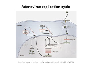
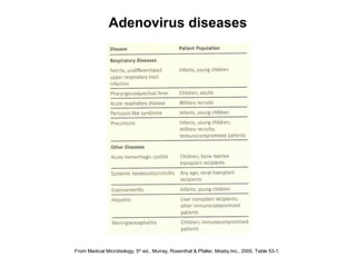
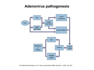
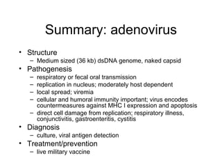
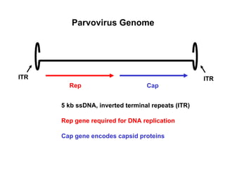
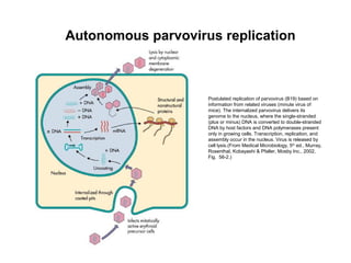
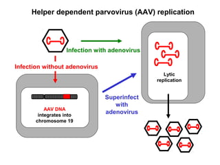
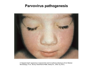
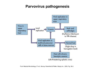
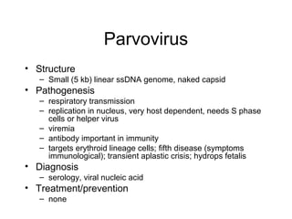
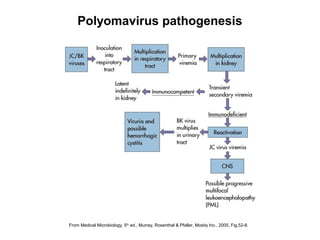
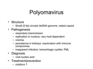
Adenovirus, parvovirus,
polyomavirus
Adenovirus
From Medical Microbiology, 5th
ed., Murray, Rosenthal & Pfaller, Mosby Inc., 2005, Fig. 53-1.
Adenovirus Structure
E1
E2 E4
E3
Late genes
Adenovirus Genome
36 kb linear ds DNA
Early genes for host and viral transcription control,
viral DNA replication
Late genes for virion structure
(From Fields Virology, 4th ed, Knipe & Howley, eds, Lippincott Williams & Wilkins, 2001, Fig. 67-5.)
Adenovirus replication cycle
From Medical Microbiology, 5th
ed., Murray, Rosenthal & Pfaller, Mosby Inc., 2005, Table 53-1.
Adenovirus diseases
From Medical Microbiology, 5th
ed., Murray, Rosenthal & Pfaller, Mosby Inc., 2005,, Fig. 50-4.
Adenovirus pathogenesis
Summary: adenovirus
• Structure
– Medium sized (36 kb) dsDNA genome, naked capsid
• Pathogenesis
– respiratory or fecal oral transmission
– replication in nucleus; moderately host dependent
– local spread; viremia
– cellular and humoral immunity important; virus encodes
countermeasures against MHC I expression and apoptosis
– direct cell damage from replication; respiratory illness,
conjunctivitis, gastroenteritis, cystitis
• Diagnosis
– culture, viral antigen detection
• Treatment/prevention
– live military vaccine
Parvovirus
Parvovirus structure
From Medical Microbiology, 4th
ed., Murray, Rosenthal, Kobayashi & Pfaller, Mosby Inc., 2002, Fig. 53-1.
Parvovirus Genome
Rep Cap
5 kb ssDNA, inverted terminal repeats (ITR)
Rep gene required for DNA replication
Cap gene encodes capsid proteins
ITR ITR
Autonomous parvovirus replication
Postulated replication of parvovirus (B19) based on
information from related viruses (minute virus of
mice). The internalized parvovirus delivers its
genome to the nucleus, where the single-stranded
(plus or minus) DNA is converted to double-stranded
DNA by host factors and DNA polymerases present
only in growing cells. Transcription, replication, and
assembly occur in the nucleus. Virus is released by
cell lysis.(From Medical Microbiology, 5th
ed., Murray,
Rosenthal, Kobayashi & Pfaller, Mosby Inc., 2002,
Fig. 56-2.)
Helper dependent parvovirus (AAV) replication
AAV DNA
integrates into
chromosome 19
Infection without adenovirus
Infection with adenovirus
Superinfect
with
adenovirus
Lytic
replication
A "slapped-cheek" appearance is typical of the rash for erythema infectiosum.(From Medical
Microbiology, 5th
ed., Murray, Rosenthal & Pfaller, Mosby Inc., 2005, Fig. 56-5.)
Parvovirus pathogenesis
From Medical Microbiology, 5th
ed., Murray, Rosenthal & Pfaller, Mosby Inc., 2005, Fig. 56-3.
Parvovirus pathogenesis
Parvovirus
• Structure
– Small (5 kb) linear ssDNA genome, naked capsid
• Pathogenesis
– respiratory transmission
– replication in nucleus, very host dependent, needs S phase
cells or helper virus
– viremia
– antibody important in immunity
– targets erythroid lineage cells; fifth disease (symptoms
immunological); transient aplastic crisis; hydrops fetalis
• Diagnosis
– serology, viral nucleic acid
• Treatment/prevention
– none
Polyomavirus
Polyomavirus genome
Genome of the SV40 virus. The genome
is a prototype of other polyomaviruses
and contains early, late, and noncoding
regions. The noncoding region contains
the start sequence for the early and late
genes and for DNA replication (ori). The
individual early and late messenger RNAs
are processed from the larger nested
transcripts.(From Medical Microbiology,
5th
ed., Murray, Rosenthal & Pfaller,
Mosby Inc., 2005, Fig. 52-7.)
Replication cycle of polyomaviruses. Steps in the replication cycle are indicated by numbers as follows: 1, adsorption of virions to the cell surface;
2, entry by endocytosis; 3, transport to the cell nucleus (route and mechanism not yet known); 4, uncoating; 5, transcription to produce early
region mRNAs; 6, translation to produce early proteins (T antigens); 7, viral DNA replication; 8, transcription to produce late region mRNAs; 9,
translation to produce late proteins (capsid proteins); 10, assembly of progeny virions in the nucleus; 11, entry of virions into cytoplasmic vesicles
(mechanism unknown); 12, release of virions from the cell by fusion of membrane vesicles with the plasma membrane; 13, released virion. (From
Fields Virology, 4th ed, Knipe & Howley, eds, Lippincott Williams & Wilkins, 2001, Fig. 63-4.)
Polyomavirus replication
From Medical Microbiology, 5th
ed., Murray, Rosenthal & Pfaller, Mosby Inc., 2005, Fig.52-8.
Polyomavirus pathogenesis
Polyomavirus
• Structure
– Small (5 kb) circular dsDNA genome, naked capsid
• Pathogenesis
– respiratory transmission
– replication in nucleus; very host dependent
– viremia
– persistence in kidneys; reactivation with immune
compromise
– inapparent infection; hemorrhagic cystitis; PML
• Diagnosis
– viral nucleic acid
• Treatment/prevention
– cidofovir ?

Adenovirus parvovirus polyomavirus

  • 1.
  • 2.
  • 3.
    From Medical Microbiology,5th ed., Murray, Rosenthal & Pfaller, Mosby Inc., 2005, Fig. 53-1. Adenovirus Structure
  • 4.
    E1 E2 E4 E3 Late genes AdenovirusGenome 36 kb linear ds DNA Early genes for host and viral transcription control, viral DNA replication Late genes for virion structure
  • 5.
    (From Fields Virology,4th ed, Knipe & Howley, eds, Lippincott Williams & Wilkins, 2001, Fig. 67-5.) Adenovirus replication cycle
  • 6.
    From Medical Microbiology,5th ed., Murray, Rosenthal & Pfaller, Mosby Inc., 2005, Table 53-1. Adenovirus diseases
  • 7.
    From Medical Microbiology,5th ed., Murray, Rosenthal & Pfaller, Mosby Inc., 2005,, Fig. 50-4. Adenovirus pathogenesis
  • 8.
    Summary: adenovirus • Structure –Medium sized (36 kb) dsDNA genome, naked capsid • Pathogenesis – respiratory or fecal oral transmission – replication in nucleus; moderately host dependent – local spread; viremia – cellular and humoral immunity important; virus encodes countermeasures against MHC I expression and apoptosis – direct cell damage from replication; respiratory illness, conjunctivitis, gastroenteritis, cystitis • Diagnosis – culture, viral antigen detection • Treatment/prevention – live military vaccine
  • 9.
  • 10.
    Parvovirus structure From MedicalMicrobiology, 4th ed., Murray, Rosenthal, Kobayashi & Pfaller, Mosby Inc., 2002, Fig. 53-1.
  • 11.
    Parvovirus Genome Rep Cap 5kb ssDNA, inverted terminal repeats (ITR) Rep gene required for DNA replication Cap gene encodes capsid proteins ITR ITR
  • 12.
    Autonomous parvovirus replication Postulatedreplication of parvovirus (B19) based on information from related viruses (minute virus of mice). The internalized parvovirus delivers its genome to the nucleus, where the single-stranded (plus or minus) DNA is converted to double-stranded DNA by host factors and DNA polymerases present only in growing cells. Transcription, replication, and assembly occur in the nucleus. Virus is released by cell lysis.(From Medical Microbiology, 5th ed., Murray, Rosenthal, Kobayashi & Pfaller, Mosby Inc., 2002, Fig. 56-2.)
  • 13.
    Helper dependent parvovirus(AAV) replication AAV DNA integrates into chromosome 19 Infection without adenovirus Infection with adenovirus Superinfect with adenovirus Lytic replication
  • 14.
    A "slapped-cheek" appearanceis typical of the rash for erythema infectiosum.(From Medical Microbiology, 5th ed., Murray, Rosenthal & Pfaller, Mosby Inc., 2005, Fig. 56-5.) Parvovirus pathogenesis
  • 15.
    From Medical Microbiology,5th ed., Murray, Rosenthal & Pfaller, Mosby Inc., 2005, Fig. 56-3. Parvovirus pathogenesis
  • 16.
    Parvovirus • Structure – Small(5 kb) linear ssDNA genome, naked capsid • Pathogenesis – respiratory transmission – replication in nucleus, very host dependent, needs S phase cells or helper virus – viremia – antibody important in immunity – targets erythroid lineage cells; fifth disease (symptoms immunological); transient aplastic crisis; hydrops fetalis • Diagnosis – serology, viral nucleic acid • Treatment/prevention – none
  • 17.
  • 19.
    Polyomavirus genome Genome ofthe SV40 virus. The genome is a prototype of other polyomaviruses and contains early, late, and noncoding regions. The noncoding region contains the start sequence for the early and late genes and for DNA replication (ori). The individual early and late messenger RNAs are processed from the larger nested transcripts.(From Medical Microbiology, 5th ed., Murray, Rosenthal & Pfaller, Mosby Inc., 2005, Fig. 52-7.)
  • 20.
    Replication cycle ofpolyomaviruses. Steps in the replication cycle are indicated by numbers as follows: 1, adsorption of virions to the cell surface; 2, entry by endocytosis; 3, transport to the cell nucleus (route and mechanism not yet known); 4, uncoating; 5, transcription to produce early region mRNAs; 6, translation to produce early proteins (T antigens); 7, viral DNA replication; 8, transcription to produce late region mRNAs; 9, translation to produce late proteins (capsid proteins); 10, assembly of progeny virions in the nucleus; 11, entry of virions into cytoplasmic vesicles (mechanism unknown); 12, release of virions from the cell by fusion of membrane vesicles with the plasma membrane; 13, released virion. (From Fields Virology, 4th ed, Knipe & Howley, eds, Lippincott Williams & Wilkins, 2001, Fig. 63-4.) Polyomavirus replication
  • 21.
    From Medical Microbiology,5th ed., Murray, Rosenthal & Pfaller, Mosby Inc., 2005, Fig.52-8. Polyomavirus pathogenesis
  • 22.
    Polyomavirus • Structure – Small(5 kb) circular dsDNA genome, naked capsid • Pathogenesis – respiratory transmission – replication in nucleus; very host dependent – viremia – persistence in kidneys; reactivation with immune compromise – inapparent infection; hemorrhagic cystitis; PML • Diagnosis – viral nucleic acid • Treatment/prevention – cidofovir ?