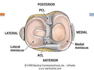
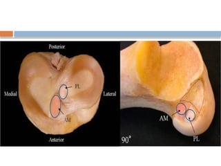
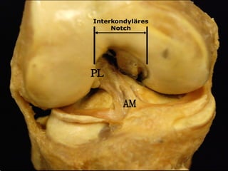
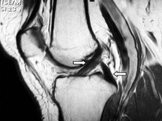
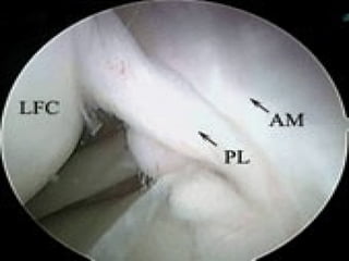
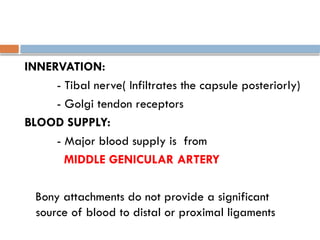
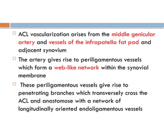
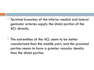
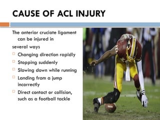
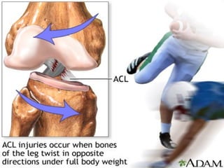
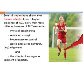
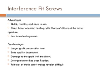
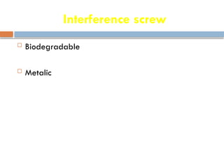
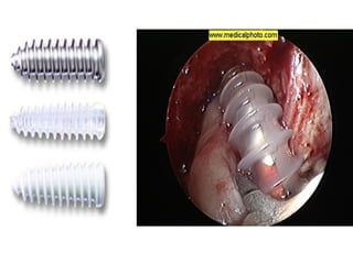
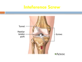
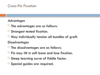
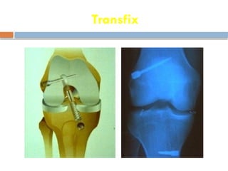
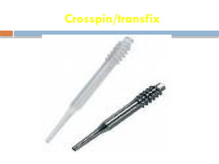
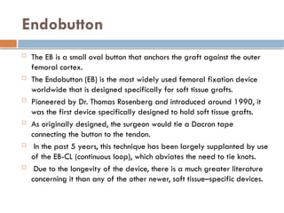
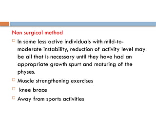
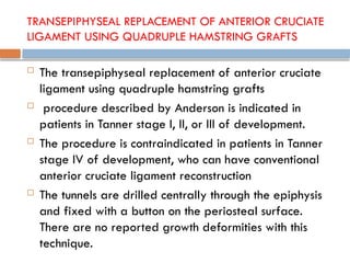
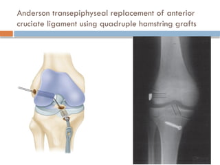
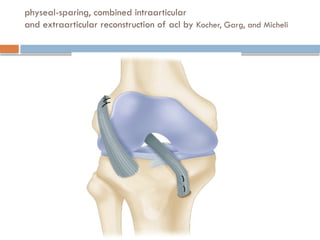
ANTERIOR CRUCIATE
LIGAMENT-INJURY &
MANAGEMENT
Dr.Akshay
ANATOMY
 ACL is composed of multiple collagen fascicles
surrounded by an endotendineum which is
grouped into fibers measuring around
38mm in length (range 25 to 41 mm) and
10 mm in width (range 7 to 12 mm)
 Microspocially composed of interlacing fibrils
(150 to 250 Nanometer in diamter)
 synovial membrane envelope the ACL
 ORIGIN
- From the posteromedial corner of medial
aspect of lateral femoral condyle in the
intercondylar notch
 INSERTION
- Fossa in front of lateral to anterior spine of
tibia
 ACL is composed of two principal parts
1. Small Anteromedial band
and
2. Larger bulky posterolateral portion
CLINICAL IMPORTANCE
- Anteromedial bundle is tight in flexion and the
posterolateral bundle is tight in extension
- In extension both bundles are parallel
- In flexion both bundles are crossed
Action
These attachments allow the ACL to resist anterior
translation and medial rotation of the tibia, in
relation to the femur.
INNERVATION:
- Tibal nerve( Infiltrates the capsule posteriorly)
- Golgi tendon receptors
BLOOD SUPPLY:
- Major blood supply is from
MIDDLE GENICULAR ARTERY
Bony attachments do not provide a significant
source of blood to distal or proximal ligaments
 ACL vascularization arises from the middle genicular
artery and vessels of the infrapatella fat pad and
adjacent synovium
 The artery gives rise to periligamentous vessels
which form a web-like network within the synovial
membrane
 These periligamentous vessels give rise to
penetrating branches which transversely cross the
ACL and anastomose with a network of
longitudinally oriented endoligamentous vessels
 Terminal branches of the inferior medial and lateral
genicular arteries supply the distal portion of the
ACL directly.
 The extremities of the ACL seem to be better
vascularized than the middle part, and the proximal
portion seems to have a greater vascular density
than the distal portion
CAUSE OF ACL INJURY
The anterior cruciate ligament
can be injured in
several ways
 Changing direction rapidly
 Stopping suddenly
 Slowing down while running
 Landing from a jump
incorrectly
 Direct contact or collision,
such as a football tackle
 Several studies have shown that
female athletes have a higher
incidence of ACL injury than male
athletes because of Differences in
- Physical conditioning
- Muscular strength
- Neuromuscular control
- pelvis and lower extremity
(leg) alignment
and
- the effects of estrogen on
ligament properties.
 ACL injuries occur in combination with damage to
-The meniscus
-Articular cartilage or
-Other ligaments
 ACL Injury Triad (Unhappy Triad) : ACL Tear, MCL Tear, and
Medial Meniscus Tear.
 Secondary damage may occur in patients who have
repeated episodes of instability due to ACL injury.
 With chronic instability, up to 90 percent of patients
will have meniscus damage when reassessed 10 or
more years after the initial injury.
 Similarly, the prevalence of articular cartilage
lesions increases up to 70 percent in patients who
have a 10-year-old ACL deficiency
GRADING
 Partial tears of the anterior cruciate ligament are
rare
 Most ACL injuries are complete or near complete
tears
 Injured ligaments are considered "sprains" and are
graded on a severity scale.
 Grade 1 Sprains.
The ligament is mildly damaged . It has been slightly
stretched, but is still able to keep the knee joint stable.
 Grade 2 Sprains.
The ligament is stretched to the point where it becomes
loose. This is often referred to as a partial tear of the
ligament.
 Grade 3 Sprains.
This type of sprain is most commonly referred to as a
complete tear of the ligament. The ligament has been
split into two pieces, and the knee joint is unstable.
SYMPTOMS
 When ACL is injured , pt might hear a "popping"
noise.
 Other typical symptoms include:
-Pain with swelling.
-Loss of full range of motion
-Tenderness along the joint line
-Discomfort while walking
PHYSICAL EXAMINATION
INCLUDE
 ANTERIOR DRAWER TEST
 LACHMAN’S TEST
 PIVOT SHIFT TEST
 KT-2000 ARTHROMETER TEST
ANTERIOR DRAWER TEST
 To perform anterior drawer test, examiner grasps
pt's tibia & pulls it forward when the affected leg is
flexed at 90 degree while noting degree of
anterior tibial displacement
LACHMAN’S TEST
 This is a variant of the anterior drawer test
 The examination is carried out with the knee in 15 deg of
flexion, and external rotation (relaxes IT band)
 For a right knee, the examiner's right hand grips the inner
aspect of the calf and the left hand grasps outer aspect of
the distal thigh
 Attempt to quantify the displacement in mm is done by
comparing this displacement to the normal side
 End point should be graded as hard or soft
- End point is said to be hard when the ACL
abruptly halts the forward motion of the tibia on
the femur
- End point is soft when there is no ACL &
restraints are more elastic secondary stabilizers;
PIVOT SHIFT TEST
 The patient lies supine with legs relaxed
 Therapist presses against the head of the fibula with one
hand, and grasps the ankle with the other hand.
 The lower leg is passively internally rotated, while the
knee is kept in full extension.
 The hip is flexed to 30 degrees, while the knee is flexed.
 A valgus force and axial load are applied to the knee at
the same time.
 Upon damage to the ACL, there will be subluxation of the
lateral tibial plateau in the fully extended position.
 When the knee is flexed between 20 and 40 degrees, the
lateral tibial plateau will reduce itself, and a palpable
shift or clunk will be noticed.
 GRADES:
Grade 0 - Normal
Grade 1 - Glide
Grade 2 - Clunk
Grade 3 - Locked Subluxation
KT-2000 ARTHROMETER TEST
KT-2000 Arthrometer
 The KT-2000 arthrometer is a device used to measure the anterior-posterior
laxity of the knee, specifically to assess the integrity of the anterior cruciate
ligament (ACL). It provides objective data on the degree of knee laxity by
applying a standardized force to the tibia and measuring the displacement
relative to the femur.
Procedure:
1. Positioning: The patient lies supine with the knee flexed at about 20-30
degrees.
2. Device Application: The KT-2000 arthrometer is placed on the anterior aspect
of the knee with pads positioned on the tibia and femur.
3. Calibration: The device is calibrated to zero to ensure accurate measurements.
4. Measurement: The clinician applies an anterior force to the tibia, and the device
measures the displacement. The procedure is repeated several times to ensure
consistency.
KT-2000 Arthrometer
 Interpretation:
Normal ACL: Displacement of less than 3 mm.
Partial Tear: Displacement between 3 mm and 5 mm.
Complete Tear: Displacement greater than 5 mm.
 Advantages:
- Provides quantitative data.
- Helps in the diagnosis of ACL tears.
- Useful for pre- and post-surgical evaluation.
INVESTIGATIONS
 MRI
 Arthroscopy
INVESTIGATION
TREATMENT
 NON-SURGICAL METHOD
 SURGICAL METHOD
TREATMENT
 Immediately after injury
 R.I.C.E ( Rest Ice Compression Elevation )
 This is reserved for Grade I and II tears and
consists of rest, long leg casts for 4-6 weeks,
NSAIDs, physiotherapy
 Exercise (after swelling decreases and weight-bearing
progresses)
 Braces
 Rehabilitation Brace
 Functional Brace
Nonsurgical Treatment
 Nonsurgical management is indicated in patients
with
- partial tears and no instability symptoms
- complete tears and no symptoms of knee
instability
- Who do light manual work or live sedentary
lifestyles
- Whose growth plates are still open (children)
Precautions
 Modification of active lifestyle to avoid high
demand activities
 Muscle strengthening exercises
 May require knee brace
 Despite above precautions ,secondary damage to
knee cartilage & meniscus leading to premature
arthritis
Surgical Treatment
 Timing of Surgery
 1) Swelling in the knee must go down to near-normal
levels
 2) Range-of-motion (bending and straightening) of the
injured knee must be nearly equal to the uninjured knee
 3) Good Quadriceps muscle strength must be present.
 Usually it takes a couple of weeks after injury before
ACL reconstruction can be performed.
 The presence of any associated injuries to the knee joint
involving cartilage, meniscus, or other ligaments may
change the time-frame for surgery.
Surgical Treatment
 ACL tears are not usually repaired using suture to
sew it back together, because repaired ACLs have
generally been shown to fail over time
 Therefore, the torn ACL is generally replaced by a
substitute graft made of tendon
The grafts commonly used to replace the ACL include
 Patellar tendon
 Hamstring tendon
 Quadriceps tendon
 patellar tendon,
 Achilles tendon,
 semitendinosus,
 gracilis, or posterior
tibialis tendon
autograft Allograft
 Patients treated with surgical reconstruction of the
ACL have long-term success rates of 82 %- 95%
 The goal of the ACL reconstruction surgery is to
prevent instability and restore the function of the
torn ligament, creating a stable knee.
 Recurrent instability and graft failure are seen in
approximately 8% of patients.
PATIENT CONSIDERATIONS
 Active adult patients involved in sports or jobs that
require pivoting, turning or hard-cutting as well as
heavy manual work are encouraged to consider
surgical treatment.
 Activity, not age, should determine if surgical
intervention should be considered.
 In young children or adolescents with ACL tears,
early ACL reconstruction creates a possible risk of
growth plate injury, leading to bone growth
problems. The surgeon can delay ACL surgery until
the child is closer to skeletal maturity or the surgeon
may modify the ACL surgery technique to decrease
the risk of growth plate injury.
 A patient with a torn ACL and significant functional
instability has a high risk of developing secondary
knee damage and should therefore consider ACL
reconstruction.
 It is common to see ACL injuries combined with
damage to the menisci (50 %), articular cartilage
(30 %), collateral ligaments (30 %), joint capsule, or
a combination of the above.
Surgical Choices
1.PATELLAR TENDON AUTOGRAFT.
 The middle third of the patellar tendon of the patient, along
with a bone plug from the shin and the patella is used in the
patellar tendon autograft. Occasionally referred to by some
surgeons as the "gold standard" for ACL reconstruction,
recommended for high-demand athletes and patients whose
jobs do not require a significant amount of kneeling.
 In studies comparing outcomes of ACL reconstruction, the rate
of graft failure was lower in the patellar tendon group
 In addition, most studies show equal or better
outcomes in terms of postoperative tests for knee
laxity (Lachman's, anterior drawer and instrumented
tests) when this graft is compared to others.
DISADVANTAGES
The Disadvantages of the patellar tendon
autograft are:
-Postoperative patellofemoral pain
-Pain with kneeling
-increased risk of postoperative stiffness
-risk of patella fracture
-Quadriceps Weakness
-Persistent Tendon Defect
HAMSTRING TENDON AUTOGRAFT
2.Hamstring tendon autograft.
 The semitendinosus hamstring tendon on the inner
side of the knee is used in creating the hamstring
tendon autograft for ACL reconstruction.
 Some use an additional tendon, the gracilis, which
is attached below the knee in the same area. This
creates a two- or four-strand tendon graft.
 Hamstring graft proponents claim there are fewer
problems associated with harvesting of the graft
compared to the patellar tendon autograft
including:
- Fewer problems with anterior knee pain after
surgery
- Less postoperative stiffness problems
- Smaller incision
- Faster recovery
 The graft function may be limited by the strength and
type of fixation in the bone tunnels, as the graft does
not have bone plugs.
 There have been conflicting results in research studies
as to whether hamstring grafts are slightly more
susceptible to graft elongation (stretching), which may
lead to increased laxity during objective testing. Recently,
some studies have demonstrated decreased hamstring
strength in patients after surgery.
 There are some indications that patients who have
intrinsic ligamentous laxity and knee hyperextension
of 10 degrees or more may have increased risk of
postoperative hamstring graft laxity on clinical exam.
Therefore, some clinicians recommend the use of
patellar tendon autografts in these hypermobile
patients.
 chronic or residual medial collateral ligament
laxity (grade 2 or more) at the time of ACL
reconstruction may be a contra-indication for
use of the patient's own semitendinosus and
gracilis tendons as an ACL graft.
QUADRICEPS TENDON AUTOGRAFT
3.QUADRICEPS TENDON AUTOGRAFT.
 The quadriceps tendon autograft is often used for
patients who have already failed ACL
reconstruction.
 Middle third of the patient's quadriceps tendon and
a bone plug from the upper end of the patella
are used.
 This yields a larger graft for taller and heavier
patients. Because there is a bone plug on one side
only, the fixation is not as solid as for the patellar
tendon graft.
 There is a high association with postoperative
anterior knee pain and a low risk of patella
fracture. Patients may find the incision is not
cosmetically appealing
ALLOGRAFTS.
 Allografts are grafts taken from cadavers and are
becoming increasingly popular.
 These grafts are also used for patients who have failed
ACL reconstruction before and in surgery to repair or
reconstruct more than one knee ligament.
 Advantages of using allograft tissue include
- Elimination of pain caused by obtaining the graft
from the patient
- Decreased surgery time and smaller incisions.
The PATELLAR TENDON ALLOGRAFT allows for
strong bony fixation in the tibial and femoral bone
tunnels with screws.
 However, allografts are associated with
- Risk of infection, including viral transmission (HIV
and Hepatitis C)
- There have also been conflicting results in
research studies as to whether allografts are
slightly more susceptible to graft elongation
(stretching), which may lead to increased laxity
during testing.
 Recently published literature may point to a higher
failure rate with the use of allografts for ACL
reconstruction.
 Failure rates ranging from 23% to 34.4% have
been reported in young, active patients returning to
high-demand sporting activities after ACL
reconstruction with allografts.
 This is compared to autograft failure rates ranging
from 5% to 10%.
Meta-analysis of Patellar vs. Hamstring tendons in ACL
reconstruction
 • Controlled trials with minimum 2 year follow-up
• Evaluated; return to pre-injury level of activity, KT
testing, Lachman scores, pivot shift scores, ROM,
complications, failures • 4 studies fulfilled inclusion
criteria • B-T-B (bone-tendon-bone) showed a
>20% chance return to pre-injury activity level
versus hamstring, (p value = 0.01)
 Yunes, M. et al “Patellar Versus Hamstring Tendons
in ACL reconstruction; A Meta-analysis” Arthroscopy
Vol. 17, No. 3 (March) 2001; pp248-257
SYNTHETIC GRAFTS
 The best scenario for the use of the synthetic graft is
when the graft can be buried in soft tissue, such as
in extra-articular reconstruction.
 This allows for collagen in growth and ensures the
long-term viability of the synthetic graft.
 It will be sure to fail early if it is laid into a joint
bare, especially going around tunnel edges, and is
unprotected by soft tissue.
Common Synthetic Grafts:
1. Ligament Augmentation and Reconstruction System
(LARS): Made from polyethylene terephthalate
(PET) fibers.
2. Leeds-Keio Ligament: Also made from PET fibers,
used in some regions.
3. Carbon Fiber Ligaments: Less commonly used due
to issues with durability and biocompatibility.
4. Gore-Tex graft : It is made from “expanded
polytetrafluroethylene (ePTFE)”
Disadvantages
 The main disadvantage is that all the long-term studies have
shown high failure rate. There is the potential for reaction to the
graft material with synovitis, as seen with the use of the Gore-
Tex graft.
 With the Gore-Tex graft, there was also the increased risk of
late hematogenous joint infection.
 The results that have been reported with the use of the Gore-Tex
graft suggest that it should not be used for ACL reconstruction.
 Unacceptable failure rates have also been reported with the use
of the Stryker Dacron ligament and the Leeds-Keio ligament.
GRAFT FIXATION
FEMORAL FIXATION DEVICES
Ultimate load to failure of femoral fixation devices.
 Mitek 600N
 BioScrew 400N
 Endo-button: tape 500N
 BioScrew: Endo-pearl 700N
 Bone mulch screw 900N
 Cross pin 900N
 Endo-button with closed loop tape 1300N
Interference Fit Screws
Advantages
 Quick, familiar, and easy to use.
 Direct bone to tendon healing, with Sharpey’s fibers at the tunnel
aperture.
 Less tunnel enlargement.
Disadvantages
 Longer graft preparation time.
 Bone quality dependent.
 Damage to the graft with the screw.
 Divergent screw has poor fixation.
 Removal of metal screw makes revision difficult
Interference screw
 Biodegradable
 Metalic
Inteference Screw
Cross-Pin Fixation
Advantages
 The advantages are as follows:
 Strongest tested fixation.
 May individually tension all bundles of graft.
Disadvantages
 The disadvantages are as follows:
 Pin may tilt in soft bone and lose fixation.
 Steep learning curve of fiddle factor.
 Special guides are required.
Transfix
Crosspin/transfix
Endobutton
 The EB is a small oval button that anchors the graft against the outer
femoral cortex.
 The Endobutton (EB) is the most widely used femoral fixation device
worldwide that is designed specifically for soft tissue grafts.
 Pioneered by Dr. Thomas Rosenberg and introduced around 1990, it
was the first device specifically designed to hold soft tissue grafts.
 As originally designed, the surgeon would tie a Dacron tape
connecting the button to the tendon.
 In the past 5 years, this technique has been largely supplanted by use
of the EB-CL (continuous loop), which obviates the need to tie knots.
 Due to the longevity of the device, there is a much greater literature
concerning it than any of the other newer, soft tissue–specific devices.
ENDOBUTTON
Advantages
 The Endo-button with closed loop tape is strong,
 The plastic button is cheap, available and easy to do
Disadvantages
 Fixation site is distant with increase in laxity, with the
bungee cord effect.
 Increased in tunnel widening.
 Plastic button has low pullout strength, dependent on
the sutures
Endobuttom Loop
Clinical Results
 In the largest meta-analysis of anterior cruciate
ligament reconstruction (ACLR) autografts, the EB-
hamstring combination was found to have the
highest stability rates of any graft-fixation construct
when paired with modern tibial fixation. Morbidity
has been minimal.
MILAGRO
 Milagro (Beta-Tricalcium Phosphate, Polylactide
Co-Glycolide Biocomposite)
The Milagro screw can be used for femoral or tibial
fixation for soft tissue or bone–tendon–bone (BTB)
autografts or allografts. It is available in various
diameters from 7 to 12mm and in 23-, 30-, and
35-mm lengths. The Milagro screw is made from a
polymer composite, Biocryl Rapide.
 EZLoc Femoral Fixation of a Soft Tissue Graft
 The EZLoc (Arthrotek, Warsaw, IN) is a cortical femoral fixation
device for a soft tissue anterior cruciate ligament (ACL)
reconstruction that combines superior fixation properties (high
resistance to slippage, infinite stiffness, and 1427N strength)
with a simple surgical technique.
 The EZLoc consists of a deployable lever arm connected to an
axle in a slotted body through which the ACL graft is looped.
 The EZLoc comes sterilely package with a sharp-tip passing pin
that is secured in the slotted body with a suture tied under
tension. The passing pin is passed through the tunnels, the gold
lever arm is positioned lateral, and the soft tissue graft is
looped through the slot in the EZLoc.
Tibial Fixing Devices
Ultimate load to failure of tibial fixation devices.
 Single staple 100N
 Double staple 500N
 Screw post 600N
 Button 400N
 RCI 300N
 BioScrew 400N
 BioScrew and button 600N
 Intrafix 700N
 Screw and washer 800N
 Washer Loc 900N
One bundle or two bundle ACL
reconstruction
 What is an “Anatomic” ACL reconstruction?
 Every person is different; some people are short, others
are tall. Similarly, each person has a different size and
shape of the ACL. In order to properly reconstruct the ACL it
is important to reproduce each persons individual anatomy.
The goals of anatomic ACL reconstruction are to:
 Restore 60 – 80% of normal ACL anatomy
 Regain stability and return to pre-injury activity level
 Maintain long term knee health
 What is anatomic Double-Bundle ACL
reconstruction?
 In a “double-bundle” ACL reconstruction, the ACL is
restored using two bundles. Just like the normal ACL,
there will be an AM and a PL bundle.
 In a “single-bundle” reconstruction, the ACL is
restored using one bundle. There are some benefits
of a “double-bundle” reconstruction, when
compared to a “single-bundle” reconstruction.
 Anatomic double-bundle reconstruction better
restores knee stability compared to single-bundle
reconstruction.
 Because anatomic double-bundle reconstruction
uses two bundles to restore the ACL, it allows for a
replacement of a larger size ACL
Pre requisite for single-bundle/double-bundle
reconstruction
 An ACL insertion site greater than 18 mm allows for
double-bundle reconstruction.
 If the insertion site is less than 14 mm, there is only
space available for a single-bundle procedure.
 Between 14 – 18 mm, we can perform either
double- or single-bundle reconstruction.
Indications for single bundle recon.
 The patient is still growing and his or her growth
plate is not closed.
 The patient has severe arthritis of the knee.
 The patient has multiple knee ligament injuries or a
knee dislocation and multiple other ligaments need
to be reconstructed at the same time.
 The patient has bone that is severely bruised.
 The patient has a small intercondylar notch.
 A prospective comparative cohort study was carried out with 72
consecutive patients with chronic ACL deficiency to compare three
ACL reconstruction procedures using hamstring tendon grafts.
 The first 24 patients underwent a single-bundle procedure using
a six-strand hamstring tendon graft.
 The next 24 patients underwent a non anatomical double-bundle
procedure using four-strand and two-strand hamstring tendon
grafts.
 The final 24 patients underwent the anatomical double-bundle
procedure using the same four-strand and two-strand hamstring
tendon grafts. All 72 patients underwent postoperative
management with the same rehabilitation protocol. There were no
significant differences among the background factors.
Conclusion
 The postoperative anterior laxity measured was
significantly less after the anatomical double-
bundle reconstruction than after the single-bundle
reconstruction.
 Outcome of Arthroscopic Single-Bundle Versus Double-
Bundle Reconstruction of the Anterior Cruciate Ligament: A
Preliminary 2-Year Prospective Study
 Se-Jin Park, M.D., Young-Bok Jung, M.D., Hwa-Jae Jung,
M.D., Ho-Joong Jung, M.D., Hun Kyu Shin, M.D., Eugene
Kim, M.D., Kwang-Sup Song, M.D., Gwang-Sin Kim,
M.D., Hye-Young Cheon, P.A., Seonwoo Kim, Ph.D.Received:
December 29, 2008; Accepted: September 9, 2009;
Published Online: February 22, 2010
 Arthroscopy Volume 26, Issue 5, Pages 630–636, May
2010
 113 were included in this study. They serially obtained
clinical and radiologic data preoperatively and
postoperatively. They compared preoperative data and
data at 2 years postoperatively in patients who had
undergone single-bundle ACL reconstruction versus
patients who had undergone double-bundle ACL
reconstruction.
 There were 50 single-bundle reconstructions and 63
double-bundle reconstructions. Antero-posterior stability
was assessed objectively by anterior stress radiographs
with the telos device (telos, Marburg, Germany) and the
maximal manual test with the KT-2000 arthrometer
Conclusions
 Double-bundle reconstruction of the ACL by a
method using 2 femoral tunnel and 2 tibial tunnels
showed no differences in stability results or any
other clinical aspects or in terms of patient
satisfaction.
COMPLICATIONS
Skeletally immature patients
 Anterior cruciate ligament injuries in skeletally
immature adolescents are being diagnosed with
increasing frequency.
 Nonoperative management of midsubstance ACL
injuries in adolescent athletes frequently
results in a high incidence of giving-way episodes,
recurrent meniscal tears, and early onset of
osteoarthritis
 The concern about ACL reconstruction in the athlete
with open growth plates is that there will be
 premature fusion of the plate, growth arrest, and
potential for angular deformities.
Skeletally immature patients
 Non surgical methods
or
 surgical methods
Non surgical method
 In some less active individuals with mild-to-
moderate instability, reduction of activity level may
be all that is necessary until they have had an
appropriate growth spurt and maturing of the
physes.
 Muscle strengthening exercises
 knee brace
 Away from sports activities
TRANSEPIPHYSEAL REPLACEMENT OF ANTERIOR CRUCIATE
LIGAMENT USING QUADRUPLE HAMSTRING GRAFTS
 The transepiphyseal replacement of anterior cruciate
ligament using quadruple hamstring grafts
 procedure described by Anderson is indicated in
patients in Tanner stage I, II, or III of development.
 The procedure is contraindicated in patients in Tanner
stage IV of development, who can have conventional
anterior cruciate ligament reconstruction
 The tunnels are drilled centrally through the epiphysis
and fixed with a button on the periosteal surface.
There are no reported growth deformities with this
technique.
Anderson transepiphyseal replacement of anterior
cruciate ligament using quadruple hamstring grafts
physeal-sparing, combined intraarticular
and extraarticular reconstruction of acl by Kocher, Garg, and Micheli
Anterior Cruciate Ligament Reconstruction in
Skeletally Immature Patients With Transphyseal
Tunnels
 Lauren H. Redler, M.D., Rebecca T. Brafman, B.A., Natasha
Trentacosta, M.D., Christopher S. Ahmad, M.D.(Department of
Orthopaedic Surgery, Columbia University Medical Center, New
York, New York, U.S.A.)
 Arthroscopy Volume 28, Issue 11, Pages 1710–1717,
November 2012
 Moises Cohen, M.D., Ph.D., Mario Ferretti, M.D., Ph.D., Marcelo
Quarteiro, M.D., Frank B. Marcondes, M.D., João P.B. de
Hollanda, M.D., Joicemar T. Amaro, M.D., Rene J. Abdalla, M.D.,
Ph.D.(Orthopedic Sports Medicine Division, Department of
Orthopaedic Surgery and Traumatology, Universidade Federal
de São Paulo–Escola Paulista de Medicina, São Paulo, Brazil)
 Arthroscopy Volume 25, Issue 8, Pages 831–838, August 2009
Conclusions
 ACL reconstruction by use of the transphyseal
technique in an immature skeleton with a hamstring
autograft, with careful attention being paid to the
technique, resulted in good clinical outcomes and no
growth abnormalities.

ACL Injury.pptx. . .

  • 1.
  • 2.
    ANATOMY  ACL iscomposed of multiple collagen fascicles surrounded by an endotendineum which is grouped into fibers measuring around 38mm in length (range 25 to 41 mm) and 10 mm in width (range 7 to 12 mm)  Microspocially composed of interlacing fibrils (150 to 250 Nanometer in diamter)  synovial membrane envelope the ACL
  • 3.
     ORIGIN - Fromthe posteromedial corner of medial aspect of lateral femoral condyle in the intercondylar notch  INSERTION - Fossa in front of lateral to anterior spine of tibia
  • 4.
     ACL iscomposed of two principal parts 1. Small Anteromedial band and 2. Larger bulky posterolateral portion CLINICAL IMPORTANCE - Anteromedial bundle is tight in flexion and the posterolateral bundle is tight in extension - In extension both bundles are parallel - In flexion both bundles are crossed
  • 10.
    Action These attachments allowthe ACL to resist anterior translation and medial rotation of the tibia, in relation to the femur.
  • 11.
    INNERVATION: - Tibal nerve(Infiltrates the capsule posteriorly) - Golgi tendon receptors BLOOD SUPPLY: - Major blood supply is from MIDDLE GENICULAR ARTERY Bony attachments do not provide a significant source of blood to distal or proximal ligaments
  • 12.
     ACL vascularizationarises from the middle genicular artery and vessels of the infrapatella fat pad and adjacent synovium  The artery gives rise to periligamentous vessels which form a web-like network within the synovial membrane  These periligamentous vessels give rise to penetrating branches which transversely cross the ACL and anastomose with a network of longitudinally oriented endoligamentous vessels
  • 13.
     Terminal branchesof the inferior medial and lateral genicular arteries supply the distal portion of the ACL directly.  The extremities of the ACL seem to be better vascularized than the middle part, and the proximal portion seems to have a greater vascular density than the distal portion
  • 14.
    CAUSE OF ACLINJURY The anterior cruciate ligament can be injured in several ways  Changing direction rapidly  Stopping suddenly  Slowing down while running  Landing from a jump incorrectly  Direct contact or collision, such as a football tackle
  • 16.
     Several studieshave shown that female athletes have a higher incidence of ACL injury than male athletes because of Differences in - Physical conditioning - Muscular strength - Neuromuscular control - pelvis and lower extremity (leg) alignment and - the effects of estrogen on ligament properties.
  • 17.
     ACL injuriesoccur in combination with damage to -The meniscus -Articular cartilage or -Other ligaments  ACL Injury Triad (Unhappy Triad) : ACL Tear, MCL Tear, and Medial Meniscus Tear.  Secondary damage may occur in patients who have repeated episodes of instability due to ACL injury.
  • 18.
     With chronicinstability, up to 90 percent of patients will have meniscus damage when reassessed 10 or more years after the initial injury.  Similarly, the prevalence of articular cartilage lesions increases up to 70 percent in patients who have a 10-year-old ACL deficiency
  • 19.
    GRADING  Partial tearsof the anterior cruciate ligament are rare  Most ACL injuries are complete or near complete tears  Injured ligaments are considered "sprains" and are graded on a severity scale.
  • 20.
     Grade 1Sprains. The ligament is mildly damaged . It has been slightly stretched, but is still able to keep the knee joint stable.  Grade 2 Sprains. The ligament is stretched to the point where it becomes loose. This is often referred to as a partial tear of the ligament.  Grade 3 Sprains. This type of sprain is most commonly referred to as a complete tear of the ligament. The ligament has been split into two pieces, and the knee joint is unstable.
  • 21.
    SYMPTOMS  When ACLis injured , pt might hear a "popping" noise.  Other typical symptoms include: -Pain with swelling. -Loss of full range of motion -Tenderness along the joint line -Discomfort while walking
  • 22.
    PHYSICAL EXAMINATION INCLUDE  ANTERIORDRAWER TEST  LACHMAN’S TEST  PIVOT SHIFT TEST  KT-2000 ARTHROMETER TEST
  • 23.
    ANTERIOR DRAWER TEST To perform anterior drawer test, examiner grasps pt's tibia & pulls it forward when the affected leg is flexed at 90 degree while noting degree of anterior tibial displacement
  • 24.
    LACHMAN’S TEST  Thisis a variant of the anterior drawer test  The examination is carried out with the knee in 15 deg of flexion, and external rotation (relaxes IT band)  For a right knee, the examiner's right hand grips the inner aspect of the calf and the left hand grasps outer aspect of the distal thigh  Attempt to quantify the displacement in mm is done by comparing this displacement to the normal side
  • 25.
     End pointshould be graded as hard or soft - End point is said to be hard when the ACL abruptly halts the forward motion of the tibia on the femur - End point is soft when there is no ACL & restraints are more elastic secondary stabilizers;
  • 26.
    PIVOT SHIFT TEST The patient lies supine with legs relaxed  Therapist presses against the head of the fibula with one hand, and grasps the ankle with the other hand.  The lower leg is passively internally rotated, while the knee is kept in full extension.  The hip is flexed to 30 degrees, while the knee is flexed.  A valgus force and axial load are applied to the knee at the same time.
  • 27.
     Upon damageto the ACL, there will be subluxation of the lateral tibial plateau in the fully extended position.  When the knee is flexed between 20 and 40 degrees, the lateral tibial plateau will reduce itself, and a palpable shift or clunk will be noticed.  GRADES: Grade 0 - Normal Grade 1 - Glide Grade 2 - Clunk Grade 3 - Locked Subluxation
  • 28.
  • 29.
    KT-2000 Arthrometer  TheKT-2000 arthrometer is a device used to measure the anterior-posterior laxity of the knee, specifically to assess the integrity of the anterior cruciate ligament (ACL). It provides objective data on the degree of knee laxity by applying a standardized force to the tibia and measuring the displacement relative to the femur. Procedure: 1. Positioning: The patient lies supine with the knee flexed at about 20-30 degrees. 2. Device Application: The KT-2000 arthrometer is placed on the anterior aspect of the knee with pads positioned on the tibia and femur. 3. Calibration: The device is calibrated to zero to ensure accurate measurements. 4. Measurement: The clinician applies an anterior force to the tibia, and the device measures the displacement. The procedure is repeated several times to ensure consistency.
  • 30.
    KT-2000 Arthrometer  Interpretation: NormalACL: Displacement of less than 3 mm. Partial Tear: Displacement between 3 mm and 5 mm. Complete Tear: Displacement greater than 5 mm.  Advantages: - Provides quantitative data. - Helps in the diagnosis of ACL tears. - Useful for pre- and post-surgical evaluation.
  • 31.
  • 32.
  • 34.
  • 35.
    TREATMENT  Immediately afterinjury  R.I.C.E ( Rest Ice Compression Elevation )  This is reserved for Grade I and II tears and consists of rest, long leg casts for 4-6 weeks, NSAIDs, physiotherapy  Exercise (after swelling decreases and weight-bearing progresses)  Braces  Rehabilitation Brace  Functional Brace
  • 36.
    Nonsurgical Treatment  Nonsurgicalmanagement is indicated in patients with - partial tears and no instability symptoms - complete tears and no symptoms of knee instability - Who do light manual work or live sedentary lifestyles - Whose growth plates are still open (children)
  • 37.
    Precautions  Modification ofactive lifestyle to avoid high demand activities  Muscle strengthening exercises  May require knee brace  Despite above precautions ,secondary damage to knee cartilage & meniscus leading to premature arthritis
  • 38.
    Surgical Treatment  Timingof Surgery  1) Swelling in the knee must go down to near-normal levels  2) Range-of-motion (bending and straightening) of the injured knee must be nearly equal to the uninjured knee  3) Good Quadriceps muscle strength must be present.  Usually it takes a couple of weeks after injury before ACL reconstruction can be performed.  The presence of any associated injuries to the knee joint involving cartilage, meniscus, or other ligaments may change the time-frame for surgery.
  • 39.
    Surgical Treatment  ACLtears are not usually repaired using suture to sew it back together, because repaired ACLs have generally been shown to fail over time  Therefore, the torn ACL is generally replaced by a substitute graft made of tendon
  • 40.
    The grafts commonlyused to replace the ACL include  Patellar tendon  Hamstring tendon  Quadriceps tendon  patellar tendon,  Achilles tendon,  semitendinosus,  gracilis, or posterior tibialis tendon autograft Allograft
  • 41.
     Patients treatedwith surgical reconstruction of the ACL have long-term success rates of 82 %- 95%  The goal of the ACL reconstruction surgery is to prevent instability and restore the function of the torn ligament, creating a stable knee.  Recurrent instability and graft failure are seen in approximately 8% of patients.
  • 42.
    PATIENT CONSIDERATIONS  Activeadult patients involved in sports or jobs that require pivoting, turning or hard-cutting as well as heavy manual work are encouraged to consider surgical treatment.  Activity, not age, should determine if surgical intervention should be considered.
  • 43.
     In youngchildren or adolescents with ACL tears, early ACL reconstruction creates a possible risk of growth plate injury, leading to bone growth problems. The surgeon can delay ACL surgery until the child is closer to skeletal maturity or the surgeon may modify the ACL surgery technique to decrease the risk of growth plate injury.
  • 44.
     A patientwith a torn ACL and significant functional instability has a high risk of developing secondary knee damage and should therefore consider ACL reconstruction.  It is common to see ACL injuries combined with damage to the menisci (50 %), articular cartilage (30 %), collateral ligaments (30 %), joint capsule, or a combination of the above.
  • 45.
    Surgical Choices 1.PATELLAR TENDONAUTOGRAFT.  The middle third of the patellar tendon of the patient, along with a bone plug from the shin and the patella is used in the patellar tendon autograft. Occasionally referred to by some surgeons as the "gold standard" for ACL reconstruction, recommended for high-demand athletes and patients whose jobs do not require a significant amount of kneeling.  In studies comparing outcomes of ACL reconstruction, the rate of graft failure was lower in the patellar tendon group
  • 46.
     In addition,most studies show equal or better outcomes in terms of postoperative tests for knee laxity (Lachman's, anterior drawer and instrumented tests) when this graft is compared to others.
  • 47.
    DISADVANTAGES The Disadvantages ofthe patellar tendon autograft are: -Postoperative patellofemoral pain -Pain with kneeling -increased risk of postoperative stiffness -risk of patella fracture -Quadriceps Weakness -Persistent Tendon Defect
  • 48.
    HAMSTRING TENDON AUTOGRAFT 2.Hamstringtendon autograft.  The semitendinosus hamstring tendon on the inner side of the knee is used in creating the hamstring tendon autograft for ACL reconstruction.  Some use an additional tendon, the gracilis, which is attached below the knee in the same area. This creates a two- or four-strand tendon graft.
  • 49.
     Hamstring graftproponents claim there are fewer problems associated with harvesting of the graft compared to the patellar tendon autograft including: - Fewer problems with anterior knee pain after surgery - Less postoperative stiffness problems - Smaller incision - Faster recovery
  • 50.
     The graftfunction may be limited by the strength and type of fixation in the bone tunnels, as the graft does not have bone plugs.  There have been conflicting results in research studies as to whether hamstring grafts are slightly more susceptible to graft elongation (stretching), which may lead to increased laxity during objective testing. Recently, some studies have demonstrated decreased hamstring strength in patients after surgery.
  • 51.
     There aresome indications that patients who have intrinsic ligamentous laxity and knee hyperextension of 10 degrees or more may have increased risk of postoperative hamstring graft laxity on clinical exam. Therefore, some clinicians recommend the use of patellar tendon autografts in these hypermobile patients.
  • 52.
     chronic orresidual medial collateral ligament laxity (grade 2 or more) at the time of ACL reconstruction may be a contra-indication for use of the patient's own semitendinosus and gracilis tendons as an ACL graft.
  • 53.
    QUADRICEPS TENDON AUTOGRAFT 3.QUADRICEPSTENDON AUTOGRAFT.  The quadriceps tendon autograft is often used for patients who have already failed ACL reconstruction.  Middle third of the patient's quadriceps tendon and a bone plug from the upper end of the patella are used.
  • 54.
     This yieldsa larger graft for taller and heavier patients. Because there is a bone plug on one side only, the fixation is not as solid as for the patellar tendon graft.  There is a high association with postoperative anterior knee pain and a low risk of patella fracture. Patients may find the incision is not cosmetically appealing
  • 57.
    ALLOGRAFTS.  Allografts aregrafts taken from cadavers and are becoming increasingly popular.  These grafts are also used for patients who have failed ACL reconstruction before and in surgery to repair or reconstruct more than one knee ligament.  Advantages of using allograft tissue include - Elimination of pain caused by obtaining the graft from the patient - Decreased surgery time and smaller incisions.
  • 58.
    The PATELLAR TENDONALLOGRAFT allows for strong bony fixation in the tibial and femoral bone tunnels with screws.
  • 59.
     However, allograftsare associated with - Risk of infection, including viral transmission (HIV and Hepatitis C) - There have also been conflicting results in research studies as to whether allografts are slightly more susceptible to graft elongation (stretching), which may lead to increased laxity during testing.
  • 60.
     Recently publishedliterature may point to a higher failure rate with the use of allografts for ACL reconstruction.  Failure rates ranging from 23% to 34.4% have been reported in young, active patients returning to high-demand sporting activities after ACL reconstruction with allografts.  This is compared to autograft failure rates ranging from 5% to 10%.
  • 61.
    Meta-analysis of Patellarvs. Hamstring tendons in ACL reconstruction  • Controlled trials with minimum 2 year follow-up • Evaluated; return to pre-injury level of activity, KT testing, Lachman scores, pivot shift scores, ROM, complications, failures • 4 studies fulfilled inclusion criteria • B-T-B (bone-tendon-bone) showed a >20% chance return to pre-injury activity level versus hamstring, (p value = 0.01)  Yunes, M. et al “Patellar Versus Hamstring Tendons in ACL reconstruction; A Meta-analysis” Arthroscopy Vol. 17, No. 3 (March) 2001; pp248-257
  • 62.
    SYNTHETIC GRAFTS  Thebest scenario for the use of the synthetic graft is when the graft can be buried in soft tissue, such as in extra-articular reconstruction.  This allows for collagen in growth and ensures the long-term viability of the synthetic graft.  It will be sure to fail early if it is laid into a joint bare, especially going around tunnel edges, and is unprotected by soft tissue.
  • 63.
    Common Synthetic Grafts: 1.Ligament Augmentation and Reconstruction System (LARS): Made from polyethylene terephthalate (PET) fibers. 2. Leeds-Keio Ligament: Also made from PET fibers, used in some regions. 3. Carbon Fiber Ligaments: Less commonly used due to issues with durability and biocompatibility. 4. Gore-Tex graft : It is made from “expanded polytetrafluroethylene (ePTFE)”
  • 64.
    Disadvantages  The maindisadvantage is that all the long-term studies have shown high failure rate. There is the potential for reaction to the graft material with synovitis, as seen with the use of the Gore- Tex graft.  With the Gore-Tex graft, there was also the increased risk of late hematogenous joint infection.  The results that have been reported with the use of the Gore-Tex graft suggest that it should not be used for ACL reconstruction.  Unacceptable failure rates have also been reported with the use of the Stryker Dacron ligament and the Leeds-Keio ligament.
  • 65.
  • 66.
    FEMORAL FIXATION DEVICES Ultimateload to failure of femoral fixation devices.  Mitek 600N  BioScrew 400N  Endo-button: tape 500N  BioScrew: Endo-pearl 700N  Bone mulch screw 900N  Cross pin 900N  Endo-button with closed loop tape 1300N
  • 67.
    Interference Fit Screws Advantages Quick, familiar, and easy to use.  Direct bone to tendon healing, with Sharpey’s fibers at the tunnel aperture.  Less tunnel enlargement. Disadvantages  Longer graft preparation time.  Bone quality dependent.  Damage to the graft with the screw.  Divergent screw has poor fixation.  Removal of metal screw makes revision difficult
  • 68.
  • 70.
  • 71.
    Cross-Pin Fixation Advantages  Theadvantages are as follows:  Strongest tested fixation.  May individually tension all bundles of graft. Disadvantages  The disadvantages are as follows:  Pin may tilt in soft bone and lose fixation.  Steep learning curve of fiddle factor.  Special guides are required.
  • 72.
  • 73.
  • 74.
    Endobutton  The EBis a small oval button that anchors the graft against the outer femoral cortex.  The Endobutton (EB) is the most widely used femoral fixation device worldwide that is designed specifically for soft tissue grafts.  Pioneered by Dr. Thomas Rosenberg and introduced around 1990, it was the first device specifically designed to hold soft tissue grafts.  As originally designed, the surgeon would tie a Dacron tape connecting the button to the tendon.  In the past 5 years, this technique has been largely supplanted by use of the EB-CL (continuous loop), which obviates the need to tie knots.  Due to the longevity of the device, there is a much greater literature concerning it than any of the other newer, soft tissue–specific devices.
  • 75.
    ENDOBUTTON Advantages  The Endo-buttonwith closed loop tape is strong,  The plastic button is cheap, available and easy to do Disadvantages  Fixation site is distant with increase in laxity, with the bungee cord effect.  Increased in tunnel widening.  Plastic button has low pullout strength, dependent on the sutures
  • 76.
  • 77.
    Clinical Results  Inthe largest meta-analysis of anterior cruciate ligament reconstruction (ACLR) autografts, the EB- hamstring combination was found to have the highest stability rates of any graft-fixation construct when paired with modern tibial fixation. Morbidity has been minimal.
  • 78.
    MILAGRO  Milagro (Beta-TricalciumPhosphate, Polylactide Co-Glycolide Biocomposite) The Milagro screw can be used for femoral or tibial fixation for soft tissue or bone–tendon–bone (BTB) autografts or allografts. It is available in various diameters from 7 to 12mm and in 23-, 30-, and 35-mm lengths. The Milagro screw is made from a polymer composite, Biocryl Rapide.
  • 79.
     EZLoc FemoralFixation of a Soft Tissue Graft  The EZLoc (Arthrotek, Warsaw, IN) is a cortical femoral fixation device for a soft tissue anterior cruciate ligament (ACL) reconstruction that combines superior fixation properties (high resistance to slippage, infinite stiffness, and 1427N strength) with a simple surgical technique.  The EZLoc consists of a deployable lever arm connected to an axle in a slotted body through which the ACL graft is looped.  The EZLoc comes sterilely package with a sharp-tip passing pin that is secured in the slotted body with a suture tied under tension. The passing pin is passed through the tunnels, the gold lever arm is positioned lateral, and the soft tissue graft is looped through the slot in the EZLoc.
  • 80.
    Tibial Fixing Devices Ultimateload to failure of tibial fixation devices.  Single staple 100N  Double staple 500N  Screw post 600N  Button 400N  RCI 300N  BioScrew 400N  BioScrew and button 600N  Intrafix 700N  Screw and washer 800N  Washer Loc 900N
  • 81.
    One bundle ortwo bundle ACL reconstruction
  • 83.
     What isan “Anatomic” ACL reconstruction?  Every person is different; some people are short, others are tall. Similarly, each person has a different size and shape of the ACL. In order to properly reconstruct the ACL it is important to reproduce each persons individual anatomy. The goals of anatomic ACL reconstruction are to:  Restore 60 – 80% of normal ACL anatomy  Regain stability and return to pre-injury activity level  Maintain long term knee health
  • 84.
     What isanatomic Double-Bundle ACL reconstruction?  In a “double-bundle” ACL reconstruction, the ACL is restored using two bundles. Just like the normal ACL, there will be an AM and a PL bundle.  In a “single-bundle” reconstruction, the ACL is restored using one bundle. There are some benefits of a “double-bundle” reconstruction, when compared to a “single-bundle” reconstruction.
  • 85.
     Anatomic double-bundlereconstruction better restores knee stability compared to single-bundle reconstruction.  Because anatomic double-bundle reconstruction uses two bundles to restore the ACL, it allows for a replacement of a larger size ACL
  • 86.
    Pre requisite forsingle-bundle/double-bundle reconstruction  An ACL insertion site greater than 18 mm allows for double-bundle reconstruction.  If the insertion site is less than 14 mm, there is only space available for a single-bundle procedure.  Between 14 – 18 mm, we can perform either double- or single-bundle reconstruction.
  • 87.
    Indications for singlebundle recon.  The patient is still growing and his or her growth plate is not closed.  The patient has severe arthritis of the knee.  The patient has multiple knee ligament injuries or a knee dislocation and multiple other ligaments need to be reconstructed at the same time.  The patient has bone that is severely bruised.  The patient has a small intercondylar notch.
  • 90.
     A prospectivecomparative cohort study was carried out with 72 consecutive patients with chronic ACL deficiency to compare three ACL reconstruction procedures using hamstring tendon grafts.  The first 24 patients underwent a single-bundle procedure using a six-strand hamstring tendon graft.  The next 24 patients underwent a non anatomical double-bundle procedure using four-strand and two-strand hamstring tendon grafts.  The final 24 patients underwent the anatomical double-bundle procedure using the same four-strand and two-strand hamstring tendon grafts. All 72 patients underwent postoperative management with the same rehabilitation protocol. There were no significant differences among the background factors.
  • 91.
    Conclusion  The postoperativeanterior laxity measured was significantly less after the anatomical double- bundle reconstruction than after the single-bundle reconstruction.
  • 92.
     Outcome ofArthroscopic Single-Bundle Versus Double- Bundle Reconstruction of the Anterior Cruciate Ligament: A Preliminary 2-Year Prospective Study  Se-Jin Park, M.D., Young-Bok Jung, M.D., Hwa-Jae Jung, M.D., Ho-Joong Jung, M.D., Hun Kyu Shin, M.D., Eugene Kim, M.D., Kwang-Sup Song, M.D., Gwang-Sin Kim, M.D., Hye-Young Cheon, P.A., Seonwoo Kim, Ph.D.Received: December 29, 2008; Accepted: September 9, 2009; Published Online: February 22, 2010  Arthroscopy Volume 26, Issue 5, Pages 630–636, May 2010
  • 93.
     113 wereincluded in this study. They serially obtained clinical and radiologic data preoperatively and postoperatively. They compared preoperative data and data at 2 years postoperatively in patients who had undergone single-bundle ACL reconstruction versus patients who had undergone double-bundle ACL reconstruction.  There were 50 single-bundle reconstructions and 63 double-bundle reconstructions. Antero-posterior stability was assessed objectively by anterior stress radiographs with the telos device (telos, Marburg, Germany) and the maximal manual test with the KT-2000 arthrometer
  • 94.
    Conclusions  Double-bundle reconstructionof the ACL by a method using 2 femoral tunnel and 2 tibial tunnels showed no differences in stability results or any other clinical aspects or in terms of patient satisfaction.
  • 95.
  • 96.
    Skeletally immature patients Anterior cruciate ligament injuries in skeletally immature adolescents are being diagnosed with increasing frequency.  Nonoperative management of midsubstance ACL injuries in adolescent athletes frequently results in a high incidence of giving-way episodes, recurrent meniscal tears, and early onset of osteoarthritis
  • 97.
     The concernabout ACL reconstruction in the athlete with open growth plates is that there will be  premature fusion of the plate, growth arrest, and potential for angular deformities.
  • 98.
    Skeletally immature patients Non surgical methods or  surgical methods
  • 99.
    Non surgical method In some less active individuals with mild-to- moderate instability, reduction of activity level may be all that is necessary until they have had an appropriate growth spurt and maturing of the physes.  Muscle strengthening exercises  knee brace  Away from sports activities
  • 100.
    TRANSEPIPHYSEAL REPLACEMENT OFANTERIOR CRUCIATE LIGAMENT USING QUADRUPLE HAMSTRING GRAFTS  The transepiphyseal replacement of anterior cruciate ligament using quadruple hamstring grafts  procedure described by Anderson is indicated in patients in Tanner stage I, II, or III of development.  The procedure is contraindicated in patients in Tanner stage IV of development, who can have conventional anterior cruciate ligament reconstruction  The tunnels are drilled centrally through the epiphysis and fixed with a button on the periosteal surface. There are no reported growth deformities with this technique.
  • 101.
    Anderson transepiphyseal replacementof anterior cruciate ligament using quadruple hamstring grafts
  • 102.
    physeal-sparing, combined intraarticular andextraarticular reconstruction of acl by Kocher, Garg, and Micheli
  • 103.
    Anterior Cruciate LigamentReconstruction in Skeletally Immature Patients With Transphyseal Tunnels  Lauren H. Redler, M.D., Rebecca T. Brafman, B.A., Natasha Trentacosta, M.D., Christopher S. Ahmad, M.D.(Department of Orthopaedic Surgery, Columbia University Medical Center, New York, New York, U.S.A.)  Arthroscopy Volume 28, Issue 11, Pages 1710–1717, November 2012  Moises Cohen, M.D., Ph.D., Mario Ferretti, M.D., Ph.D., Marcelo Quarteiro, M.D., Frank B. Marcondes, M.D., João P.B. de Hollanda, M.D., Joicemar T. Amaro, M.D., Rene J. Abdalla, M.D., Ph.D.(Orthopedic Sports Medicine Division, Department of Orthopaedic Surgery and Traumatology, Universidade Federal de São Paulo–Escola Paulista de Medicina, São Paulo, Brazil)  Arthroscopy Volume 25, Issue 8, Pages 831–838, August 2009
  • 104.
    Conclusions  ACL reconstructionby use of the transphyseal technique in an immature skeleton with a hamstring autograft, with careful attention being paid to the technique, resulted in good clinical outcomes and no growth abnormalities.