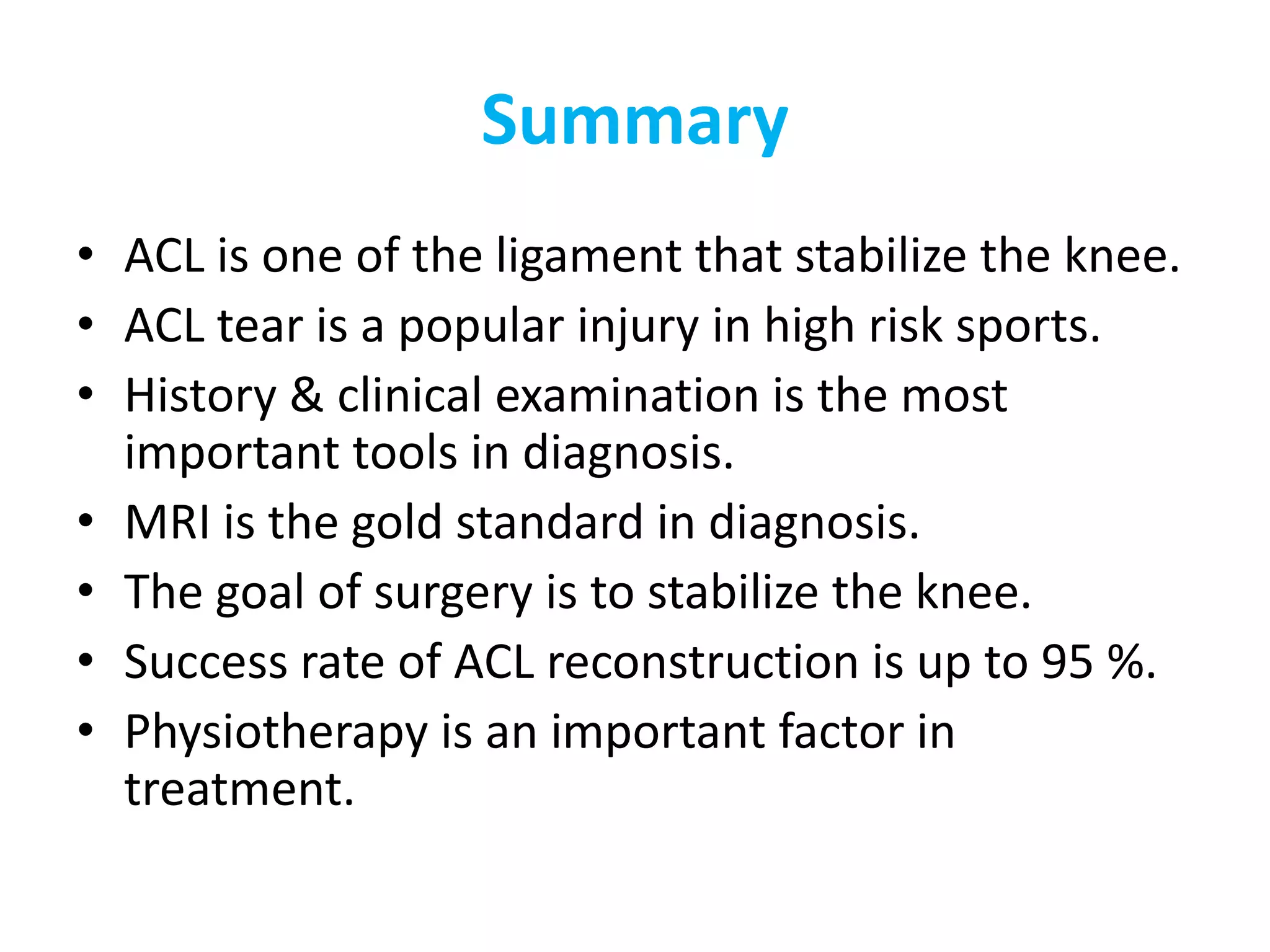
ACL TEAR
Dr. M.A.ROSHAN ZAMEER
1st Year,Pg
• Stability of knee.
• Anatomy of the ACL.
• Functions of ACL.
• Risk Factors to ACL tear.
• Clinical picture.
• Examinations.
• Investigations.
• Treatment.
• complications
Stability of knee
Anatomy
• The ACL is composed of densely organized,
fibrous collagenous connective tissue that
attaches the femur to the tibia.
• 2 groups:
• -Antromedia band
• -Postrolateral band
Attachment
On the Femur, the ACL is attached to:
a fossa on the posteromedial edge of the
lateral femoral condyle.
On the Tibia, the ACL is inserted to:
a fossa that is anterior to the anterior
tibialspine
Attachment
Function of ACL
• primary (85%) restraint to limit anterior
translation of the tibia.
• secondary restraint to tibial rotation and
varus/valgus angulation at full extension.
• The average tensile strength for the ACL is
2160 N.
Biomechanics
• Primary- Anterior translation of tibia
• Secondary – internal rotation of tibia
BIOMECHANICS
Mechanism of Injury
• Contact :
• Non contact:
Rapid deceleration
Jumping
Cutting
• Contact and high-energy traumatic injuries:
• -often are associated with other ligamentous
and meniscal injuries.
SYMPTOMS
• Acute :
• h/o injury
• Knee popped out and reduced
• Not able to getup and walk immediately
• Within hours knee swells
• Chronic :
• Giving way &instabiity
SIGNS
• Knee effusion (haemarthrosis) – A/C cases
• Quadriceps wasting – chronic cases
• Lachman test – most sensitive test
• Anterior drawer test
• Pivot shift test
Investigations
• Imaging Studies:
• Plain radiographs. Usually -ve
• Arthrograms. replaced by MRI
• MRI
* Gold standard
* 90-98% sensitivity.
* identify bone bruising.
MRI
TREATMENT
• Conservative
• Surgical
• Acute Phase
• Recovery Phase
• Maintenance Phase
Non operative
• who are willing to make lifestyle changes and
avoid the activities that cause recurrent
instability.
• Aggressive rehabilitation program and
counseling about activity level.
• functional knee brace - is controversial and
has not been shown to reduce the incidence
of reinjury significantly if a patient returns to
highlevel sports.
Arthroscopic Reconstruction
• Indications
• Atheletes - jumping, pivoting, cutting
• Recurrent episodes of giving way, recurrent effusions.
• Children
• Persistant anterior knee pain
• Associated Meniscal or collateral ligament injuries
Timing of surgery
• No swelling
• Full range of knee motion
• 3 weeks
Graft options
• Autografts
• Allografts
• synthetic
Autografts
– Hamstrings –
semitendinosus &
gracilis
– Bone patellar tendon
bone
– Quadriceps
Technique of Reconstruction
• Diagnostic arthroscopy
• Address meniscal pathology
• Clear the remanent of ACL
Graft harvesting and preparation
Tibial tunnel
Femoral tunnel
Femoral tunnel
Passing the graft
• Fixation of graft
Post op x ray
Recovery Phase
• Physical Therapy:
• Therapy protocols divided into the following 4 categories:
• Phase I: preoperative period when the goal is to maintain
full ROM.
• Phase II (0-2 wk): The goal is to achieve full extension,
maintain quadriceps control, minimize swelling, and
achieve flexion to 90o.
• Phase III (3-5 wk): Maintain full extension and increase
flexion up to full ROM.
• Phase IV (6 wk): Increase strength and agility, progressive
return to sports.
• Return to all sports without activity may take 6-9 months
Maintenance Phase
• Physical Therapy
• Once quadricepsstrengthreaches 65%of the
opposite leg, sports-specific activities may be
performed
• The athlete may return to activity when the
quadricepsstrengthhas reached 80%>>>
• Re-growth to takes time, it may be need
>>>>5-8 weeks3-4 month
• 6 months
complications
• Stiffness / loss of motion
• Loss of fixation
• Infection
• Graft site complications – patella fracture,
numbness over lateral aspect of leg
• DVT
• Recurrent instability
Summary
• ACL is one of the ligament that stabilize the knee.
• ACL tear is a popular injury in high risk sports.
• History & clinical examination is the most
important tools in diagnosis.
• MRI is the gold standard in diagnosis.
• The goal of surgery is to stabilize the knee.
• Success rate of ACL reconstruction is up to 95 %.
• Physiotherapy is an important factor in
treatment.
reference
• Campbell’s
Thank u

Acl tear

  • 1.
    ACL TEAR Dr. M.A.ROSHANZAMEER 1st Year,Pg
  • 2.
    • Stability ofknee. • Anatomy of the ACL. • Functions of ACL. • Risk Factors to ACL tear. • Clinical picture. • Examinations. • Investigations. • Treatment. • complications
  • 3.
  • 4.
    Anatomy • The ACLis composed of densely organized, fibrous collagenous connective tissue that attaches the femur to the tibia. • 2 groups: • -Antromedia band • -Postrolateral band
  • 5.
    Attachment On the Femur,the ACL is attached to: a fossa on the posteromedial edge of the lateral femoral condyle.
  • 6.
    On the Tibia,the ACL is inserted to: a fossa that is anterior to the anterior tibialspine Attachment
  • 7.
    Function of ACL •primary (85%) restraint to limit anterior translation of the tibia. • secondary restraint to tibial rotation and varus/valgus angulation at full extension. • The average tensile strength for the ACL is 2160 N.
  • 8.
    Biomechanics • Primary- Anteriortranslation of tibia • Secondary – internal rotation of tibia
  • 9.
  • 10.
    Mechanism of Injury •Contact : • Non contact: Rapid deceleration Jumping Cutting
  • 11.
    • Contact andhigh-energy traumatic injuries: • -often are associated with other ligamentous and meniscal injuries.
  • 12.
    SYMPTOMS • Acute : •h/o injury • Knee popped out and reduced • Not able to getup and walk immediately • Within hours knee swells • Chronic : • Giving way &instabiity
  • 13.
    SIGNS • Knee effusion(haemarthrosis) – A/C cases • Quadriceps wasting – chronic cases • Lachman test – most sensitive test • Anterior drawer test • Pivot shift test
  • 14.
    Investigations • Imaging Studies: •Plain radiographs. Usually -ve • Arthrograms. replaced by MRI • MRI * Gold standard * 90-98% sensitivity. * identify bone bruising.
  • 15.
  • 16.
    TREATMENT • Conservative • Surgical •Acute Phase • Recovery Phase • Maintenance Phase
  • 17.
    Non operative • whoare willing to make lifestyle changes and avoid the activities that cause recurrent instability. • Aggressive rehabilitation program and counseling about activity level. • functional knee brace - is controversial and has not been shown to reduce the incidence of reinjury significantly if a patient returns to highlevel sports.
  • 18.
    Arthroscopic Reconstruction • Indications •Atheletes - jumping, pivoting, cutting • Recurrent episodes of giving way, recurrent effusions. • Children • Persistant anterior knee pain • Associated Meniscal or collateral ligament injuries
  • 19.
    Timing of surgery •No swelling • Full range of knee motion • 3 weeks
  • 20.
    Graft options • Autografts •Allografts • synthetic
  • 21.
    Autografts – Hamstrings – semitendinosus& gracilis – Bone patellar tendon bone – Quadriceps
  • 22.
    Technique of Reconstruction •Diagnostic arthroscopy
  • 23.
    • Address meniscalpathology • Clear the remanent of ACL
  • 24.
  • 25.
  • 26.
  • 27.
  • 28.
  • 29.
  • 30.
  • 31.
    Recovery Phase • PhysicalTherapy: • Therapy protocols divided into the following 4 categories: • Phase I: preoperative period when the goal is to maintain full ROM. • Phase II (0-2 wk): The goal is to achieve full extension, maintain quadriceps control, minimize swelling, and achieve flexion to 90o. • Phase III (3-5 wk): Maintain full extension and increase flexion up to full ROM. • Phase IV (6 wk): Increase strength and agility, progressive return to sports. • Return to all sports without activity may take 6-9 months
  • 32.
    Maintenance Phase • PhysicalTherapy • Once quadricepsstrengthreaches 65%of the opposite leg, sports-specific activities may be performed • The athlete may return to activity when the quadricepsstrengthhas reached 80%>>> • Re-growth to takes time, it may be need >>>>5-8 weeks3-4 month • 6 months
  • 33.
    complications • Stiffness /loss of motion • Loss of fixation • Infection • Graft site complications – patella fracture, numbness over lateral aspect of leg • DVT • Recurrent instability
  • 34.
    Summary • ACL isone of the ligament that stabilize the knee. • ACL tear is a popular injury in high risk sports. • History & clinical examination is the most important tools in diagnosis. • MRI is the gold standard in diagnosis. • The goal of surgery is to stabilize the knee. • Success rate of ACL reconstruction is up to 95 %. • Physiotherapy is an important factor in treatment.
  • 35.
  • 36.

Editor's Notes