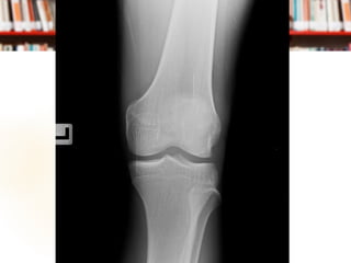
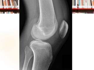
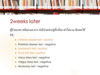
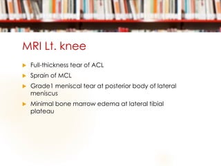
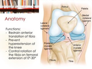
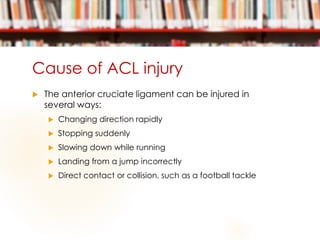
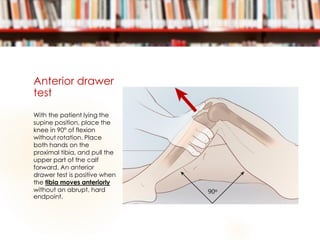
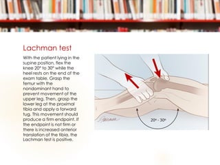
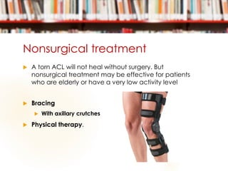
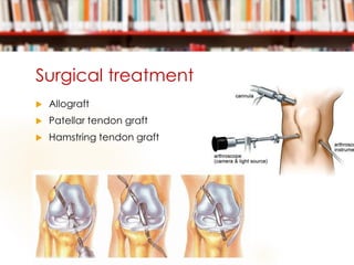
Case presentation
ORTHOPEADICS KORAT| EXT. POONYAPORN PHANKOSOL
Cheif complaint
ผู้ป่วยชายอายุ 19 ปี
มาด้วย เข่าซ้ายบวม 1 ชั่วโมงก่อนมารพ.
Present illness
 1 ชั่วโมงก่อนมารพ. ผู้ป่วยเตะฟุตบอล กระโดดลงพื้น เข่า
ซ้ายบิดแล้วล้มลง เจ็บหัวเข่าด้านซ้ายทันที ลงน้าหนักได้ไม่
สุด กดเจ็บด้านนอกหัวเข่า ไม่มีขาผิดรูป เดินได้แต่กระเผลก
ไม่มีขาชา ไม่มีบาดแผลภายนอก pain score 8/10 เข่า
ซ้ายบวมและปวดมากขึ้น จึงมาโรงพยาบาล
Past history
 ปฏิเสธโรคประจาตัว
 ปฏิเสธแพ้ยา,แพ้อาหาร
 ปฏิเสธยาที่รับประทานประจา
 ปฏิเสธประวัติผ่าตัด
Physical examination
 V/S: BT 37°C PR 84 bpm RR 18 /min BP126/68 mmHg
 GA: A teenage Thai male, good consciousness
 HEENT: Not pale conjunctivae, anicteric sclerae
 Heart: Normal S1S2, No murmur
 Lung: Clear, no adventitious sound
 Abdomen: Normoactive bowel sound, soft, not tender
 Affected part:
 no deformity, swelling and tender Lt. knee, limit active
movement due to pain, full passive ROM, ballottement
test positive
Differential diagnosis
 ACL injury
 Meniscus injury
 Fracture distal femur/proximal tibia
Management at ER
 Film Knee AP, Lateral
Management at ER
 Immobilization: On posterior long leg slab
 D/C
 Pain control
 F/U 2 weeks
2weeks later
ผู้ป่วยมาตรวจติดตามอาการ ยังมีปวดเข่าอยู่เล็กน้อย เข่าไม่บวม เดินพอได้
PE:
 Anterior drawer test : positive
 Posterior drawer test : negative
 Lachman's test : positive
 Pivot shift test : positive
 Varus stress test : negative
 Valgus stress test : negative
 McMurrey test : negative
Investigation
 MRI Lt. knee
MRI Lt. knee
 Full-thickness tear of ACL
 Sprain of MCL
 Grade1 meniscal tear at posterior body of lateral
meniscus
 Minimal bone marrow edema at lateral tibial
plateau
ACL injury
Anatomy
 ORIGIN
 posterior aspect of the
lateral femoral condyle
 INSERTION
 middle of the tibial plateau
 Blood supply
 Middle geniculate artery
 Innervation
 Tibial nerve
 Golgi tendon receptor
Anatomy
Functions:
- Restrain anterior
translation of tibia
- Prevent
hyperextension of
the knee
- Control rotation of
the tibia on femoral
extension of 0°-30°
Cause of ACL injury
 The anterior cruciate ligament can be injured in
several ways:
 Changing direction rapidly
 Stopping suddenly
 Slowing down while running
 Landing from a jump incorrectly
 Direct contact or collision, such as a football tackle
Anterior drawer
test
With the patient lying the
supine position, place the
knee in 90° of flexion
without rotation. Place
both hands on the
proximal tibia, and pull the
upper part of the calf
forward. An anterior
drawer test is positive when
the tibia moves anteriorly
without an abrupt, hard
endpoint.
Lachman test
With the patient lying in the
supine position, flex the
knee 20° to 30° while the
heel rests on the end of the
exam table. Grasp the
femur with the
nondominant hand to
prevent movement of the
upper leg. Then, grasp the
lower leg at the proximal
tibia and apply a forward
tug. This movement should
produce a firm endpoint. If
the endpoint is not firm or
there is increased anterior
translation of the tibia, the
Lachman test is positive.
Pivot shift test
When the lower leg is
stabilized in near full
extension. With increasing
flexion, a palpable
springlike reduction should
be observed. A positive
pivot shift test usually
produces a thud or jerk
around 10° to 20° of flexion.
During a positive exam, the
force created by the
examiner will cause the
knee joint to slip, giving a
positive visual for
identifying rotational knee
instability.
Associated injury
 Meniscus (50%)
 Articular cartilage (30%)
 Collateral ligament (30%)
Grading
 Grade 1 Sprains. The ligament is mildly
damaged in a Grade 1 Sprain. It has
been slightly stretched, but is still able to
help keep the knee joint stable.
 Grade 2 Sprains. A Grade 2 Sprain
stretches the ligament to the point where
it becomes loose. This is often referred to
as a partial tear of the ligament.
 Grade 3 Sprains. This type of sprain is
most commonly referred to as a
complete tear of the ligament. The
ligament has been split into two pieces,
and the knee joint is unstable.
Symptoms
 Popping sound
 Pain with swelling (within 24hr)
 Loss of full range of motion
 Tender along joint line
 Discomfort while walking
Treatment
 Initial treatment
 R.I.C.E
 Non surgical treatment
 Surgical treatment
Nonsurgical treatment
 A torn ACL will not heal without surgery. But
nonsurgical treatment may be effective for patients
who are elderly or have a very low activity level
 Bracing
 With axillary crutches
 Physical therapy.
Surgical treatment
 Allograft
 Patellar tendon graft
 Hamstring tendon graft
THANK YOU

Acl injury

  • 1.
    Case presentation ORTHOPEADICS KORAT|EXT. POONYAPORN PHANKOSOL
  • 2.
    Cheif complaint ผู้ป่วยชายอายุ 19ปี มาด้วย เข่าซ้ายบวม 1 ชั่วโมงก่อนมารพ.
  • 3.
    Present illness  1ชั่วโมงก่อนมารพ. ผู้ป่วยเตะฟุตบอล กระโดดลงพื้น เข่า ซ้ายบิดแล้วล้มลง เจ็บหัวเข่าด้านซ้ายทันที ลงน้าหนักได้ไม่ สุด กดเจ็บด้านนอกหัวเข่า ไม่มีขาผิดรูป เดินได้แต่กระเผลก ไม่มีขาชา ไม่มีบาดแผลภายนอก pain score 8/10 เข่า ซ้ายบวมและปวดมากขึ้น จึงมาโรงพยาบาล
  • 4.
    Past history  ปฏิเสธโรคประจาตัว ปฏิเสธแพ้ยา,แพ้อาหาร  ปฏิเสธยาที่รับประทานประจา  ปฏิเสธประวัติผ่าตัด
  • 5.
    Physical examination  V/S:BT 37°C PR 84 bpm RR 18 /min BP126/68 mmHg  GA: A teenage Thai male, good consciousness  HEENT: Not pale conjunctivae, anicteric sclerae  Heart: Normal S1S2, No murmur  Lung: Clear, no adventitious sound  Abdomen: Normoactive bowel sound, soft, not tender  Affected part:  no deformity, swelling and tender Lt. knee, limit active movement due to pain, full passive ROM, ballottement test positive
  • 6.
    Differential diagnosis  ACLinjury  Meniscus injury  Fracture distal femur/proximal tibia
  • 7.
    Management at ER Film Knee AP, Lateral
  • 10.
    Management at ER Immobilization: On posterior long leg slab  D/C  Pain control  F/U 2 weeks
  • 11.
    2weeks later ผู้ป่วยมาตรวจติดตามอาการ ยังมีปวดเข่าอยู่เล็กน้อยเข่าไม่บวม เดินพอได้ PE:  Anterior drawer test : positive  Posterior drawer test : negative  Lachman's test : positive  Pivot shift test : positive  Varus stress test : negative  Valgus stress test : negative  McMurrey test : negative
  • 12.
  • 13.
    MRI Lt. knee Full-thickness tear of ACL  Sprain of MCL  Grade1 meniscal tear at posterior body of lateral meniscus  Minimal bone marrow edema at lateral tibial plateau
  • 14.
  • 15.
    Anatomy  ORIGIN  posterioraspect of the lateral femoral condyle  INSERTION  middle of the tibial plateau  Blood supply  Middle geniculate artery  Innervation  Tibial nerve  Golgi tendon receptor
  • 16.
    Anatomy Functions: - Restrain anterior translationof tibia - Prevent hyperextension of the knee - Control rotation of the tibia on femoral extension of 0°-30°
  • 17.
    Cause of ACLinjury  The anterior cruciate ligament can be injured in several ways:  Changing direction rapidly  Stopping suddenly  Slowing down while running  Landing from a jump incorrectly  Direct contact or collision, such as a football tackle
  • 18.
    Anterior drawer test With thepatient lying the supine position, place the knee in 90° of flexion without rotation. Place both hands on the proximal tibia, and pull the upper part of the calf forward. An anterior drawer test is positive when the tibia moves anteriorly without an abrupt, hard endpoint.
  • 19.
    Lachman test With thepatient lying in the supine position, flex the knee 20° to 30° while the heel rests on the end of the exam table. Grasp the femur with the nondominant hand to prevent movement of the upper leg. Then, grasp the lower leg at the proximal tibia and apply a forward tug. This movement should produce a firm endpoint. If the endpoint is not firm or there is increased anterior translation of the tibia, the Lachman test is positive.
  • 20.
    Pivot shift test Whenthe lower leg is stabilized in near full extension. With increasing flexion, a palpable springlike reduction should be observed. A positive pivot shift test usually produces a thud or jerk around 10° to 20° of flexion. During a positive exam, the force created by the examiner will cause the knee joint to slip, giving a positive visual for identifying rotational knee instability.
  • 21.
    Associated injury  Meniscus(50%)  Articular cartilage (30%)  Collateral ligament (30%)
  • 22.
    Grading  Grade 1Sprains. The ligament is mildly damaged in a Grade 1 Sprain. It has been slightly stretched, but is still able to help keep the knee joint stable.  Grade 2 Sprains. A Grade 2 Sprain stretches the ligament to the point where it becomes loose. This is often referred to as a partial tear of the ligament.  Grade 3 Sprains. This type of sprain is most commonly referred to as a complete tear of the ligament. The ligament has been split into two pieces, and the knee joint is unstable.
  • 23.
    Symptoms  Popping sound Pain with swelling (within 24hr)  Loss of full range of motion  Tender along joint line  Discomfort while walking
  • 24.
    Treatment  Initial treatment R.I.C.E  Non surgical treatment  Surgical treatment
  • 25.
    Nonsurgical treatment  Atorn ACL will not heal without surgery. But nonsurgical treatment may be effective for patients who are elderly or have a very low activity level  Bracing  With axillary crutches  Physical therapy.
  • 26.
    Surgical treatment  Allograft Patellar tendon graft  Hamstring tendon graft
  • 27.