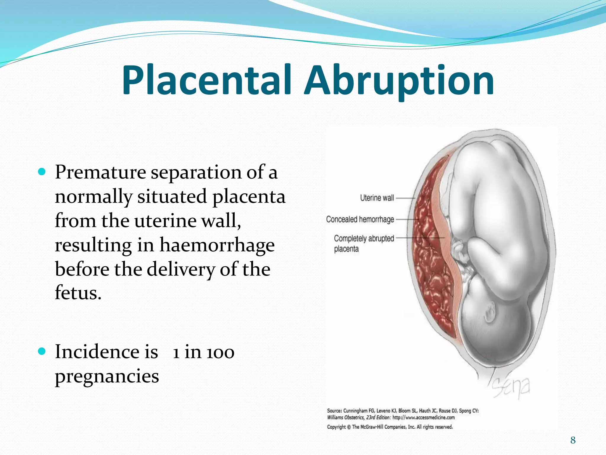
Dr .Prerit Devkota
2nd year GP/EM Resident
1
Contents
 Introduction
 Classification
 Etiopathogenesis
 Placental Abruption and Placenta Previa
 Management
 Take home messeges
2
Antepartum hemorrhage
 Bleeding from or into the genital tract, occuring after 24 weeks of
gestation and prior to birth of baby .
(RCOG 2011)
 Epidemiology:
o Affects 3-5% of all pregnancies
o 1/5th of preterm babies are in association of APH
o Occurs in 2.8/ 1000 singleton pregnancies & 3.9/1000 twin
pregnancies
3
Definition:
 Spotting – staining, streaking or blood spotting noted on
underwear or sanitary protection
 Minor hemorrhage – blood loss < 50 ml that has settled
 Major hemorrhage – blood loss of 50–1000 ml, with no
signs of clinical shock
 Massive hemorrhage – blood loss >1000 ml and/or signs
of clinical shock.
4
Etiology
 Placental bleeding
 Placenta previa (4-5/1000 pregnancies)
 Abruptio placenta (1% of all pregnancies)
 Unexplained/ Indeterminate (1-5% of all pregnancies )
 Local cause: cervical, vaginal or uterine pathology
 Cervical Polyps,erosions
 Infection/Inflammation
 Local trauma
 Varicose vein
 Blood stained cervical mucus
5
Placental bleeding:
 Placenta previa:
 Separation of placenta implanted in the immediate vicinity of cervical
canal.
 Placental abruption:
 Separation of placenta located elsewhere in the uterine cavity .
 Vasa previa
 Velamentous insertion of umbilical cord and involved placental vessels may
overlie the cervix.
6
December 6, 2019 7
Placental Abruption
 Premature separation of a
normally situated placenta
from the uterine wall,
resulting in haemorrhage
before the delivery of the
fetus.
 Incidence is 1 in 100
pregnancies
8
Risk factors for Placental Abruption:
 Increased age and parity
 Abruption in previous pregnancy( recurrence:12% in subsequent
pregnancy)
 Vascular disease: hypertension in pregnancy, SLE
 Mechanical factors: Trauma, amniocentesis, sudden decompression of
uterus, poly-hydraminos, multiple pregnancy
 Drugs: Smoking, Cocaine
 Uterine factor: uterine myoma, septum, bicornuate uterus
 Thrombophilias
9
Pathophysiology
Spasm of vessels in uteroplacental bed (decidual
spiral artery)
anoxic endothelial damage
Rupture of vessels and hemorrhage in decidua
basalis-decidua splits
Decidual hematoma(retroplacental)
Seperation and destruction of adjacent placenta
10
Types of abruption:
• Concealed
• Mixed
• Revealed
11
Grades of Placental Abruption:
Sher and Statland
Mild (grade 1):
 This is not recognized clinically before delivery and usually diagnosed
by the presence of a retro-placental clot.
 This is a retrospective diagnosis.
Moderate (grade 2):
 This is an intermediate grade in which the classical clinical signs of
abruption are present but the fetus is still alive.
 The frequency of fetal heart rate abnormalities is high.
Severe (grade 3):
 This is the severe grade in which the fetus is dead and coagulopathy
may be present.
 The volume of blood loss is appreciable in this condition.
12
Investigations:
Ultrasonography:
• To exclude placenta previa
• Detects retroplacental
hematoma/fetal viability
(Negative findings does not exclude
placental abruption)
CTG:
 fetal tachycardia/ bradycardia
 Reassuring / non – reassuring
Labs:
 CBC
 Markers of consumptive coagulopathy: Platelets, BT,CT, PT-INR, APTT
13
Complications
MATERNAL
 Hemorrhage
 Shock( out of proportion to blood
loss)
 Blood Coagulation disorders
 Renal involvement: Oligouria and
anuria
 Postpartum hemorrhage
 Puerperial sepsis
FETAL:
 Fetal death
Revealed: 25-50%
Concealed: 50-100%
14
Management
 Treatment: Varies depending on gestational age and the status of the mother and
fetus.
 Admission
 History & examination
 Assess blood loss
 IV access, X match, DIC screen
 Assess fetal well-being
 Placental localization
15
Contd.
Obstetric management:
 Viable fetus with no fetal compromise: Vaginal delivery
 Viable fetus with fetal compromise: Em LSCS (within 20
minutes)
 Severe abruption with fetal death: Vaginal delivery with
Amniotomy
(allow better spiral artery compression which serves decrease bleeding
and entry of thromboplastin into maternal circulation)
 Oxytocin : controversial
16
Coagulopathy with abruption
 Occurs in 1/3rd of fetal demise
 Usually not seen with delivery of live fetus
 Etiologies: consumption, disseminated intravascular
coagulation(DIC)
 Administer platelets, fresh frozen plasma before operative
delivery.
17
18
Placenta Previa
 Insertion of placenta partially/ completely in the lower segment
of uterus
19
Risk factors for Placenta Previa
 Previous placenta praevia (OR 9.7)
 Previous caesarean sections
 1 previous CS OR 2.2 (95% CI (1.4–3.4) with a background rate of 1%)
 2 previous CS OR 4.1 (95% CI 1.9–8.8)
 3 previous caesarean sections OR 22.4 (95% CI 6.4–78.3)
 Multiparity
 Advanced maternal age (>40 years)
20
Contd.
 Multiple pregnancy
 Smoking
 Deficient endometrium:
 uterine scar
 endometritis
 manual removal of placenta
 Assisted conception
( RCOG guidelines 2011)
21
Types of Placenta Previa
Four types:
Type I(low lying):
 placenta encroaches lower
segment but does not reach
internal OS
Type II( Marginal)
 Reaches internal OS but does not
cover it
Type III (incomplete)
 Covers parts of internal OS
Type IV(central/total)
 Completely cover the OS
22
Placent Accreta spectrum
23
Pathogenesis
 Pathogenesis unknown
 Hypothesis:
 presence of areas of sub optimally vascularised decidua in
upper uterine cavity due to previous surgeries or multiple
pregnancies promote implantation of trophoblast in, or
undirectional growth toward the LUS(placental trophotropism)
 Another hypothesis:
 large placental size directly closes the Os
24
Clinical features
 Sudden onset, painless, apparently causeless and recurrent vaginal
bleeding
 Abdominal findings:
 Uterus: soft, relaxed, non tender and proportionate to POG
 Malpresentation(breech/ transverse or unstable lie)
 Head is floating
 FHS: good
 Vulval inspection: presence of bleeding, character of blood
(PV examination is containdicated)
25
Confirmation of diagnosis
clinical Localization of placenta
By internal examination
(double set up)
Transabdominal ultrasonography
Direct visualization during CS Transvaginal(TVS)
Examination of placenta following
delivery
Transperineal(TPS)
Colour doppler flow study
MRI
26
December 6, 2019 27
Management:
 Prematurity-major complication, the main therapeutic strategy is to
prolong pregnancy until fetal lung maturity is achieved.
1. Assess general condition
2. Blood for Hb, Grouping, X-matching
3. I/V line with Crystalloid
4. Gentle abd. Examination
5. No P/V examination
6. USG for placentation, fetal maturity and viability
28
Management:
 Active bleedng:
 If preterm, consider maternal transfer to appropriate level of care
 Antenatal corticosteroids and tocolysis
 No active bleeding :
 expectant management
 No intercourse in the third trimester
 Avoid digital examinations
 Ultrasound at 36 weeks can help determine mode of delivery
29
Management
 Expectant management:
 No active bleeding
 Preterm<37weeks
 Normal fetus
 Normal Hb>10gm/dl
 If bleeding stops, can consider outpatient management
 Assurance of current maternal and fetal well‐being
 Patient lives in close proximity to hospital
 Immediate evaluation can occur if new bleeding episode starts or with onset of labor
 Current guidelines from the Royal College of Obstetricians and Gynecologists
(RCOG) recommend caesarean delivery for women with placental edge – internal
os distance of less than 2.0 cm.
30
December 6, 2019 31
Complications:
Maternal:
During pregnancy:
 Antepartum hemorrhage and varying degree of shock
 Malpresentation
 Premature labour
During labour:
 Early rupture of membrane
 Cord prolapse
 Intrapartum hemorrhage, increased operative interference
Postpartum:
 Postpartum hemorrhage
 Puerperial sepsis
 subinvolution
32
Fetal complications:
 Low birth weight
 Perinatal asphyxia
 Intrauterine death
 Congenital malformation
33
Vasa previa
 Fetal blood vessels from
placenta or umbilcal cord cross
the internal OS beneath the
baby
 Rupture of membrane lead to
damage of the fetal vessels
leading to exsanguination and
death
 High fetal mortality (50-75%)
34
Risk factors of vasa previa:
 Eccentric(velamentous cord insertion)
 Bilobed or succenturiate lobe of placenta
 Multiple gestation
 Placenta previa
 Invitro fertilization(IVF) pregnancies
 History of uterine surgery or D&C
35
 Diagnosis:
 Moderate vaginal bleeding with fetal distress
 Vessels may be palpable through dilated cervix
 Vessels may be visible on doppler ultrasound
 CTG: Tachycardia/ bradycardia
 Management:
 Caeserean section
36
Take Home Messeges
 Placenta previa and placental abruption are major causes of
antepartum hemorrhage
 Rapid clinical diagnosis is imperative and resuscitation is utmost
 Always remember CAB in case of APH
 No vaginal digital examination until placental location is known
 Ultrasound can help determine the cause of bleeding
37
References
 UpToDate 2019
 Arias’ Practical Guide to High-Risk Pregnancy and
Delivery
 Royal College of Obstetricians and Gynaecologists
guidelines
38
THANK YOU !!!!
39

Antepartum hemorrhage prerit (2)

  • 1.
    Dr .Prerit Devkota 2ndyear GP/EM Resident 1
  • 2.
    Contents  Introduction  Classification Etiopathogenesis  Placental Abruption and Placenta Previa  Management  Take home messeges 2
  • 3.
    Antepartum hemorrhage  Bleedingfrom or into the genital tract, occuring after 24 weeks of gestation and prior to birth of baby . (RCOG 2011)  Epidemiology: o Affects 3-5% of all pregnancies o 1/5th of preterm babies are in association of APH o Occurs in 2.8/ 1000 singleton pregnancies & 3.9/1000 twin pregnancies 3
  • 4.
    Definition:  Spotting –staining, streaking or blood spotting noted on underwear or sanitary protection  Minor hemorrhage – blood loss < 50 ml that has settled  Major hemorrhage – blood loss of 50–1000 ml, with no signs of clinical shock  Massive hemorrhage – blood loss >1000 ml and/or signs of clinical shock. 4
  • 5.
    Etiology  Placental bleeding Placenta previa (4-5/1000 pregnancies)  Abruptio placenta (1% of all pregnancies)  Unexplained/ Indeterminate (1-5% of all pregnancies )  Local cause: cervical, vaginal or uterine pathology  Cervical Polyps,erosions  Infection/Inflammation  Local trauma  Varicose vein  Blood stained cervical mucus 5
  • 6.
    Placental bleeding:  Placentaprevia:  Separation of placenta implanted in the immediate vicinity of cervical canal.  Placental abruption:  Separation of placenta located elsewhere in the uterine cavity .  Vasa previa  Velamentous insertion of umbilical cord and involved placental vessels may overlie the cervix. 6
  • 7.
  • 8.
    Placental Abruption  Prematureseparation of a normally situated placenta from the uterine wall, resulting in haemorrhage before the delivery of the fetus.  Incidence is 1 in 100 pregnancies 8
  • 9.
    Risk factors forPlacental Abruption:  Increased age and parity  Abruption in previous pregnancy( recurrence:12% in subsequent pregnancy)  Vascular disease: hypertension in pregnancy, SLE  Mechanical factors: Trauma, amniocentesis, sudden decompression of uterus, poly-hydraminos, multiple pregnancy  Drugs: Smoking, Cocaine  Uterine factor: uterine myoma, septum, bicornuate uterus  Thrombophilias 9
  • 10.
    Pathophysiology Spasm of vesselsin uteroplacental bed (decidual spiral artery) anoxic endothelial damage Rupture of vessels and hemorrhage in decidua basalis-decidua splits Decidual hematoma(retroplacental) Seperation and destruction of adjacent placenta 10
  • 11.
    Types of abruption: •Concealed • Mixed • Revealed 11
  • 12.
    Grades of PlacentalAbruption: Sher and Statland Mild (grade 1):  This is not recognized clinically before delivery and usually diagnosed by the presence of a retro-placental clot.  This is a retrospective diagnosis. Moderate (grade 2):  This is an intermediate grade in which the classical clinical signs of abruption are present but the fetus is still alive.  The frequency of fetal heart rate abnormalities is high. Severe (grade 3):  This is the severe grade in which the fetus is dead and coagulopathy may be present.  The volume of blood loss is appreciable in this condition. 12
  • 13.
    Investigations: Ultrasonography: • To excludeplacenta previa • Detects retroplacental hematoma/fetal viability (Negative findings does not exclude placental abruption) CTG:  fetal tachycardia/ bradycardia  Reassuring / non – reassuring Labs:  CBC  Markers of consumptive coagulopathy: Platelets, BT,CT, PT-INR, APTT 13
  • 14.
    Complications MATERNAL  Hemorrhage  Shock(out of proportion to blood loss)  Blood Coagulation disorders  Renal involvement: Oligouria and anuria  Postpartum hemorrhage  Puerperial sepsis FETAL:  Fetal death Revealed: 25-50% Concealed: 50-100% 14
  • 15.
    Management  Treatment: Variesdepending on gestational age and the status of the mother and fetus.  Admission  History & examination  Assess blood loss  IV access, X match, DIC screen  Assess fetal well-being  Placental localization 15
  • 16.
    Contd. Obstetric management:  Viablefetus with no fetal compromise: Vaginal delivery  Viable fetus with fetal compromise: Em LSCS (within 20 minutes)  Severe abruption with fetal death: Vaginal delivery with Amniotomy (allow better spiral artery compression which serves decrease bleeding and entry of thromboplastin into maternal circulation)  Oxytocin : controversial 16
  • 17.
    Coagulopathy with abruption Occurs in 1/3rd of fetal demise  Usually not seen with delivery of live fetus  Etiologies: consumption, disseminated intravascular coagulation(DIC)  Administer platelets, fresh frozen plasma before operative delivery. 17
  • 18.
  • 19.
    Placenta Previa  Insertionof placenta partially/ completely in the lower segment of uterus 19
  • 20.
    Risk factors forPlacenta Previa  Previous placenta praevia (OR 9.7)  Previous caesarean sections  1 previous CS OR 2.2 (95% CI (1.4–3.4) with a background rate of 1%)  2 previous CS OR 4.1 (95% CI 1.9–8.8)  3 previous caesarean sections OR 22.4 (95% CI 6.4–78.3)  Multiparity  Advanced maternal age (>40 years) 20
  • 21.
    Contd.  Multiple pregnancy Smoking  Deficient endometrium:  uterine scar  endometritis  manual removal of placenta  Assisted conception ( RCOG guidelines 2011) 21
  • 22.
    Types of PlacentaPrevia Four types: Type I(low lying):  placenta encroaches lower segment but does not reach internal OS Type II( Marginal)  Reaches internal OS but does not cover it Type III (incomplete)  Covers parts of internal OS Type IV(central/total)  Completely cover the OS 22
  • 23.
  • 24.
    Pathogenesis  Pathogenesis unknown Hypothesis:  presence of areas of sub optimally vascularised decidua in upper uterine cavity due to previous surgeries or multiple pregnancies promote implantation of trophoblast in, or undirectional growth toward the LUS(placental trophotropism)  Another hypothesis:  large placental size directly closes the Os 24
  • 25.
    Clinical features  Suddenonset, painless, apparently causeless and recurrent vaginal bleeding  Abdominal findings:  Uterus: soft, relaxed, non tender and proportionate to POG  Malpresentation(breech/ transverse or unstable lie)  Head is floating  FHS: good  Vulval inspection: presence of bleeding, character of blood (PV examination is containdicated) 25
  • 26.
    Confirmation of diagnosis clinicalLocalization of placenta By internal examination (double set up) Transabdominal ultrasonography Direct visualization during CS Transvaginal(TVS) Examination of placenta following delivery Transperineal(TPS) Colour doppler flow study MRI 26
  • 27.
  • 28.
    Management:  Prematurity-major complication,the main therapeutic strategy is to prolong pregnancy until fetal lung maturity is achieved. 1. Assess general condition 2. Blood for Hb, Grouping, X-matching 3. I/V line with Crystalloid 4. Gentle abd. Examination 5. No P/V examination 6. USG for placentation, fetal maturity and viability 28
  • 29.
    Management:  Active bleedng: If preterm, consider maternal transfer to appropriate level of care  Antenatal corticosteroids and tocolysis  No active bleeding :  expectant management  No intercourse in the third trimester  Avoid digital examinations  Ultrasound at 36 weeks can help determine mode of delivery 29
  • 30.
    Management  Expectant management: No active bleeding  Preterm<37weeks  Normal fetus  Normal Hb>10gm/dl  If bleeding stops, can consider outpatient management  Assurance of current maternal and fetal well‐being  Patient lives in close proximity to hospital  Immediate evaluation can occur if new bleeding episode starts or with onset of labor  Current guidelines from the Royal College of Obstetricians and Gynecologists (RCOG) recommend caesarean delivery for women with placental edge – internal os distance of less than 2.0 cm. 30
  • 31.
  • 32.
    Complications: Maternal: During pregnancy:  Antepartumhemorrhage and varying degree of shock  Malpresentation  Premature labour During labour:  Early rupture of membrane  Cord prolapse  Intrapartum hemorrhage, increased operative interference Postpartum:  Postpartum hemorrhage  Puerperial sepsis  subinvolution 32
  • 33.
    Fetal complications:  Lowbirth weight  Perinatal asphyxia  Intrauterine death  Congenital malformation 33
  • 34.
    Vasa previa  Fetalblood vessels from placenta or umbilcal cord cross the internal OS beneath the baby  Rupture of membrane lead to damage of the fetal vessels leading to exsanguination and death  High fetal mortality (50-75%) 34
  • 35.
    Risk factors ofvasa previa:  Eccentric(velamentous cord insertion)  Bilobed or succenturiate lobe of placenta  Multiple gestation  Placenta previa  Invitro fertilization(IVF) pregnancies  History of uterine surgery or D&C 35
  • 36.
     Diagnosis:  Moderatevaginal bleeding with fetal distress  Vessels may be palpable through dilated cervix  Vessels may be visible on doppler ultrasound  CTG: Tachycardia/ bradycardia  Management:  Caeserean section 36
  • 37.
    Take Home Messeges Placenta previa and placental abruption are major causes of antepartum hemorrhage  Rapid clinical diagnosis is imperative and resuscitation is utmost  Always remember CAB in case of APH  No vaginal digital examination until placental location is known  Ultrasound can help determine the cause of bleeding 37
  • 38.
    References  UpToDate 2019 Arias’ Practical Guide to High-Risk Pregnancy and Delivery  Royal College of Obstetricians and Gynaecologists guidelines 38
  • 39.

Editor's Notes

  • #21 An odds ratio (OR) is a statistic that quantifies the strength of the association between two events, A and B
  • #23 Typically, the distance from the lower placental edge and internal os increases with growth of the lower uterine segment in late pregnancy (often referred to as ‘formation of the lower uterine segment and ‘placental migration’).
  • #36 A succenturiate (accessory) lobe is a second or third placental lobe that is much smaller than the largest lobe. Unlike bipartite lobes, the smaller succenturiate lobe often has areas of infarction or atrophy. Velamentous cord insertion is an abnormal cord insertion in which the umbilical vessels diverge as they traverse between the amnion and chorion before reaching the placenta