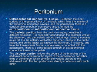
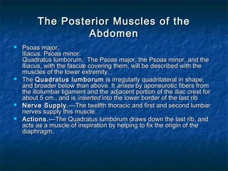
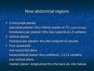
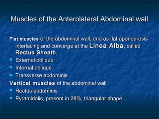
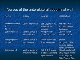
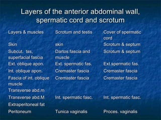
The document discusses the abdominal region and its anatomy. It describes the 9 abdominal regions defined by horizontal and vertical planes. It then discusses the 4 abdominal quadrants and lists the organs located in each quadrant. The rest of the document discusses the muscles of the abdominal wall, nerves of the abdominal wall, layers of the anterior abdominal wall and spermatic cord, arteries of the abdominal wall, and the superficial fascia of the abdomen.


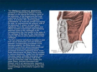

![Inguinal ligamentsInguinal ligaments
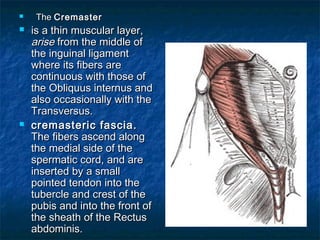
The Inguinal LigamentThe Inguinal Ligament
((ligamentum inguinaleligamentum inguinale
[[PoupartiPouparti];]; Poupart’sPoupart’s
ligamentligament))The inguinal ligament is theThe inguinal ligament is the
lower border of the aponeurosis of thelower border of the aponeurosis of the
Obliquus externus,Obliquus externus,
The Lacunar LigamentThe Lacunar Ligament
((ligamentum lacunareligamentum lacunare
[[GimbernatiGimbernati];]; Gimbernat’sGimbernat’s
ligamentligament)) The lacunar ligament isThe lacunar ligament is
that part of the aponeurosis of thethat part of the aponeurosis of the
Obliquus externus which is reflectedObliquus externus which is reflected
backward and lateralward, and isbackward and lateralward, and is
attached to the pectineal line.attached to the pectineal line.
It is about 1.25 cm. long, larger in theIt is about 1.25 cm. long, larger in the
male than in the female, almostmale than in the female, almost
horizontal in direction in the erecthorizontal in direction in the erect
posture, and of a triangular form withposture, and of a triangular form with
the base directed lateralward.the base directed lateralward.](https://image.slidesharecdn.com/abdominalanatomy-101027111151-phpapp01/85/Abdominal-anatomy-16-320.jpg)
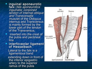
![ ((ligamentum inguinale reflexumligamentum inguinale reflexum [[CollesiCollesi];]; triangulartriangular
fasciafascia).).—The reflected inguinal ligament is a layer of tendinous—The reflected inguinal ligament is a layer of tendinous
fibers of a triangular shape, formed by an expansion from thefibers of a triangular shape, formed by an expansion from the
lacunar ligament and the inferior crus of the subcutaneous inguinallacunar ligament and the inferior crus of the subcutaneous inguinal
ring.ring.
interlaces with the ligament of the other side of the linea albainterlaces with the ligament of the other side of the linea alba
Ligament of Cooper.Ligament of Cooper. ——
It extends lateralward from the base of the lacunar ligament alongIt extends lateralward from the base of the lacunar ligament along
the pectineal line, to which it is attached. It is strengthened by thethe pectineal line, to which it is attached. It is strengthened by the
pectineal fascia, and by a lateral expansion from the lowerpectineal fascia, and by a lateral expansion from the lower
attachment of the linea alba (attachment of the linea alba (adminiculum lineæ albæadminiculum lineæ albæ).).
Variations.Variations.—The Obliquus externus may show decrease or—The Obliquus externus may show decrease or
doubling of its attachments to the ribs; addition slips from lumbardoubling of its attachments to the ribs; addition slips from lumbar
aponeurosis; doubling between lower ribs and ilium or inguinalaponeurosis; doubling between lower ribs and ilium or inguinal
ligament. Rarely tendinous inscriptions occur.ligament. Rarely tendinous inscriptions occur.](https://image.slidesharecdn.com/abdominalanatomy-101027111151-phpapp01/85/Abdominal-anatomy-17-320.jpg)