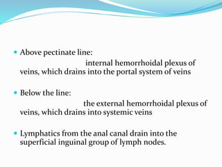
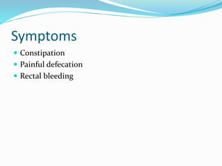
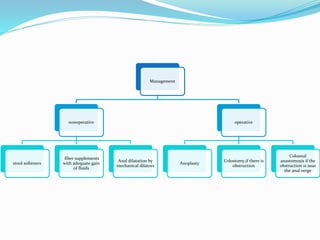
DR.R.SARAVANAN M.S
Thanjavur medical college,
Tamilnadu,India
ANATOMY
 The anal canal is the most terminal part of the lower
GI tract/large intestine
 The demarcation between the rectum above and the
anal canal below is the anorectal ring or anorectal
flexure, where the puborectalis muscle forms a sling
around the posterior aspect of the anorectal junction
 The anal canal is completely extraperitoneal. The
length of the anal canal is about 4 cm (range, 3-5 cm),
with two thirds of this being above the pectinate line
(also known as the dentate line) and one third below
the pectinate line.
Embryology
 The anal canal below the pectinate line develops from
the proctodeum (ectoderm), while that above the
pectinate line develops from the endoderm of the
hindgut.
 The epithelium of the anal canal between the anal
verge below and the pectinate line above is variously
described as anal mucosa or anal skin. As it looks like
(pigmented) skin, is sensitive like skin (why a fissure-
in-ano is very painful), and is keratinized (but does not
have skin appendages).
 The pectinate line is the site of transition of the
proctodeum below and the postallantoic gut above. It
is a scalloped demarcation formed by the anal valves
(transverse folds of mucosa) at the inferior-most ends
of the anal columns. Anal glands open above the anal
valves into the anal sinuses. The pectinate line is not
seen on inspection in clinical practice, but under
anesthesia the anal canal descends down, and the
pectinate line can be seen on slight retraction of the
anal canal skin.
 The anal canal just above the pectinate line for about 1-
2 cm is called the anal pecten or transitional zone.
Above this transitional zone, the anal canal is lined
with columnar epithelium (which is insensitive to
cutting). Anal columns (of Morgagni) are 6-10
longitudinal (vertical) mucosal folds in the upper part
of the anal canal.
 At the bottom of these columns are anal sinuses or crypts,
into which open the anal glands and anal papillae.
 Infection of the anal glands is likely the initial event in
causation of perianal abscess and fistula-in-ano.
 Three of these columns (left lateral, right posterior, and
right anterior, at 3-, 7-, and 11-o’clock positions in supine
position) are prominent; they are called anal cushions and
contain branches and tributaries of superior rectal
(hemorrhoidal) artery and vein. When prominent, veins in
these cushions form the internal hemorrhoids.
 The anorectal junction or anorectal ring is situated
about 5 cm from the anus.
 At the anorectal flexure or angle, the anorectal
junction is pulled anterosuperiorly by the puborectal
sling to continue below as the anal canal.
 The anal canal is surrounded by several perianal
spaces: subcutaneous, submucosal, intersphincteric,
ischioanal (rectal) and pelvirectal.
Blood supply AND lymphatic
drainage
 The anal canal above the pectinate line is supplied by
the terminal branches of the superior rectal
(hemorrhoidal) artery, which is the terminal branch of
the inferior mesenteric artery.
 The middle rectal artery (a branch of the internal iliac
artery) and
 The inferior rectal artery (a branch of the internal
pudendal artery) supply the lower anal canal.
 Above pectinate line:
internal hemorrhoidal plexus of
veins, which drains into the portal system of veins
 Below the line:
the external hemorrhoidal plexus of
veins, which drains into systemic veins
 Lymphatics from the anal canal drain into the
superficial inguinal group of lymph nodes.
CAUSES
 Surgery of the anal canal, (90 % of anal stenosis is caused
by overzealous hemorrhoidectomy)
 Anal malignancy
 trauma,
 inflammatory bowel disease,particularly in crohn’s
 Radiation therapy for pelvic tumors
 venereal disease,(LGV,Gonorrhea,AIDS)
 tubercolosis, and
 Chronic laxative abuse,scleroderma.
 chronic abuse of ergotamine tartrate for the treatment of
migraine headache attack may lead to anorectal stricture
Symptoms
 Constipation
 Painful defecation
 Rectal bleeding
Diagnosis
 Visual examination of the anal canal and perianal skin,
 Digital rectal examination,
 Proctoscopic examination
 Anorectal manometry is an objective method for
assessing anal musculature tone, rectal
compliance,anorectal sensation, and verifying the
integrity of the rectoanal inhibitory reflex( widely
used to document sphincter function prior to
procedures)
 It is important to ascertain the cause of the stricture
in order to determine proper therapy; a malignant
disease must be treated by excision or resection, and
anal Crohn’s disease is an absolute contraindication to
anoplasty
Classification
 On the basis of severity,
 Mild (tight anal canal can be examinated by a well-
lubricated index finger or a medium Hill-Ferguson
retractor),
 Moderate (forceful dilatation is required to insert
either the index finger or a medium Hill-Ferguson
retractor),
 severe anal stenosis (neither the little finger nor a
small Hill-Ferguson retractor can be inserted unless a
forceful dilatation is employed).
 Furthermore, stenosis may be
 diaphragmatic (after inflammatory bowel disease,
characterized by a thin strip of constrictor tissue)
 ring-like or anular (after surgical or traumatic lesions,
of length less than 2 cm), and
 tubular (length more than 2 cm).
 On the basis of the anal canal levels,
 low stenosis (distal anal canal at least 0.5 cm below the
dentate line, 65% of patients),
 middle (0.5 cm proximal to 0.5 cm distal to the dentate
line, 18.5%),
 high (proximal to 0.5 cm above the dentate line, 8.5%),
and diffuse (all anal canal, 6.5% of cases)
 90 % due to excess skin removal in
haemorrhoidectomy
 Prevented by preservation of adequate muco-
cutaneous bridges, which prevents anal stenosis.
 Treatment is based on severity, cause and localization
Management
nonoperative
stool softeners
fiber supplements
with adequate gain
of fluids
Anal dilatation by
mechanical dilators
operative
Anoplasty
Colostomy,if there is
obstruction
Coloanal
anastomosis if the
obstruction is near
the anal verge
A B C D
E
F G H
Operative procedure for the surgical treatment of anal stenosis. A: Martin’s
anoplasty; B: Y-V advancement flap; C: V-Y advancement flap; D: Diamondshaped
flap; E: House-shaped flap; F: U-shaped flap; G: Rotational S-flap.
THANK YOU

Anal stenosis

  • 1.
    DR.R.SARAVANAN M.S Thanjavur medicalcollege, Tamilnadu,India
  • 2.
    ANATOMY  The analcanal is the most terminal part of the lower GI tract/large intestine  The demarcation between the rectum above and the anal canal below is the anorectal ring or anorectal flexure, where the puborectalis muscle forms a sling around the posterior aspect of the anorectal junction  The anal canal is completely extraperitoneal. The length of the anal canal is about 4 cm (range, 3-5 cm), with two thirds of this being above the pectinate line (also known as the dentate line) and one third below the pectinate line.
  • 3.
    Embryology  The analcanal below the pectinate line develops from the proctodeum (ectoderm), while that above the pectinate line develops from the endoderm of the hindgut.  The epithelium of the anal canal between the anal verge below and the pectinate line above is variously described as anal mucosa or anal skin. As it looks like (pigmented) skin, is sensitive like skin (why a fissure- in-ano is very painful), and is keratinized (but does not have skin appendages).
  • 4.
     The pectinateline is the site of transition of the proctodeum below and the postallantoic gut above. It is a scalloped demarcation formed by the anal valves (transverse folds of mucosa) at the inferior-most ends of the anal columns. Anal glands open above the anal valves into the anal sinuses. The pectinate line is not seen on inspection in clinical practice, but under anesthesia the anal canal descends down, and the pectinate line can be seen on slight retraction of the anal canal skin.
  • 5.
     The analcanal just above the pectinate line for about 1- 2 cm is called the anal pecten or transitional zone. Above this transitional zone, the anal canal is lined with columnar epithelium (which is insensitive to cutting). Anal columns (of Morgagni) are 6-10 longitudinal (vertical) mucosal folds in the upper part of the anal canal.
  • 6.
     At thebottom of these columns are anal sinuses or crypts, into which open the anal glands and anal papillae.  Infection of the anal glands is likely the initial event in causation of perianal abscess and fistula-in-ano.  Three of these columns (left lateral, right posterior, and right anterior, at 3-, 7-, and 11-o’clock positions in supine position) are prominent; they are called anal cushions and contain branches and tributaries of superior rectal (hemorrhoidal) artery and vein. When prominent, veins in these cushions form the internal hemorrhoids.
  • 7.
     The anorectaljunction or anorectal ring is situated about 5 cm from the anus.  At the anorectal flexure or angle, the anorectal junction is pulled anterosuperiorly by the puborectal sling to continue below as the anal canal.  The anal canal is surrounded by several perianal spaces: subcutaneous, submucosal, intersphincteric, ischioanal (rectal) and pelvirectal.
  • 8.
    Blood supply ANDlymphatic drainage  The anal canal above the pectinate line is supplied by the terminal branches of the superior rectal (hemorrhoidal) artery, which is the terminal branch of the inferior mesenteric artery.  The middle rectal artery (a branch of the internal iliac artery) and  The inferior rectal artery (a branch of the internal pudendal artery) supply the lower anal canal.
  • 9.
     Above pectinateline: internal hemorrhoidal plexus of veins, which drains into the portal system of veins  Below the line: the external hemorrhoidal plexus of veins, which drains into systemic veins  Lymphatics from the anal canal drain into the superficial inguinal group of lymph nodes.
  • 10.
    CAUSES  Surgery ofthe anal canal, (90 % of anal stenosis is caused by overzealous hemorrhoidectomy)  Anal malignancy  trauma,  inflammatory bowel disease,particularly in crohn’s  Radiation therapy for pelvic tumors  venereal disease,(LGV,Gonorrhea,AIDS)  tubercolosis, and  Chronic laxative abuse,scleroderma.  chronic abuse of ergotamine tartrate for the treatment of migraine headache attack may lead to anorectal stricture
  • 11.
    Symptoms  Constipation  Painfuldefecation  Rectal bleeding
  • 12.
    Diagnosis  Visual examinationof the anal canal and perianal skin,  Digital rectal examination,  Proctoscopic examination  Anorectal manometry is an objective method for assessing anal musculature tone, rectal compliance,anorectal sensation, and verifying the integrity of the rectoanal inhibitory reflex( widely used to document sphincter function prior to procedures)
  • 13.
     It isimportant to ascertain the cause of the stricture in order to determine proper therapy; a malignant disease must be treated by excision or resection, and anal Crohn’s disease is an absolute contraindication to anoplasty
  • 14.
    Classification  On thebasis of severity,  Mild (tight anal canal can be examinated by a well- lubricated index finger or a medium Hill-Ferguson retractor),  Moderate (forceful dilatation is required to insert either the index finger or a medium Hill-Ferguson retractor),  severe anal stenosis (neither the little finger nor a small Hill-Ferguson retractor can be inserted unless a forceful dilatation is employed).
  • 15.
     Furthermore, stenosismay be  diaphragmatic (after inflammatory bowel disease, characterized by a thin strip of constrictor tissue)  ring-like or anular (after surgical or traumatic lesions, of length less than 2 cm), and  tubular (length more than 2 cm).
  • 16.
     On thebasis of the anal canal levels,  low stenosis (distal anal canal at least 0.5 cm below the dentate line, 65% of patients),  middle (0.5 cm proximal to 0.5 cm distal to the dentate line, 18.5%),  high (proximal to 0.5 cm above the dentate line, 8.5%), and diffuse (all anal canal, 6.5% of cases)
  • 17.
     90 %due to excess skin removal in haemorrhoidectomy  Prevented by preservation of adequate muco- cutaneous bridges, which prevents anal stenosis.  Treatment is based on severity, cause and localization
  • 18.
    Management nonoperative stool softeners fiber supplements withadequate gain of fluids Anal dilatation by mechanical dilators operative Anoplasty Colostomy,if there is obstruction Coloanal anastomosis if the obstruction is near the anal verge
  • 19.
    A B CD E F G H Operative procedure for the surgical treatment of anal stenosis. A: Martin’s anoplasty; B: Y-V advancement flap; C: V-Y advancement flap; D: Diamondshaped flap; E: House-shaped flap; F: U-shaped flap; G: Rotational S-flap.
  • 20.