Human skeleton
Humanskeletal system consist of 206 bones
Parts of the skeletal system include
-Bones (skeleton)
-Joints
-Cartilages
-Ligaments
skeletal system can be divided into 2 parts
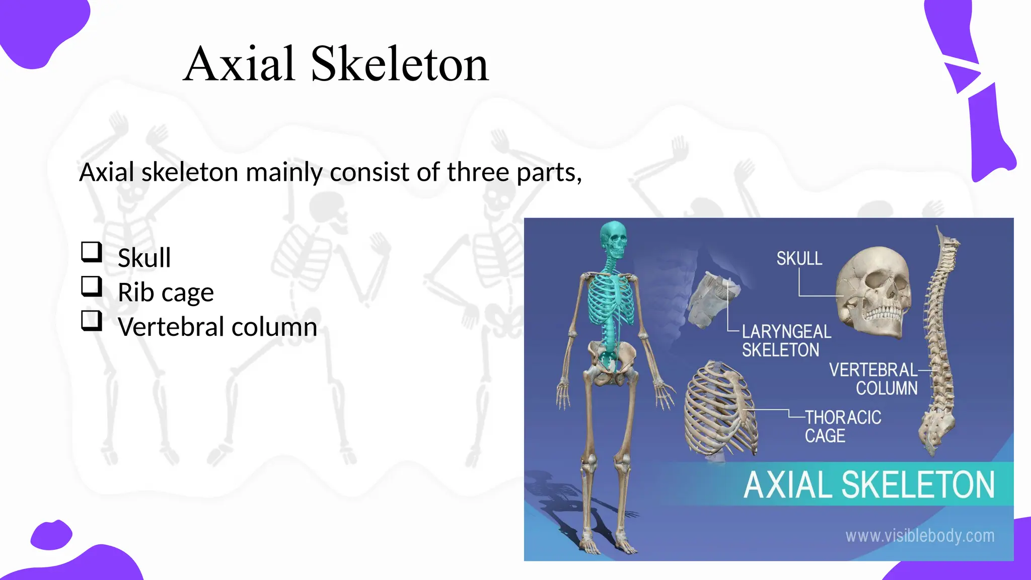
Axial skeleton
Appendicular skeleton
Upper limb
Thearm is formed by a single bone
Humerus
The forearm has two bones
Ulna
Radius
The hand
Carpals – wrist
Metacarpals – palm
Phalanges – fingers
Lower limb
Thethigh has one bone
Femur – thigh bone
The leg has two bones
Tibia
Fibula
The foot
Tarsus – ankle
Metatarsals – sole
Phalanges – toes
Compact bone (Corticalbone)
Osseous tissue is arranged in osteons/ Haversian systems (structural
unit of compact bone)
Osteons
Each osteon is a compact cylinder of concentric bone layers- lamellae
surrounds a central canal – Harversian canal
Osteocytes are found on the edges of each lamella
Cells in - lacunae
25.
Cyptoplasmic extensions- in canaliculi
Central canal /Haversian canal contains blood vessels, lymph vessels, and
nerves
Volkmann’s canals / perforating canals run at right angles connect the
adjacent osteons together (Also contain blood vessels, lymph vessels &
nerves)
No osteons at the outer edges of compact bone
Osseous tissue arranged in circumferential lamellae
27.
Spongy bone (Cancellousbone)
Light, porous bone enclosing numerous large spaces
Osseous tissue is arranged into trabeculae
(3dimensional latticework of bony processes)
Within a single trabecular:
concentric lamellae , with osteocytes in lacunae connected to one
another via in canaliculi
28.
The spacesbetween trabeculae are often filled with bone marrow and
blood vessels
Found in most areas of bone that are not subject to great mechanical
stress
ends of long bones (epiphyses)
near joints
interior of vertebrae
Cancellous bone is usually surrounded by a shell of compact bone
provides greater strength and rigidity
30.
Bone cells
Osteogeniccells
Stem cells that give rise to other bone cells
Osteoblasts
Bone forming cells
Synthesize soft organic bone matrix
Hardens later by deposition of minerals
[calcification/ ossification)
31.
Osteoclasts
Responsible forbone resorption
Large, multi nucleated cells
Osteocytes
Capable of bone deposition and resorption
Able to sense mechanical stresses to bone
Control the activity of osteoblasts and osteoclasts as necessary
Bone remodeling
levels in blood
Helps in regulating Calcium and phosphate levels in the body
32.
Bone formation &growth
In embryos, the skeleton is primarily hyaline
cartilage
During development, much of this cartilage is
replaced by bone.
Cartilage remains in isolated areas
-Bridge of the nose
-Parts of ribs
-Joints
Bones form later by 2 mechanisms;
1. Intramembranous ossification
2. Endochondral ossification
33.
1.Bone develops directlyfrom sheets of mesenchymal (undifferentiated) connective tissue
2.Differentiate into capillaries & osteogenic cells
3.Early osteoblasts appear in a cluster (ossification centre)
4.Secrete uncalcified matrix (osteoid )
5. Osteoid
6.Undergo calcification
7.Network of bone trabeculae is formed (Woven bone)
8.Mesenchyme condenses to form the periosteum
9.Trabeculae just deep to the periosteum thicken & converted to compact bone
Intramembranous ossification
35.
Bones formed byIntramembranous ossification (Intramembranous bone)
Flat bones of the face
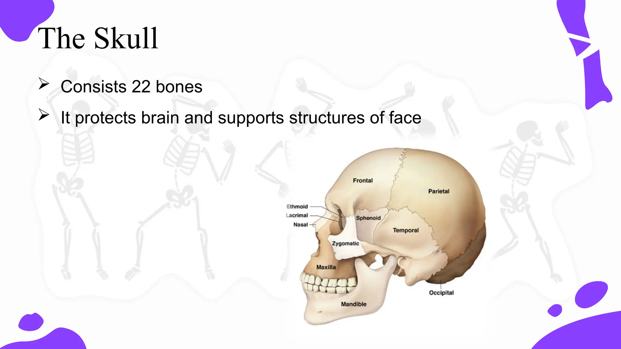
Most of the cranial bones
Clavicles
36.
Endochondral ossification
Templateof the bone is laid down by hyaline cartilage
Bone develops by replacing hyaline cartilage Most of the long bones are endochondral bones
Mesenchymal Form cells differentiate into chondroblasts
Form the hyaline cartilaginous precursors of the Bones
Blood vessels in the perichondrium bring osteoblasts to the edges of the structure
Deposit bone in a ring around the diaphysis (Bone collar)
Chondrocyte Blood death and disintegration the of cartilage in the centre of the structure
Blood Vessels penetrate the resulting Spaces
Spacrs enlarge form medullary cavity
Invaded by bone forming cells
37.
Deposit bonein the medullary cavity (Primary ossification centre)
Cartilage continue to grows at ends of the structure (the future epiphyses)
Increase the length of the structure
After birth, same process takes place in the epiphyseal regions (Secondary ossification
centre)
Thin plate of hyaline cartilage remains between the diaphysis and epiphysis throughout the
childhood and adolescence – epiphyseal plate
Venus has abeautiful name and is the
second planet from the Sun. It’s terribly hot,
even hotter than Mercury, and its
atmosphere is extremely poisonous. It’s the
second-brightest natural object in the night
sky after the Moon
Fibrous joint
The two bony surfaces involved are separated by
fibrous tissue
Movement is negligible
Types,
Sutures of the skull
Distal tibiofibular join- syndesmosis
Gomphosis
41.
Cartilaginous joints
Bonesare united by fibrocartilage or hyaline cartilage
Primary cartilaginous
(synchondrosis)
Bone and hyaline cartilage meet
Completely immobile
Types,
– Ossifying hyaline cartilage in the epiphysis
– Junctions between the ribs and the costal
You can entera subtitle here if you need it
Type of synovial joint Features Examples
Plane joints Two articular
surfaces are flat or
slightly convex or
concave
Joints between the
articular processes of
the vertebrae
Some carpal joints
Some tarsal joints
Hinge joints Permits movements
in one plane
Interphalangeal joints
Elbow joint
Knee joint
Ankle joint
Pivot joints Formed by a central
bony pivot
surrounded by a bony
ligamentous ring
Proximal and distal
radio ulna joint
Atlantoaxial joint
Saddle joints Articular surfaces are
concave-covex
Carpometacapal joint
of thumb
Ellipsoidal joints Ovoid articular
surface or condyle,
articulates with a
elliptical cavity
Wrist joint
Metacarpophalangeal
joint
Metatarsophalangeal
joint
Ball and socket Globular head in toa
cup–like cavity
Hip joint
Shoulder joint
44.
Common Skeletal SystemDisorders
A number of disorders affect the skeletal system, including bone
fractures and bone cancers.
However, the two most common disorders of the skeletal system
are osteoporosis and osteoarthritis
45.
Abnormal Spinal Curvatures
Abnormal spinal curvatures can result from disease,
weakness or paralysis of the trunk muscles, poor
posture, pregnancy, or congenital defects in vertebral
anatomy