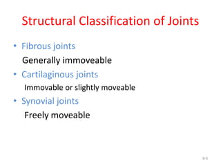
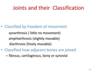
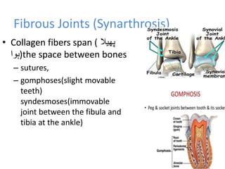
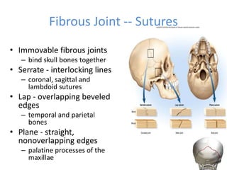
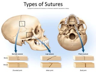
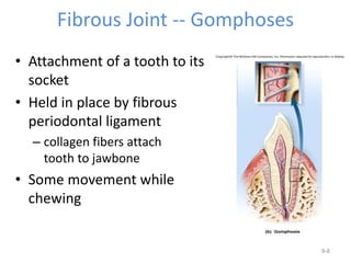
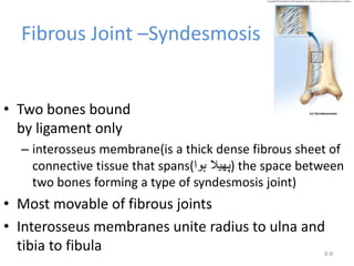
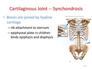
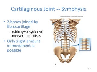
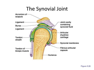
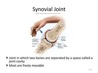
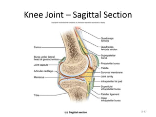
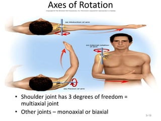
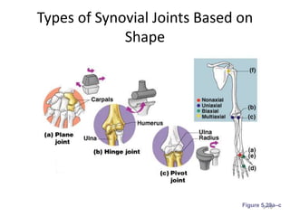
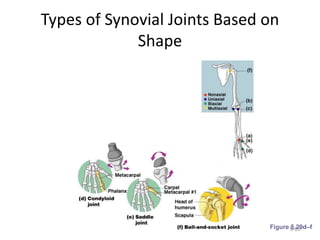
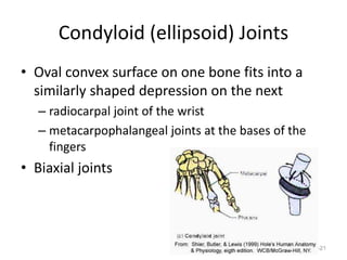
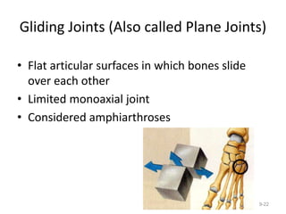
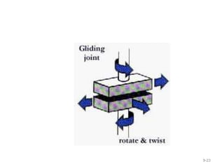
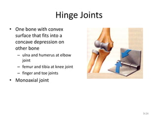
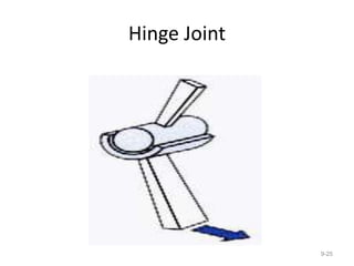
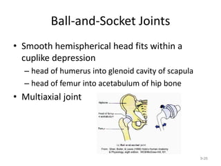
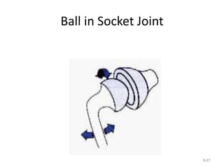
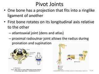
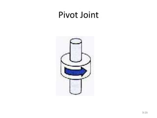
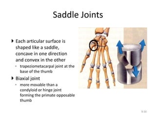
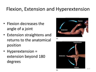
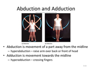
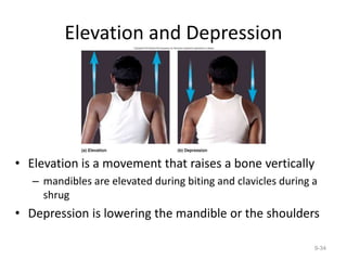
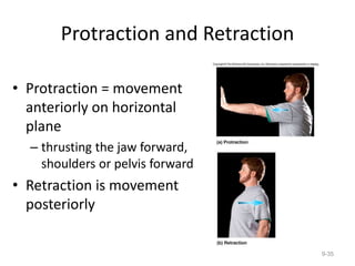
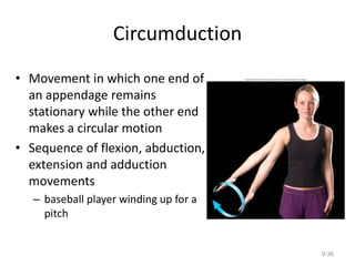
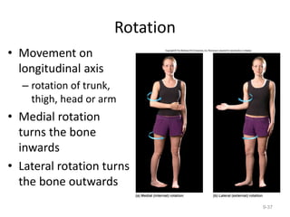
Joints
SCK
9-1
Definition of Joint
the point of connection between two bones or
elements of a skeleton (especially if it allows
motion)
9-2
Structural Classification of Joints
• Fibrous joints
Generally immoveable
• Cartilaginous joints
Immovable or slightly moveable
• Synovial joints
Freely moveable
9-3
Joints and their Classification
• Classified by freedom of movement
synarthrosis ( little no movement)
amphiarthrosis (slightly movable)
diarthrosis (freely movable)
• Classified how adjacent bones are joined
– fibrous, cartilaginous, bony or synovial
9-4
Fibrous Joints (Synarthrosis)
• Collagen fibers span ( ‫پھیال‬
‫)ہوا‬the space between bones
– sutures,
– gomphoses(slight movable
teeth)
syndesmoses(immovable
joint between the fibula and
tibia at the ankle)
9-5
Fibrous Joint -- Sutures
• Immovable fibrous joints
– bind skull bones together
• Serrate - interlocking lines
– coronal, sagittal and
lambdoid sutures
• Lap - overlapping beveled
edges
– temporal and parietal
bones
• Plane - straight,
nonoverlapping edges
– palatine processes of the
maxillae 9-6
Types of Sutures
9-7
Fibrous Joint -- Gomphoses
• Attachment of a tooth to its
socket
• Held in place by fibrous
periodontal ligament
– collagen fibers attach
tooth to jawbone
• Some movement while
chewing
9-8
Fibrous Joint –Syndesmosis
• Two bones bound
by ligament only
– interosseus membrane(is a thick dense fibrous sheet of
connective tissue that spans(‫ہوا‬ ‫)پھیال‬ the space between
two bones forming a type of syndesmosis joint)
• Most movable of fibrous joints
• Interosseus membranes unite radius to ulna and
tibia to fibula 9-9
Cartilaginous Joint -- Synchondrosis
• Bones are joined by hyaline
cartilage
– rib attachment to sternum
– epiphyseal plate in children
binds epiphysis and diaphysis
9-10
Cartilaginous Joint -- Symphysis
• 2 bones joined by
fibrocartilage
– pubic symphysis and
intervertebral discs
• Only slight amount
of movement is
possible
9-11
The Synovial Joint
Figure 5.28
Synovial Joint
 Joint in which two bones are separated by a space called a
joint cavity
 Most are freely movable
9-13
Features of Synovial Joints
• Articular cartilage (hyaline cartilage) covers
the ends of bones
• Joint surfaces are enclosed by a fibrous
articular capsule
• Have a joint cavity filled with synovial fluid
• Ligaments reinforce the joint
General Anatomy
• Articular capsule encloses joint cavity
– continuous with periosteum
– lined by synovial membrane
• Synovial fluid = slippery fluid; feeds cartilages
• Articular cartilage = hyaline cartilage covering the joint surfaces
• Articular discs and menisci
– jaw, wrist, sternoclavicular and knee joints
– absorbs shock, guides bone movements and distributes forces
• Tendon attaches b to bone
• Ligament attaches m to bone
9-15
What is a Bursar?
 is a small fluid-filled sac lined
by synovial membrane with a
inner capillary layer of slimy
fluid (similar to that of an egg
white).
 It provides a cushion
between bones and tendons
and/or muscles around a
joint. This helps to reduce any
friction between the bones
and allows free movement.
 Bursae are filled with
synovial fluid and are found
around almost every major
joint of the body; when they
become inflamed, the
condition is called bursitis. 9-16
Knee Joint – Sagittal Section
9-17
Axes of Rotation
• Shoulder joint has 3 degrees of freedom =
multiaxial joint
• Other joints – monoaxial or biaxial 9-18
Types of Synovial Joints Based on
Shape
9-19
Figure 5.29a–c
Types of Synovial Joints Based on
Shape
9-20
Figure 5.29d–f
Condyloid (ellipsoid) Joints
• Oval convex surface on one bone fits into a
similarly shaped depression on the next
– radiocarpal joint of the wrist
– metacarpophalangeal joints at the bases of the
fingers
• Biaxial joints
9-21
Gliding Joints (Also called Plane Joints)
• Flat articular surfaces in which bones slide
over each other
• Limited monoaxial joint
• Considered amphiarthroses
9-22
9-23
Hinge Joints
• One bone with convex
surface that fits into a
concave depression on
other bone
– ulna and humerus at elbow
joint
– femur and tibia at knee joint
– finger and toe joints
• Monoaxial joint
9-24
Hinge Joint
9-25
Ball-and-Socket Joints
• Smooth hemispherical head fits within a
cuplike depression
– head of humerus into glenoid cavity of scapula
– head of femur into acetabulum of hip bone
• Multiaxial joint
9-26
Ball in Socket Joint
9-27
Pivot Joints
• One bone has a projection that fits into a ringlike
ligament of another
• First bone rotates on its longitudinal axis relative
to the other
– atlantoaxial joint (dens and atlas)
– proximal radioulnar joint allows the radius during
pronation and supination
9-28
Pivot Joint
9-29
Saddle Joints
 Each articular surface is
shaped like a saddle,
concave in one direction
and convex in the other
◦ trapeziometacarpal joint at the
base of the thumb
 Biaxial joint
◦ more movable than a
condyloid or hinge joint
forming the primate opposable
thumb
9-30
Flexion, Extension and Hyperextension
• Flexion decreases the
angle of a joint
• Extension straightens and
returns to the anatomical
position
• Hyperextension =
extension beyond 180
degrees
9-31
Flexion, Extension and Hyperextension
9-32
Abduction and Adduction
• Abduction is movement of a part away from the midline
– hyperabduction – raise arm over back or front of head
• Adduction is movement towards the midline
– hyperadduction – crossing fingers
9-33
Elevation and Depression
• Elevation is a movement that raises a bone vertically
– mandibles are elevated during biting and clavicles during a
shrug
• Depression is lowering the mandible or the shoulders
9-34
Protraction and Retraction
• Protraction = movement
anteriorly on horizontal
plane
– thrusting the jaw forward,
shoulders or pelvis forward
• Retraction is movement
posteriorly
9-35
Circumduction
• Movement in which one end of
an appendage remains
stationary while the other end
makes a circular motion
• Sequence of flexion, abduction,
extension and adduction
movements
– baseball player winding up for a
pitch
9-36
Rotation
• Movement on
longitudinal axis
– rotation of trunk,
thigh, head or arm
• Medial rotation
turns the bone
inwards
• Lateral rotation turns
the bone outwards
9-37
Supination and Pronation
 In the forearm and foot
 Supination
◦ rotation of forearm so that the palm
faces forward
◦ inversion and abduction of foot
(raising the medial edge of the foot)
 Pronation
◦ rotation of forearm so the palm faces
to the rear
◦ eversion and abduction of foot
(raising the lateral edge of the foot)
9-38
Movements of Head and Trunk
• Flexion, hyperextension and lateral flexion of vertebral
column 9-39
Rotation of Trunk and Head
• Right rotation of trunk; rotation of head
9-40
Movements of Mandible
 Lateral excursion = sideways movement
 Medial excursion = movement back to the midline
◦ side-to-side grinding during chewing
 Protraction – retraction of mandible
9-41
Movement of Hand and Digits
• Radial and ulnar
flexion
• Abduction of fingers
and thumb
• Opposition is
movement of the
thumb to approach or
touch the fingertips
• Reposition is
movement back to the
anatomical position
9-42
Movements of the Foot
 Dorsiflexion is raising of the toes as when you swing the foot
forward to take a step (heel strike)
 Plantarflexion is extension of the foot so that the toes point
downward as in standing on tiptoe
 Inversion is a movement in which the soles are turned medially
 Eversion is a turning of the soles to face laterally
9-43

7th joints.ppt

  • 1.
  • 2.
    Definition of Joint thepoint of connection between two bones or elements of a skeleton (especially if it allows motion) 9-2
  • 3.
    Structural Classification ofJoints • Fibrous joints Generally immoveable • Cartilaginous joints Immovable or slightly moveable • Synovial joints Freely moveable 9-3
  • 4.
    Joints and theirClassification • Classified by freedom of movement synarthrosis ( little no movement) amphiarthrosis (slightly movable) diarthrosis (freely movable) • Classified how adjacent bones are joined – fibrous, cartilaginous, bony or synovial 9-4
  • 5.
    Fibrous Joints (Synarthrosis) •Collagen fibers span ( ‫پھیال‬ ‫)ہوا‬the space between bones – sutures, – gomphoses(slight movable teeth) syndesmoses(immovable joint between the fibula and tibia at the ankle) 9-5
  • 6.
    Fibrous Joint --Sutures • Immovable fibrous joints – bind skull bones together • Serrate - interlocking lines – coronal, sagittal and lambdoid sutures • Lap - overlapping beveled edges – temporal and parietal bones • Plane - straight, nonoverlapping edges – palatine processes of the maxillae 9-6
  • 7.
  • 8.
    Fibrous Joint --Gomphoses • Attachment of a tooth to its socket • Held in place by fibrous periodontal ligament – collagen fibers attach tooth to jawbone • Some movement while chewing 9-8
  • 9.
    Fibrous Joint –Syndesmosis •Two bones bound by ligament only – interosseus membrane(is a thick dense fibrous sheet of connective tissue that spans(‫ہوا‬ ‫)پھیال‬ the space between two bones forming a type of syndesmosis joint) • Most movable of fibrous joints • Interosseus membranes unite radius to ulna and tibia to fibula 9-9
  • 10.
    Cartilaginous Joint --Synchondrosis • Bones are joined by hyaline cartilage – rib attachment to sternum – epiphyseal plate in children binds epiphysis and diaphysis 9-10
  • 11.
    Cartilaginous Joint --Symphysis • 2 bones joined by fibrocartilage – pubic symphysis and intervertebral discs • Only slight amount of movement is possible 9-11
  • 12.
  • 13.
    Synovial Joint  Jointin which two bones are separated by a space called a joint cavity  Most are freely movable 9-13
  • 14.
    Features of SynovialJoints • Articular cartilage (hyaline cartilage) covers the ends of bones • Joint surfaces are enclosed by a fibrous articular capsule • Have a joint cavity filled with synovial fluid • Ligaments reinforce the joint
  • 15.
    General Anatomy • Articularcapsule encloses joint cavity – continuous with periosteum – lined by synovial membrane • Synovial fluid = slippery fluid; feeds cartilages • Articular cartilage = hyaline cartilage covering the joint surfaces • Articular discs and menisci – jaw, wrist, sternoclavicular and knee joints – absorbs shock, guides bone movements and distributes forces • Tendon attaches b to bone • Ligament attaches m to bone 9-15
  • 16.
    What is aBursar?  is a small fluid-filled sac lined by synovial membrane with a inner capillary layer of slimy fluid (similar to that of an egg white).  It provides a cushion between bones and tendons and/or muscles around a joint. This helps to reduce any friction between the bones and allows free movement.  Bursae are filled with synovial fluid and are found around almost every major joint of the body; when they become inflamed, the condition is called bursitis. 9-16
  • 17.
    Knee Joint –Sagittal Section 9-17
  • 18.
    Axes of Rotation •Shoulder joint has 3 degrees of freedom = multiaxial joint • Other joints – monoaxial or biaxial 9-18
  • 19.
    Types of SynovialJoints Based on Shape 9-19 Figure 5.29a–c
  • 20.
    Types of SynovialJoints Based on Shape 9-20 Figure 5.29d–f
  • 21.
    Condyloid (ellipsoid) Joints •Oval convex surface on one bone fits into a similarly shaped depression on the next – radiocarpal joint of the wrist – metacarpophalangeal joints at the bases of the fingers • Biaxial joints 9-21
  • 22.
    Gliding Joints (Alsocalled Plane Joints) • Flat articular surfaces in which bones slide over each other • Limited monoaxial joint • Considered amphiarthroses 9-22
  • 23.
  • 24.
    Hinge Joints • Onebone with convex surface that fits into a concave depression on other bone – ulna and humerus at elbow joint – femur and tibia at knee joint – finger and toe joints • Monoaxial joint 9-24
  • 25.
  • 26.
    Ball-and-Socket Joints • Smoothhemispherical head fits within a cuplike depression – head of humerus into glenoid cavity of scapula – head of femur into acetabulum of hip bone • Multiaxial joint 9-26
  • 27.
    Ball in SocketJoint 9-27
  • 28.
    Pivot Joints • Onebone has a projection that fits into a ringlike ligament of another • First bone rotates on its longitudinal axis relative to the other – atlantoaxial joint (dens and atlas) – proximal radioulnar joint allows the radius during pronation and supination 9-28
  • 29.
  • 30.
    Saddle Joints  Eacharticular surface is shaped like a saddle, concave in one direction and convex in the other ◦ trapeziometacarpal joint at the base of the thumb  Biaxial joint ◦ more movable than a condyloid or hinge joint forming the primate opposable thumb 9-30
  • 31.
    Flexion, Extension andHyperextension • Flexion decreases the angle of a joint • Extension straightens and returns to the anatomical position • Hyperextension = extension beyond 180 degrees 9-31
  • 32.
    Flexion, Extension andHyperextension 9-32
  • 33.
    Abduction and Adduction •Abduction is movement of a part away from the midline – hyperabduction – raise arm over back or front of head • Adduction is movement towards the midline – hyperadduction – crossing fingers 9-33
  • 34.
    Elevation and Depression •Elevation is a movement that raises a bone vertically – mandibles are elevated during biting and clavicles during a shrug • Depression is lowering the mandible or the shoulders 9-34
  • 35.
    Protraction and Retraction •Protraction = movement anteriorly on horizontal plane – thrusting the jaw forward, shoulders or pelvis forward • Retraction is movement posteriorly 9-35
  • 36.
    Circumduction • Movement inwhich one end of an appendage remains stationary while the other end makes a circular motion • Sequence of flexion, abduction, extension and adduction movements – baseball player winding up for a pitch 9-36
  • 37.
    Rotation • Movement on longitudinalaxis – rotation of trunk, thigh, head or arm • Medial rotation turns the bone inwards • Lateral rotation turns the bone outwards 9-37
  • 38.
    Supination and Pronation In the forearm and foot  Supination ◦ rotation of forearm so that the palm faces forward ◦ inversion and abduction of foot (raising the medial edge of the foot)  Pronation ◦ rotation of forearm so the palm faces to the rear ◦ eversion and abduction of foot (raising the lateral edge of the foot) 9-38
  • 39.
    Movements of Headand Trunk • Flexion, hyperextension and lateral flexion of vertebral column 9-39
  • 40.
    Rotation of Trunkand Head • Right rotation of trunk; rotation of head 9-40
  • 41.
    Movements of Mandible Lateral excursion = sideways movement  Medial excursion = movement back to the midline ◦ side-to-side grinding during chewing  Protraction – retraction of mandible 9-41
  • 42.
    Movement of Handand Digits • Radial and ulnar flexion • Abduction of fingers and thumb • Opposition is movement of the thumb to approach or touch the fingertips • Reposition is movement back to the anatomical position 9-42
  • 43.
    Movements of theFoot  Dorsiflexion is raising of the toes as when you swing the foot forward to take a step (heel strike)  Plantarflexion is extension of the foot so that the toes point downward as in standing on tiptoe  Inversion is a movement in which the soles are turned medially  Eversion is a turning of the soles to face laterally 9-43