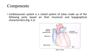
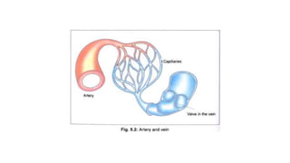
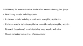
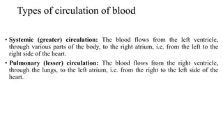
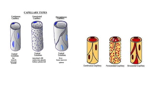
final cvs physio to reach out to the same thing with you regardingLokesh444339
Cvs received this is the same thing with you regarding the same thing with you regarding the same thing with you and your family is not able get registered
Arteries are blood vessels that carry blood away from the heart. This blood is normally oxygenated, exceptions made for the pulmonary and umbilical arteries
The first topic in the practical histology coarse for pharmacy students
In this lecture the student will be able to recognize the histological layers of the circulatory system parts such as veins and arteries and the similarities and differences between each layer
There are three kinds of blood vessels: arteries, veins, and capillaries.
Each of these plays a very specific role in the circulation process/blood circulation.
Arteries transport blood away from the heart.
Veins return blood back toward the heart.
Capillaries surround body cells and tissues to deliver and absorb oxygen, nutrients, and other substances.
The capillaries also connect the branches of arteries and to the branches of veins.
The walls of most blood vessels have three distinct layers: the tunica externa, the tunica media, and the tunica intima.
The circulatory system transports fluids throughout the body;
it consists of the cardiovascular and lymphatic systems.
The heart and blood vessels make up the blood transportation network, the cardiovascular system.
Through this system, the heart pumps blood through the body’s vast system of blood vessels.
The blood carries nutrients, oxygen, and waste products to and from the cells.
VASCULAR CIRCUITS
The heart consists of two muscular pumps dividing the circulation into two components:
pulmonary circulations
systemic circulations or circuit
Pulmonary Circulation
Rt ventricle propels low O2 blood into the lungs via the pulmonary arteries.
CO2 is exchanged for O2 in the capillaries of the lungs.
Then the O2 -rich blood is returned via the pulmonary veins to the Lft atrium.
This circuit, from the right ventricle through the lungs to the left atrium, is the pulmonary circulation.
Systemic Circulation
Left ventricle propels the O2 -rich blood through systemic arteries (the aorta and its branches),
exchanging O2 and nutrients for CO2 in the remainder of the body’s capillaries.
Low- O2 blood returns to right atrium via systemic veins (tributaries of the superior and inferior vena cava).
This circuit, from left ventricle to right atrium, is the systemic circulation.
Blood Vessels
By: Saiyed Falakaara
Assistant Professor
Department of Pharmacy
Sumandeep Vidyapeeth
Introduction
Blood vessels form a closed system of tubes that carry blood away from the heart, transport it to the tissues of the body, and then return it to the heart.
The blood vessels of human body carry blood to every tissue and organ.
Vessels decrease in size as they move away from the heart (arteries and arterioles), ending in the capillaries, and then increase in size as they move towards the heart (venules and veins)
The largest artery in the blood is Aorta.
Types of blood vessels
1. Arteries
2. Arterioles
3. Capillaries
4. Venules
5. veins
Learning Objectives:
Compare and contrast the structure and function
of
Arteries
Veins
Capillaries
ulatory
system
Arteries
Arterioles
Capillaries
Venules
Veins
3 tunics
Lume
The Vessels
Functions:
Distribution of blood
Exchange of materials with tissues
Return of blood to the heart
Structure:
Most have the same basic structure:
– 3 layers surrounding a hollow lumen
04- PT as a Patient Client manager.pptxChangezKhan33
In this lecture role of PT is defined and explained as a patient client manager, how he or she uses his or her knowledge for the betterment of patient symptoms and history.
New Directions in Targeted Therapeutic Approaches for Older Adults With Mantl...i3 Health
i3 Health is pleased to make the speaker slides from this activity available for use as a non-accredited self-study or teaching resource.
This slide deck presented by Dr. Kami Maddocks, Professor-Clinical in the Division of Hematology and
Associate Division Director for Ambulatory Operations
The Ohio State University Comprehensive Cancer Center, will provide insight into new directions in targeted therapeutic approaches for older adults with mantle cell lymphoma.
STATEMENT OF NEED
Mantle cell lymphoma (MCL) is a rare, aggressive B-cell non-Hodgkin lymphoma (NHL) accounting for 5% to 7% of all lymphomas. Its prognosis ranges from indolent disease that does not require treatment for years to very aggressive disease, which is associated with poor survival (Silkenstedt et al, 2021). Typically, MCL is diagnosed at advanced stage and in older patients who cannot tolerate intensive therapy (NCCN, 2022). Although recent advances have slightly increased remission rates, recurrence and relapse remain very common, leading to a median overall survival between 3 and 6 years (LLS, 2021). Though there are several effective options, progress is still needed towards establishing an accepted frontline approach for MCL (Castellino et al, 2022). Treatment selection and management of MCL are complicated by the heterogeneity of prognosis, advanced age and comorbidities of patients, and lack of an established standard approach for treatment, making it vital that clinicians be familiar with the latest research and advances in this area. In this activity chaired by Michael Wang, MD, Professor in the Department of Lymphoma & Myeloma at MD Anderson Cancer Center, expert faculty will discuss prognostic factors informing treatment, the promising results of recent trials in new therapeutic approaches, and the implications of treatment resistance in therapeutic selection for MCL.
Target Audience
Hematology/oncology fellows, attending faculty, and other health care professionals involved in the treatment of patients with mantle cell lymphoma (MCL).
Learning Objectives
1.) Identify clinical and biological prognostic factors that can guide treatment decision making for older adults with MCL
2.) Evaluate emerging data on targeted therapeutic approaches for treatment-naive and relapsed/refractory MCL and their applicability to older adults
3.) Assess mechanisms of resistance to targeted therapies for MCL and their implications for treatment selection
Lung Cancer: Artificial Intelligence, Synergetics, Complex System Analysis, S...Oleg Kshivets
RESULTS: Overall life span (LS) was 2252.1±1742.5 days and cumulative 5-year survival (5YS) reached 73.2%, 10 years – 64.8%, 20 years – 42.5%. 513 LCP lived more than 5 years (LS=3124.6±1525.6 days), 148 LCP – more than 10 years (LS=5054.4±1504.1 days).199 LCP died because of LC (LS=562.7±374.5 days). 5YS of LCP after bi/lobectomies was significantly superior in comparison with LCP after pneumonectomies (78.1% vs.63.7%, P=0.00001 by log-rank test). AT significantly improved 5YS (66.3% vs. 34.8%) (P=0.00000 by log-rank test) only for LCP with N1-2. Cox modeling displayed that 5YS of LCP significantly depended on: phase transition (PT) early-invasive LC in terms of synergetics, PT N0—N12, cell ratio factors (ratio between cancer cells- CC and blood cells subpopulations), G1-3, histology, glucose, AT, blood cell circuit, prothrombin index, heparin tolerance, recalcification time (P=0.000-0.038). Neural networks, genetic algorithm selection and bootstrap simulation revealed relationships between 5YS and PT early-invasive LC (rank=1), PT N0—N12 (rank=2), thrombocytes/CC (3), erythrocytes/CC (4), eosinophils/CC (5), healthy cells/CC (6), lymphocytes/CC (7), segmented neutrophils/CC (8), stick neutrophils/CC (9), monocytes/CC (10); leucocytes/CC (11). Correct prediction of 5YS was 100% by neural networks computing (area under ROC curve=1.0; error=0.0).
CONCLUSIONS: 5YS of LCP after radical procedures significantly depended on: 1) PT early-invasive cancer; 2) PT N0--N12; 3) cell ratio factors; 4) blood cell circuit; 5) biochemical factors; 6) hemostasis system; 7) AT; 8) LC characteristics; 9) LC cell dynamics; 10) surgery type: lobectomy/pneumonectomy; 11) anthropometric data. Optimal diagnosis and treatment strategies for LC are: 1) screening and early detection of LC; 2) availability of experienced thoracic surgeons because of complexity of radical procedures; 3) aggressive en block surgery and adequate lymph node dissection for completeness; 4) precise prediction; 5) adjuvant chemoimmunoradiotherapy for LCP with unfavorable prognosis.
HOT NEW PRODUCT! BIG SALES FAST SHIPPING NOW FROM CHINA!! EU KU DB BK substit...GL Anaacs
Contact us if you are interested:
Email / Skype : kefaya1771@gmail.com
Threema: PXHY5PDH
New BATCH Ku !!! MUCH IN DEMAND FAST SALE EVERY BATCH HAPPY GOOD EFFECT BIG BATCH !
Contact me on Threema or skype to start big business!!
Hot-sale products:
NEW HOT EUTYLONE WHITE CRYSTAL!!
5cl-adba precursor (semi finished )
5cl-adba raw materials
ADBB precursor (semi finished )
ADBB raw materials
APVP powder
5fadb/4f-adb
Jwh018 / Jwh210
Eutylone crystal
Protonitazene (hydrochloride) CAS: 119276-01-6
Flubrotizolam CAS: 57801-95-3
Metonitazene CAS: 14680-51-4
Payment terms: Western Union,MoneyGram,Bitcoin or USDT.
Deliver Time: Usually 7-15days
Shipping method: FedEx, TNT, DHL,UPS etc.Our deliveries are 100% safe, fast, reliable and discreet.
Samples will be sent for your evaluation!If you are interested in, please contact me, let's talk details.
We specializes in exporting high quality Research chemical, medical intermediate, Pharmaceutical chemicals and so on. Products are exported to USA, Canada, France, Korea, Japan,Russia, Southeast Asia and other countries.
MANAGEMENT OF ATRIOVENTRICULAR CONDUCTION BLOCK.pdfJim Jacob Roy
Cardiac conduction defects can occur due to various causes.
Atrioventricular conduction blocks ( AV blocks ) are classified into 3 types.
This document describes the acute management of AV block.
micro teaching on communication m.sc nursing.pdfAnurag Sharma
Microteaching is a unique model of practice teaching. It is a viable instrument for the. desired change in the teaching behavior or the behavior potential which, in specified types of real. classroom situations, tends to facilitate the achievement of specified types of objectives.
Couples presenting to the infertility clinic- Do they really have infertility...Sujoy Dasgupta
Dr Sujoy Dasgupta presented the study on "Couples presenting to the infertility clinic- Do they really have infertility? – The unexplored stories of non-consummation" in the 13th Congress of the Asia Pacific Initiative on Reproduction (ASPIRE 2024) at Manila on 24 May, 2024.
These lecture slides, by Dr Sidra Arshad, offer a quick overview of physiological basis of a normal electrocardiogram.
Learning objectives:
1. Define an electrocardiogram (ECG) and electrocardiography
2. Describe how dipoles generated by the heart produce the waveforms of the ECG
3. Describe the components of a normal electrocardiogram of a typical bipolar leads (limb II)
4. Differentiate between intervals and segments
5. Enlist some common indications for obtaining an ECG
Study Resources:
1. Chapter 11, Guyton and Hall Textbook of Medical Physiology, 14th edition
2. Chapter 9, Human Physiology - From Cells to Systems, Lauralee Sherwood, 9th edition
3. Chapter 29, Ganong’s Review of Medical Physiology, 26th edition
4. Electrocardiogram, StatPearls - https://www.ncbi.nlm.nih.gov/books/NBK549803/
5. ECG in Medical Practice by ABM Abdullah, 4th edition
6. ECG Basics, http://www.nataliescasebook.com/tag/e-c-g-basics
2. Introduction
• Cardiovascular system is the transport system of the body, through which the
nutrients are conveyed to places where these are utilized, and the metabolites
(waste products) are conveyed to appropriate places from where these are
expelled.
• The conveying medium is a liquid tissue, the blood, which flows in tubular
channels called blood vessels. The circulation is maintained by the central
pumping organ called the heart.
3. Components
• Cardiovascular system is a closed system of tubes made up of the
following parts based on their structural and topographical
characteristics (Fig. 5.1)
4. HEART
• It is a four-chambered muscular organ which pumps blood to various
parts of the body. Each half of the heart has a receiving chamber called
atrium, and a pumping chamber called ventricle.
5. Arteries
• These are distributing channels which carry blood away from the heart.
• They branch like trees on their way to different parts of the body.
• The large arteries are rich in elastic tissue, but as branching progresses there is an ever-
increasing amount of smooth muscle in their walls.
• The minute branches which are just visible to naked eye are called arterioles.
• Angeion is a Greek word, meaning a vessel (blood vessel or lymph vessel). Its word
derivatives are angiology, angiography, haemangioma and thromboangitis obliterans.
6. VEINS
These are draining channels which carry blood from different parts of the body
back to the heart.
Like rivers, the veins are formed by tributaries.
The small veins (venules) join together to form larger veins, which in turn unite
to form great veins called venae cavae.
7.
8. CAPILLARIES
• These are networks of microscopic vessels which connect arterioles with the
venules.
• These come in intimate contact with the tissues for a free exchange of nutrients
and metabolites across their walls between the blood and the tissue fluid.
• The metabolites are partly drained by the capillaries and partly by lymphatics.
• Capillaries are replaced by sinusoids in certain organs, like liver and spleen.
9. Functionally, the blood vessels can be classified into the following five groups.
• Distributing vessels, including arteries
• Resistance vessels, including arterioles and precapillary sphincters
• Exchange vessels, including capillaries, sinusoids, and post-capillary venules
• Reservoir (capacitance) vessels, including larger venules and veins
• Shunts, including various types of anastomoses.
10. Types of circulation of blood
• Systemic (greater) circulation: The blood flows from the left ventricle,
through various parts of the body, to the right atrium, i.e. from the left to the
right side of the heart.
• Pulmonary (lesser) circulation: The blood flows from the right ventricle,
through the lungs, to the left atrium, i.e. from the right to the left side of the
heart.
11. Portal circulation: It is a part of systemic circulation, which has the following characteristics.
• The blood passes through two sets of capillaries before draining into a systemic vein.
• The vein draining the first capillary network is known as portal vein which branches like an artery to
form the second set of capillaries or sinusoids.
• Examples: hepatic portal circulation, hypothalamo-hypophyseal, portal circulation and renal portal
circulation
13. CHARACTERISTIC FEATURES
1. Arteries are thick-walled, being uniformly thicker than the accompanying
veins, except for the arteries within the cranium and vertebral canal where
these are thin.
2. Their lumen is smaller than that of the accompanying veins.
3. Arteries have no valves.
4. An artery is usually accompanied by vein(s) and nerve(s), and the three of
them together form the neurovascular bundle which is surrounded and
supported by a fibro-areolar sheath.
14. TYPES OF ARTERIES AND STRUCTURES
1. Large arteries of elastic type, e.g. aorta and its main branches (brachiocephalic,
common carotid, subclavian and common iliac) and the pulmonary arteries.
2. Medium and small arteries of muscular type, e.g. temporal, occipital, radial, popliteal,
etc.
3. Smallest arteries of muscular type are called arterioles. They measure 50-100 micron in
diameter.
4. Arterioles divide into terminal arterioles with a diameter of 15-20 micron, and having
one or two layers of smooth muscle in their walls. The side branches from terminal
arterioles are called metarterioles which measure 10-15 micron at their origin and about
5 micron at their termination (Fig. 5.4).
5. The terminal narrow end of met-arteriole is surrounded by a precapillary sphincter
which regulates blood flow into the capillary bed.
15.
16.
17. It is important to know that the muscular arterioles are responsible for generating peripheral resistance, and
thereby for regulating the diastolic blood pressure.
Microscopically, all arteries are made up of three coats.
a) The inner coat is called tunica intima (Fig. 5.5).
b) The middle coat is called tunica media.
c) The outer coat is called tunica adventitia. It is strongest of all coats and merges with the perivascular sheath.
The relative thickness of the coats and the relative proportion of the muscular, elastic and fibrous tissues
vary in different types of arteries.
18. Blood Supply of Arteries
• The large arteries (of more than 1 mm diameter) are supplied with blood vessels.
• The nutrient vessels, called vasa vasorum, form a dense capillary network in the
tunica adventitia, and supply the adventitia and the outer part of tunica media.
• The rest of the vessel wall (intima + inner part of media) is nourished directly by
diffusion from the luminal blood.
• Minute veins accompanying the arteries drain the blood from the outer part of
arterial wall.
• Lymphatics are also present in the adventitia.
19. Palpable Arteries
Some arteries can be palpated through the skin. These are: common carotid,
facial, brachial, radial, abdominal aorta, femoral, posterior tibial and dorsalis
pedis (Fig. 5.6).
20. Nerve Supply of Arteries
• The nerves supplying an artery are called nervi vascularis.
• The nerves are mostly non-myelinated sympathetic fibres which are vasoconstrictor in function.
• A few fibres are myelinated, and are believed to be sensory to the outer and inner coats of the
arteries.
• Vasodilator innervation is restricted to the following sites:
a) The skeletal muscle vessels are dilated by cholinergic sympathetic nerves.
b) The exocrine gland vessels are dilated on parasympathetic stimulation.
c) The cutaneous vessels are dilated locally to produce the flare VEINS (redness) after an injury.
d) The vasodilatation is produced by the afferent impulses in the cutaneous nerves which pass
antidromically in their collaterals to the blood vessels (axon reflex).
22. CHARACTERISTIC FEATURES
1. Veins are thin-walled, being thinner than the arteries.
2. Their lumen is larger than that of the accompanying arteries.
3. Veins have valves which maintain the unidirectional flow of blood, even against gravity.
4. Since the venous pressure is low (7 mm Hg), the valves are of utmost value in the venous return.
5. However, the valves are absent: (a) In the veins of less than 2 mm diameter. (b) In the venae
cavae. (c) In the hepatic, renal, uterine, ovarian (not testicular), cerebral, spinal, pulmonary, and
umbilical veins.
6. The muscular and elastic tissue content of the venous walls is much less than that of the arteries.
This is directly related to the low venous pressure.
7. Large veins have dead space around them for their dilatation during increased venous return. The
dead space commonly contains the regional lymph nodes (Fig. 5.7).
23.
24. STRUCTURE OF VEINS
• Veins are made up of usual three coats which are found in the arteries.
• But the coats are ill-defined, and the muscle and elastic tissue content is poor.
• In poorly developed tunica media, the amount of collagen fibres is more than
the elastic and muscle fibres.
• The adventitia is thickest and best developed.
• The smooth muscle is altogether absent:
a) in the veins of maternal part of placenta;
b) in the cranial venous sinuses and pial veins;
c) in the retinal veins;
d) in the veins of cancellous bone;
e) in the venous spaces of the corpora cavernosa and corpus spongiosum.
25. BLOOD and Nerve Supply of VEINS
• The larger veins, like the arteries, are supplied with nutrient vessels called
vasa vasorum.
• But in the veins, the vessels may penetrate up to the intima, probably because
of the low venous pressure and the low oxygen tension.
• Nerves also are distributed to the veins in the same manner as to the arteries,
but are fewer in number.
26.
27.
28.
29. FACTORS HELPING IN VENOUS RETURN
1. Overflow from the capillaries, pushed from behind by the arteries (vis-a-tergo).
2. Negative intrathoracic pressure sucks the blood into the heart from all over the body.
3. Gravity helps venous return in the upper part of the body.
4. Arterial pulsations press on the venae comitantes intermittently and drive the venous
blood towards the heart.
5. Venous valves prevent any regurgitation (back flow) of the luminal blood (Fig. 5.8).
6. Muscular contractions press on the veins and form a very effective mechanism of
venous return. This becomes still more effective, within the tight sleeve of the deep
fascia, as is seen in the lower limbs. The calf muscles (soleus) for this reason are
known as the peripheral heart. Thus the muscle pumps are important factors in the
venous return.
31. INTRODUCTION
• Capillaries (capillus = hair) are networks of microscopic endothelial tubes
interposed between the metarterioles and venules (Fig. 5.4).
• The true capillaries (without any smooth muscle cell) begin after a transition
zone of 50-100 micron beyond the precapillary sphincters.
• The capillaries are replaced by cavernous (dilated) spaces in the sex organs,
splenic pulp and placenta.
32. SIZE
• The average diameter of a capillary is 6-8 micron, just sufficient to permit the red
blood cells to pass through in 'single file'.
• But the size varies from organ to organ.
• It is smallest in the brain and intestines, and is largest (20 micron) in the skin and
bone marrow.
33. TYPES AND STRUCTURE
• The capillaries are classified as continuous and fenestrated according to the type of junctions
between the endothelial cells.
1. Continuous capillaries are found in the skin, connective tissue, skeletal and smooth muscles,
lung and brain. These allow passage across their walls of small molecules (up to 10 micron size).
2. Fenestrated capillaries are found in the renal glomeruli/intestinal mucosa, endocrine glands and
pancreas. These allow passage across their walls of larger molecules (up to 20-100 micron size).
3. The capillary bed and post-capillary venules form an enormous area for the exchange of nutrients,
gases, metabolites and water, between the blood and interstitial fluid. Capillaries also allow
migration of leucocytes out of the vessels.
34.
35. SINUSOIDS
• Sinusoids, replace capillaries in certain organs, like liver, spleen, bone marrow, suprarenal glands,
parathyroid glands, carotid body, etc
CHARACTERISTICS
• Sinusoids are large, irregular, vascular spaces which are closely surrounded by the parenchyma of
the organ.
• These differ from capillaries in the following respects;
1. Their lumen is wider (upto 30 micron) and irregular.
2. Their walls are thinner and may be incomplete. They are lined by endothelium in which the
phagocytic cells (macrophages) are often distributed. The adventitial support is absent.
3. These may connect arteriole with venule (spleen, bone marrow), or venule with venule (liver).
Editor's Notes
A haemangioma is a collection of small blood vessels that form a lump under the skin. They're sometimes called 'strawberry marks' because the surface of a haemangioma can look like the surface of a strawberry.
Thromboangiitis obliterans (Buerger disease) is caused by small blood vessels that become inflamed and swollen. The blood vessels then narrow or get blocked by blood clots (thrombosis). Blood vessels of the hands and feet are mostly affected. Arteries are more affected than veins.
Angiography is a type of X-ray used to check blood vessels. Blood vessels do not show clearly on a normal X-ray, so a special dye called a contrast agent needs to be injected into your blood first. This highlights your blood vessels, allowing your doctor to see any problems.
Angiology focuses on the prevention, diagnosis of and therapy for diseases of the veins, arteries and lymphatic vessels. Angiologists perform both surgical and non-surgical treatments. Vascular diseases occur throughout the body
Sinusoids, small blood vessels between the radiating rows of hepatocytes, convey oxygen-rich hepatic arterial blood and nutrient-rich portal venous blood to the hepatocytes and eventually drain into the central vein, which drains into the hepatic vein.
A lumen (plural: lumina) is a term that describes the cavity within the tubular structure.
A metarteriole is a short microvessel in the microcirculation that links arterioles and capillaries.[1] Instead of a continuous tunica media, they have individual smooth muscle cells placed a short distance apart, each forming a precapillary sphincter that encircles the entrance to that capillary bed. Constriction of these sphincters reduces or shuts off blood flow through their respective capillary beds. This allows the blood to be diverted to elsewhere in the body.
cholinergic sympathetic nerves: Sympathetic cholinergic nerves controlled by hypothalamic thermoregulatory nuclei play an important role in producing cutaneous active vasodilation when skin and body core temperature increase in warm environments.
EXOCRINE GLANDS A gland that makes substances such as sweat, tears, saliva, milk, and digestive juices, and releases them through a duct or opening to a body surface. Examples of exocrine glands include sweat glands, lacrimal glands, salivary glands, mammary glands, and digestive glands in the stomach, pancreas, and intestines.
The axon reflex (or the flare response) is the response stimulated by peripheral nerves of the body that travels away from the nerve cell body and branches to stimulate target organs
antidromically: proceeding or conducting in a direction opposite to the usual one. used especially of a nerve impulse or fiber.
corpus spongiosum.
a mass of erectile tissue alongside the corpora cavernosa of the penis and terminating in the glans.
corpora cavernosa
either of two masses of erectile tissue forming the bulk of the penis and the clitoris.
The vasa vasorum are a specialized microvasculature that play a major role in normal vessel wall biology and pathology. Under physiological conditions, the adventitial vasa vasorum take up molecules that are transmitted from the blood to the adventitia by mass transport through the arterial wall
: a force acting from behind.
The blood enters the venules with an appreciable pressure residuum (vis a tergo) from the arterial side, after it has pressed through the capillaries. The resistance meets the pressure by flow from the veins themselves and from the heart itself (the vis afronte).
The postcapillary venules drain the blood from the capillaries but, occasionally, capillaries may join a major vein directly. The terminal arterioles and postcapillary venules are situated in an alternating pattern, with the capillary bed in between the two
A venule is a very small vein in the microcirculation that allows blood to return from the capillary beds to drain into the venous system via increasingly larger veins. Post-capillary venules are the smallest of the veins with a diameter of between 10 and 30 micrometres (μm). When the post-capillary venules increase in diameter to 50μm they can incorporate smooth muscle and are known as muscular venules.[1] Veins contain approximately 70% of total blood volume, while about 25% is contained in the venules.[2] Many venules unite to form a vein.