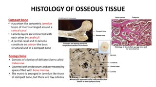
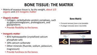
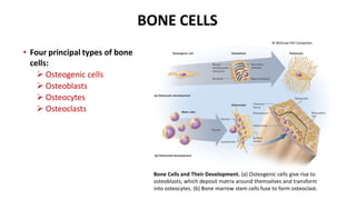
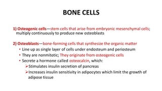
ANATOMY AND
PHYSIOLOGY
CHAPTER 7: SKELETAL SYSTEM-Part 1
BIO 343
OUTLINE
1) INTRODUCTION TO THE SKELETAL SYSTEM
2) ANATOMY OF BONES/BONE TISSUE
INTRODUCTION TO SKELETAL SYSTEM
• Skeletal system—composed of bones,
cartilages, and ligaments
 Forms strong, flexible framework of the
body
• Osteology—the study of bone
• Cartilage—forerunner of most bones
 Tissue that covers many joint surfaces of
mature bones
• Ligaments—hold bones together at the joints
• Tendons—attach muscle to bone
© The McGraw-Hill Companies, Inc.
FUNCTIONS OF THE SKELETAL SYSTEM
• Support—holds up the body, supports muscles
• Protection—brain, spinal cord, heart, lungs
• Movement—limb movements, breathing, action
of muscle on bone
• Electrolyte balance—calcium and phosphate
ions
• Acid–base balance—buffers blood against
excessive pH changes (CaCO3 and phosphate
help in blood acidosis)
• Blood formation—red bone marrow is the chief
producer of blood cells
OVERVIEW OF THE SKELETON
• Axial skeleton is colored tan
• Forms central supporting
axis of the body
• Skull, auditory ossicles, hyoid
bones (anterior neck), vertebral
column, and thoracic cage
• Appendicular skeleton is
colored green
• Bones of upper limb and
pectoral girdle
• Bones of the lower limb and
pelvic girdle
Copyright © The McGraw-Hill Companies.
GENERAL CLASSIFICATION OF BONES
a) Long bones (ex: humerus)
• Longer than wide
• Rigid levers acted upon by muscles
b) Short bones (ex: carpal of wrist)
• Equal in horizontal and vertical
dimensions (cubed shape)
• Glide across one another in multiple
directions
c) Flat bones (ex: sternum)
• Protect soft organs
• Thin, flattened and curved
d) Irregular bones (ex: vertebra)
• Elaborate shapes that do not fit into
other categories
GENERAL FEATURES OF BONES
• Compact and spongy bone (bone
tissue)
• Diaphysis and epiphysis
• Nutrient foramen
• Periosteum
• Endosteum
• Articular cartilage
• Marrow (medullary) cavity
• Red and yellow bone marrow
• Epiphyseal plate (growth plate)
• Epiphyseal line
(a) Living (b) Dried
Marrow cavity
Periosteum
Nutrient foramen
Site of endosteum
Compact bone
Spongy bone
Epiphysis
Epiphysis
Diaphysis
Articular
cartilage
Red bone marrow
Yellow bone marrow
Epiphyseal line
Articular cartilage
The bone is a composite material—
combination of two basic structural
materials, a ceramic (hydroxyapatite) and a
polymer (collagen).
Epiphyseal line
OUTLINE
1) INTRODUCTION TO THE SKELETAL SYSTEM
2) ANATOMY OF BONES/BONE TISSUE
ANATOMY OF A LONG BONE
• Compact (dense) bone—outer shell of dense
white osseous tissue
• Spongy (cancellous) bone—more loosely
organized form of osseous tissue that occupies
the central space of the bone
• Diaphysis (shaft)—cylinder of compact bone that
provides leverage
• Epiphyses—expanded head at each end;
Enlarged to strengthen the joint and provide
added surface area for the attachment of
tendons and ligaments
ANATOMY OF A LONG BONE
• Nutrient foramen—holes in the bone surface through
which blood vessels penetrate
• Endosteum—thin layer of reticular connective tissue
lining the marrow cavity. It also lines a canal system
found throughout the compact bone
• Periosteum—external layer of the bone except
the articular cartilage. It is composed of:
Outer fibrous layer of collagen
• Dense membrane of connective tissue containing
fibroblasts that produce collagen
Inner osteogenic layer of bone-forming cells
• Contains fibroblasts that produce cartilage cells
• Important to growth of bone and healing of
fractures
ANATOMY OF A LONG BONE
• Articular cartilage—layer of hyaline cartilage that
covers the joint surface where one bone meets
another
• Enables joints to move more easily
• Medullary cavity (marrow cavity)—space in the
diaphysis of a long bone that contains bone marrow
• Epiphyseal plate (growth plate)—area of hyaline
cartilage that separates the marrow spaces of the
epiphysis and diaphysis
• Enables growth in length
• Epiphyseal line—in adults, a bony scar that
marks where the epiphyseal plate used to be
BONE MARROW
• Bone marrow—general term for soft tissue that occupies the
marrow cavity of a long bone and small spaces amid the
trabeculae of spongy bone
Copyright © The McGraw-Hill Companies, Inc. Permission required for reproduction or display.
1) Red marrow (myeloid tissue)
• Found in nearly every bone in a child
• Hematopoietic tissue—produces blood cells and is composed
of multiple tissues
• In adults, found in skull, vertebrae, ribs, sternum, part of
pelvic girdle, and proximal heads of humerus and femur
2) Yellow marrow found in adults
• Most red marrow turns into fatty yellow
marrow in adults
• No longer produces blood
HISTOLOGY OF OSSEOUS TISSUE
Compact bone
Spongy bone
Bone marrow Trabecula
Lacunae
Canaliculi
Central canal
Lamella
Compact and spongy bone in a
longitudinal section of the femur
Histology of decalcified spongy bone and
red bone marrow
Microscopic appearance of a cross section of an
osteon of dried compact bone
20 µm
Compact bone
• Has onion-like concentric lamellae
layers of matrix arranged around a
central canal
• Lamella layers are connected with
each other by canaliculi
• A central canal and its lamella
constitute an osteonꟷthe basic
structural unit of a compact bone
Spongy bone
• Consists of a lattice of delicate slivers called
trabeculae
• Covered with endosteum and permeated by
spaces filled with bone marrow
• The matrix is arranged in lamellae like those
of compact bone, but there are few osteons
© McGraw-Hill Companies
BONE TISSUE: THE MATRIX
• Matrix of osseous tissue is, by dry weight, about 1/3
organic and 2/3 inorganic matter
• Organic matter
• Collagen, carbohydrate–protein complexes, such
as glycosaminoglycans, proteoglycans, and
glycoproteins
• Provide flexibility to the bones
• Inorganic matter
• 85% hydroxyapatite (crystallized calcium
phosphate salt)
• 10% calcium carbonate
• Other minerals (fluoride, sodium, potassium,
magnesium)
• Provide hardness to the bones
BONE CELLS
• Four principal types of bone
cells:
 Osteogenic cells
 Osteoblasts
 Osteocytes
 Osteoclasts
Bone Cells and Their Development. (a) Osteogenic cells give rise to
osteoblasts, which deposit matrix around themselves and transform
into osteocytes. (b) Bone marrow stem cells fuse to form osteoclast.
© McGraw-Hill Companies
BONE CELLS
1) Osteogenic cells—stem cells that arise from embryonic mesenchymal cells;
multiply continuously to produce new osteoblasts
2) Osteoblasts—bone-forming cells that synthesize the organic matter
• Line up as single layer of cells under endosteum and periosteum
• They are nonmitotic; They originate from osteogenic cells
• Secrete a hormone called osteocalcin, which:
Stimulates insulin secretion of pancreas
Increases insulin sensitivity in adipocytes which limit the growth of
adipose tissue
BONE CELLS
3) Osteocytes—former osteoblasts that have become trapped in the matrix
they
have deposited
• Reside in lacunae
• Some osteocytes reabsorb bone matrix while others deposit it
• Contribute to homeostatic mechanism of bone density and calcium and
phosphate ions
• When stressed, they produce biochemical signals that regulate bone
remodeling
4) Osteoclasts—bone-dissolving cells found on the bone surface
• Have a different origin from the rest of bone cells
• They develop from the same bone marrow stem cells that give rise to blood cells
• Are unusually large cells formed from the fusion of several stem cells
• Typically have 3 to 4 nuclei, may have up to 50
• Ruffled border to increase surface area
BONE FORMATION
 The formation of bone is called ossification
or osteogenesis
 There are two methods of ossification:
• Intramembranous: produces the flat bones of
the skull, most of the clavicle (bone between
sternum and shoulder blade), and part of the
mandible (bone of lower jaw). Cartilage is not
present during this process.
• Endochondral: process in which a bone
develops from a preexisting model composed
of cartilage. Most bones of the body develop
in this way.
Intramembranous ossification in the fetal
cranium. © McGraw-Hill
Early cartilage model
© McGraw-Hill
BONE FORMATION
 Bone formation occurs in four principal
situations:
(1) the initial formation of bones in an embryo
and fetus,
(2) the growth of bones during infancy, childhood,
and adolescence until their adult sizes are reached,
(3) the remodeling of bone (replacement of old bone
by new bone tissue throughout life), and
(4) the repair of fractures (breaks in bones)
throughout life
Fetal skeleton at 12 weeks of age. Red-
stained regions are calcified, while the remaining
translucent part is still composed of cartilage.
© McGraw-Hill
 Ossification doesn’t end at birth, but continues throughout life
with the growth and remodeling of bones
 Bones grow in two directions:
• Length
• Width
BONE GROWTH
BONE
ELONGATION
BONE WIDENING
BONE ELONGATION
 The epiphyseal plate functions as growth zone
where the bones elongate
• Consists of typical hyaline cartilage in the
middle, with a transition zone on each side
where cartilage is being replaced by bone
 Metaphysis is the zone of transition facing the
marrow cavity
© McGraw-Hill
X-RAY OF A CHILD’S HAND
 The cartilaginous epiphysial
plates are evident at the ends of
the long bones
 Long bones of the hand and
fingers develop only one
epiphysial plate
© McGraw-Hill
BONE WIDENING AND THICKENING
 Bones also continually grow in diameter and thickness
 This involves a process called appositional growth
• Occurs by intramembranous ossification at the bone surface
• Osteoblasts on deep side of the periosteum deposit osteoid tissue on the
bone surface
• Lay down matrix in layers parallel to the surface
• This process produces the surface layers of bone called circumferential
lamellae
BONE REMODELING
 In addition to their growth, bones are continually remodeled throughout
life by the absorption of old bone and the deposition of new one
• This process replaces ~10% of the skeletal tissue per year
 Wolff’s law of bone: the architecture of a bone is determined by the
mechanical stresses placed upon it, and the bone thereby adapts to
withstand it
• Remodeling is a collaborative and precise action of osteoblasts and
osteoclasts
• If a bone is little used, osteoclasts remove matrix and get rid of
unnecessary mass. If a bone is heavily used or a stress is consistently
applied to a particular region of a bone, osteoblasts deposit new
osseous tissue and thicken the bone
ACHONDROPLASTIC DWARFISM
• Occurs due to spontaneous mutations of
the DNA
• Due to failure of cartilage growth
• Long bones of the limbs stop growing in
childhood
• Growth of other bones is unaffected
• Individuals with this condition have a
short stature, but a normal-size head and
trunk
© McGraw-Hill

Chapter 7- Skeletal System-Part 1-SR.pptx

  • 1.
    ANATOMY AND PHYSIOLOGY CHAPTER 7:SKELETAL SYSTEM-Part 1 BIO 343
  • 2.
    OUTLINE 1) INTRODUCTION TOTHE SKELETAL SYSTEM 2) ANATOMY OF BONES/BONE TISSUE
  • 3.
    INTRODUCTION TO SKELETALSYSTEM • Skeletal system—composed of bones, cartilages, and ligaments  Forms strong, flexible framework of the body • Osteology—the study of bone • Cartilage—forerunner of most bones  Tissue that covers many joint surfaces of mature bones • Ligaments—hold bones together at the joints • Tendons—attach muscle to bone © The McGraw-Hill Companies, Inc.
  • 4.
    FUNCTIONS OF THESKELETAL SYSTEM • Support—holds up the body, supports muscles • Protection—brain, spinal cord, heart, lungs • Movement—limb movements, breathing, action of muscle on bone • Electrolyte balance—calcium and phosphate ions • Acid–base balance—buffers blood against excessive pH changes (CaCO3 and phosphate help in blood acidosis) • Blood formation—red bone marrow is the chief producer of blood cells
  • 5.
    OVERVIEW OF THESKELETON • Axial skeleton is colored tan • Forms central supporting axis of the body • Skull, auditory ossicles, hyoid bones (anterior neck), vertebral column, and thoracic cage • Appendicular skeleton is colored green • Bones of upper limb and pectoral girdle • Bones of the lower limb and pelvic girdle Copyright © The McGraw-Hill Companies.
  • 6.
    GENERAL CLASSIFICATION OFBONES a) Long bones (ex: humerus) • Longer than wide • Rigid levers acted upon by muscles b) Short bones (ex: carpal of wrist) • Equal in horizontal and vertical dimensions (cubed shape) • Glide across one another in multiple directions c) Flat bones (ex: sternum) • Protect soft organs • Thin, flattened and curved d) Irregular bones (ex: vertebra) • Elaborate shapes that do not fit into other categories
  • 7.
    GENERAL FEATURES OFBONES • Compact and spongy bone (bone tissue) • Diaphysis and epiphysis • Nutrient foramen • Periosteum • Endosteum • Articular cartilage • Marrow (medullary) cavity • Red and yellow bone marrow • Epiphyseal plate (growth plate) • Epiphyseal line (a) Living (b) Dried Marrow cavity Periosteum Nutrient foramen Site of endosteum Compact bone Spongy bone Epiphysis Epiphysis Diaphysis Articular cartilage Red bone marrow Yellow bone marrow Epiphyseal line Articular cartilage The bone is a composite material— combination of two basic structural materials, a ceramic (hydroxyapatite) and a polymer (collagen). Epiphyseal line
  • 8.
    OUTLINE 1) INTRODUCTION TOTHE SKELETAL SYSTEM 2) ANATOMY OF BONES/BONE TISSUE
  • 9.
    ANATOMY OF ALONG BONE • Compact (dense) bone—outer shell of dense white osseous tissue • Spongy (cancellous) bone—more loosely organized form of osseous tissue that occupies the central space of the bone • Diaphysis (shaft)—cylinder of compact bone that provides leverage • Epiphyses—expanded head at each end; Enlarged to strengthen the joint and provide added surface area for the attachment of tendons and ligaments
  • 10.
    ANATOMY OF ALONG BONE • Nutrient foramen—holes in the bone surface through which blood vessels penetrate • Endosteum—thin layer of reticular connective tissue lining the marrow cavity. It also lines a canal system found throughout the compact bone • Periosteum—external layer of the bone except the articular cartilage. It is composed of: Outer fibrous layer of collagen • Dense membrane of connective tissue containing fibroblasts that produce collagen Inner osteogenic layer of bone-forming cells • Contains fibroblasts that produce cartilage cells • Important to growth of bone and healing of fractures
  • 11.
    ANATOMY OF ALONG BONE • Articular cartilage—layer of hyaline cartilage that covers the joint surface where one bone meets another • Enables joints to move more easily • Medullary cavity (marrow cavity)—space in the diaphysis of a long bone that contains bone marrow • Epiphyseal plate (growth plate)—area of hyaline cartilage that separates the marrow spaces of the epiphysis and diaphysis • Enables growth in length • Epiphyseal line—in adults, a bony scar that marks where the epiphyseal plate used to be
  • 12.
    BONE MARROW • Bonemarrow—general term for soft tissue that occupies the marrow cavity of a long bone and small spaces amid the trabeculae of spongy bone Copyright © The McGraw-Hill Companies, Inc. Permission required for reproduction or display. 1) Red marrow (myeloid tissue) • Found in nearly every bone in a child • Hematopoietic tissue—produces blood cells and is composed of multiple tissues • In adults, found in skull, vertebrae, ribs, sternum, part of pelvic girdle, and proximal heads of humerus and femur 2) Yellow marrow found in adults • Most red marrow turns into fatty yellow marrow in adults • No longer produces blood
  • 13.
    HISTOLOGY OF OSSEOUSTISSUE Compact bone Spongy bone Bone marrow Trabecula Lacunae Canaliculi Central canal Lamella Compact and spongy bone in a longitudinal section of the femur Histology of decalcified spongy bone and red bone marrow Microscopic appearance of a cross section of an osteon of dried compact bone 20 µm Compact bone • Has onion-like concentric lamellae layers of matrix arranged around a central canal • Lamella layers are connected with each other by canaliculi • A central canal and its lamella constitute an osteonꟷthe basic structural unit of a compact bone Spongy bone • Consists of a lattice of delicate slivers called trabeculae • Covered with endosteum and permeated by spaces filled with bone marrow • The matrix is arranged in lamellae like those of compact bone, but there are few osteons © McGraw-Hill Companies
  • 14.
    BONE TISSUE: THEMATRIX • Matrix of osseous tissue is, by dry weight, about 1/3 organic and 2/3 inorganic matter • Organic matter • Collagen, carbohydrate–protein complexes, such as glycosaminoglycans, proteoglycans, and glycoproteins • Provide flexibility to the bones • Inorganic matter • 85% hydroxyapatite (crystallized calcium phosphate salt) • 10% calcium carbonate • Other minerals (fluoride, sodium, potassium, magnesium) • Provide hardness to the bones
  • 15.
    BONE CELLS • Fourprincipal types of bone cells:  Osteogenic cells  Osteoblasts  Osteocytes  Osteoclasts Bone Cells and Their Development. (a) Osteogenic cells give rise to osteoblasts, which deposit matrix around themselves and transform into osteocytes. (b) Bone marrow stem cells fuse to form osteoclast. © McGraw-Hill Companies
  • 16.
    BONE CELLS 1) Osteogeniccells—stem cells that arise from embryonic mesenchymal cells; multiply continuously to produce new osteoblasts 2) Osteoblasts—bone-forming cells that synthesize the organic matter • Line up as single layer of cells under endosteum and periosteum • They are nonmitotic; They originate from osteogenic cells • Secrete a hormone called osteocalcin, which: Stimulates insulin secretion of pancreas Increases insulin sensitivity in adipocytes which limit the growth of adipose tissue
  • 17.
    BONE CELLS 3) Osteocytes—formerosteoblasts that have become trapped in the matrix they have deposited • Reside in lacunae • Some osteocytes reabsorb bone matrix while others deposit it • Contribute to homeostatic mechanism of bone density and calcium and phosphate ions • When stressed, they produce biochemical signals that regulate bone remodeling 4) Osteoclasts—bone-dissolving cells found on the bone surface • Have a different origin from the rest of bone cells • They develop from the same bone marrow stem cells that give rise to blood cells • Are unusually large cells formed from the fusion of several stem cells • Typically have 3 to 4 nuclei, may have up to 50 • Ruffled border to increase surface area
  • 18.
    BONE FORMATION  Theformation of bone is called ossification or osteogenesis  There are two methods of ossification: • Intramembranous: produces the flat bones of the skull, most of the clavicle (bone between sternum and shoulder blade), and part of the mandible (bone of lower jaw). Cartilage is not present during this process. • Endochondral: process in which a bone develops from a preexisting model composed of cartilage. Most bones of the body develop in this way. Intramembranous ossification in the fetal cranium. © McGraw-Hill Early cartilage model © McGraw-Hill
  • 19.
    BONE FORMATION  Boneformation occurs in four principal situations: (1) the initial formation of bones in an embryo and fetus, (2) the growth of bones during infancy, childhood, and adolescence until their adult sizes are reached, (3) the remodeling of bone (replacement of old bone by new bone tissue throughout life), and (4) the repair of fractures (breaks in bones) throughout life Fetal skeleton at 12 weeks of age. Red- stained regions are calcified, while the remaining translucent part is still composed of cartilage. © McGraw-Hill
  • 20.
     Ossification doesn’tend at birth, but continues throughout life with the growth and remodeling of bones  Bones grow in two directions: • Length • Width BONE GROWTH BONE ELONGATION BONE WIDENING
  • 21.
    BONE ELONGATION  Theepiphyseal plate functions as growth zone where the bones elongate • Consists of typical hyaline cartilage in the middle, with a transition zone on each side where cartilage is being replaced by bone  Metaphysis is the zone of transition facing the marrow cavity © McGraw-Hill
  • 22.
    X-RAY OF ACHILD’S HAND  The cartilaginous epiphysial plates are evident at the ends of the long bones  Long bones of the hand and fingers develop only one epiphysial plate © McGraw-Hill
  • 23.
    BONE WIDENING ANDTHICKENING  Bones also continually grow in diameter and thickness  This involves a process called appositional growth • Occurs by intramembranous ossification at the bone surface • Osteoblasts on deep side of the periosteum deposit osteoid tissue on the bone surface • Lay down matrix in layers parallel to the surface • This process produces the surface layers of bone called circumferential lamellae
  • 24.
    BONE REMODELING  Inaddition to their growth, bones are continually remodeled throughout life by the absorption of old bone and the deposition of new one • This process replaces ~10% of the skeletal tissue per year  Wolff’s law of bone: the architecture of a bone is determined by the mechanical stresses placed upon it, and the bone thereby adapts to withstand it • Remodeling is a collaborative and precise action of osteoblasts and osteoclasts • If a bone is little used, osteoclasts remove matrix and get rid of unnecessary mass. If a bone is heavily used or a stress is consistently applied to a particular region of a bone, osteoblasts deposit new osseous tissue and thicken the bone
  • 25.
    ACHONDROPLASTIC DWARFISM • Occursdue to spontaneous mutations of the DNA • Due to failure of cartilage growth • Long bones of the limbs stop growing in childhood • Growth of other bones is unaffected • Individuals with this condition have a short stature, but a normal-size head and trunk © McGraw-Hill