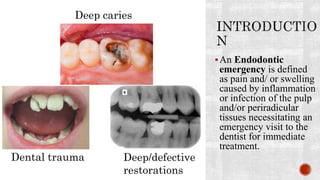
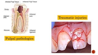
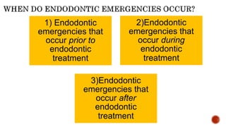
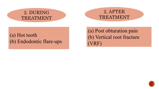
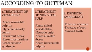
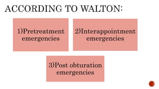
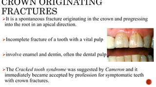
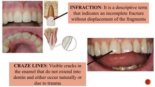
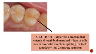
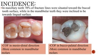
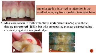
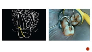
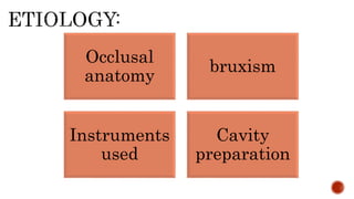
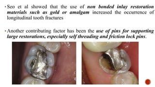
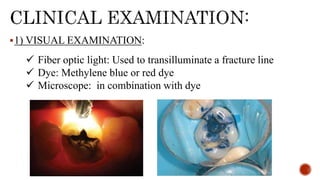
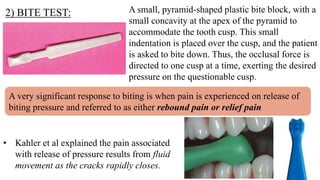
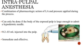
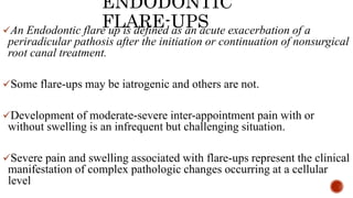
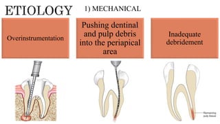
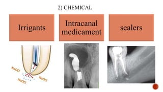
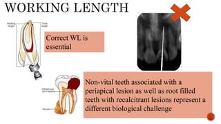
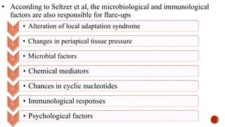
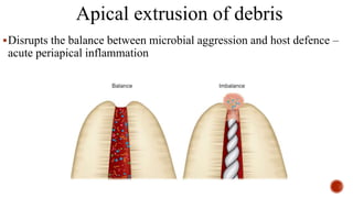
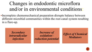
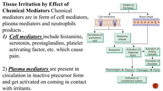
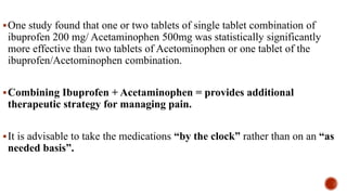
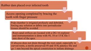
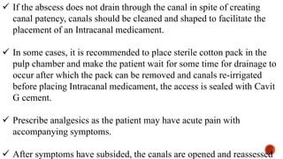
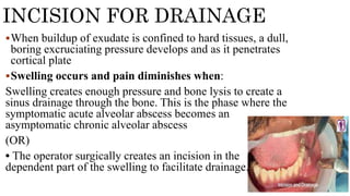
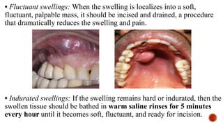
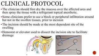
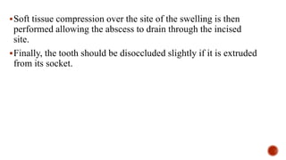
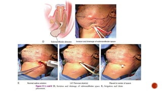
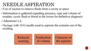
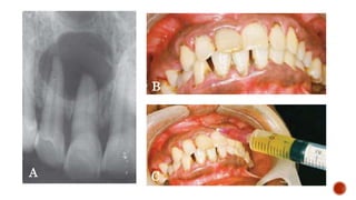
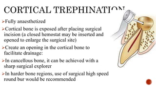
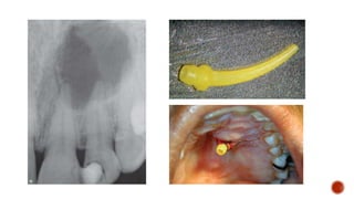
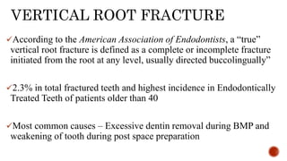
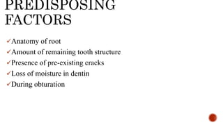
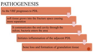
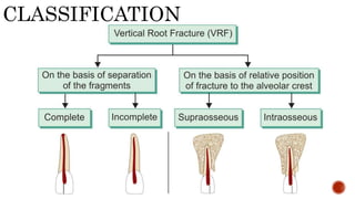
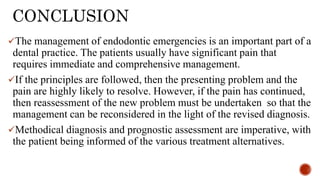
Dr. Sushmita Rane
MDS III
Seminar 10
11/12/2023
An Endodontic
emergency is defined
as pain and/ or swelling
caused by inflammation
or infection of the pulp
and/or periradicular
tissues necessitating an
emergency visit to the
dentist for immediate
treatment.
Dental trauma
Deep caries
Deep/defective
restorations
Pulpal pathologies
Traumatic injuries
1) Endodontic
emergencies that
occur prior to
endodontic
treatment
2)Endodontic
emergencies that
occur during
endodontic
treatment
3)Endodontic
emergencies that
occur after
endodontic
treatment
CLASSIFICATION OF
ENDODONTIC
EMERGENCIES
1.BEFORE
TREATMENT
(A) ENDODONTIC EMERGENCIES
PRESENTING WITH PAIN AND/OR
SWELLING
(i) Crown-originating fracture (COF)
(previously known as cracked tooth syndrome)
(ii) Symptomatic reversible pulpitis
(iii) Symptomatic irreversible pulpitis
(iv) Primary symptomatic apical periodontitis
(v) Secondary symptomatic apical periodontitis
(acute exacerbation of asymptomatic apical
periodontitis or phoenix abscess)
(vi) Symptomatic (acute) alveolar abscess
(vii) Cellulitis
(B) TRAUMATIC
INJURIES
(i) Crown/root fractures
(ii) Luxation injuries
(iii) Tooth avulsion
2. DURING
TREATMENT
2. AFTER
TREATMENT
(a) Hot tooth
(b) Endodontic flare-ups
(a) Post obturation pain
(b) Vertical root fracture
(VRF)
1.TREATMENT OF
VITAL PULP:
-Acute reversible
pulpitis
-Hypersensitivity
dentin
-Recurrent decay
-Recent restorations
-Cracked tooth
syndrome
2.TREATMENT
OF NON VITAL
PULP:
-Acute apical
periodontitis
-Necrotic pulp
-Acute alveolar
abcess
-Acute irreversible
pulpitis
3. ESTHETIC
EMERGENCY
-Fracture of crown
-Fracture of root
-Avulsed tooth
1)Pretreatment
emergencies
2)Interappointment
emergencies
3)Post obturation
emergencies
When managing a patient who presents with pain, the principles of the
“Three D’s” should be followed (Kaiser & Byrne, 2011). which is
The type and details of the treatment depend on the diagnosis of the
presenting problem and the amount of remaining tooth structure as this
will dictate whether the tooth is suitable for further restoration and how
it can be restored.
Drugs can be used intra-dentally (sedative liners, root canal
medicaments) and as systemic medications, but it is essential to
understand that systemic drugs should only be used as an adjunct to
Diagnos
is
Definitive
dental
treatment
Drugs
CROWN ORIGINATING
FRACTURES
It is a spontaneous fracture originating in the crown and progressing
into the root in an apical direction.
Incomplete fracture of a tooth with a vital pulp
involve enamel and dentin, often the dental pulp
The Cracked tooth syndrome was suggested by Cameron and it
immediately became accepted by profession for symptomatic teeth
with crown fractures.
CRAZE LINES: Visible cracks in
the enamel that do not extend into
dentin and either occur naturally or
due to trauma
INFRACTION: It is a descriptive term
that indicates an incomplete fracture
without displacement of the fragments
SPLIT TOOTH: describes a fracture that
extends through both marginal ridges usually
in a mesio-distal direction, splitting the tooth
completely into 2 separate segments
progressive in nature
discontinuity in the integrity of a tooth’s hard tissue
Some COFs are the cause of cuspal fractures and these often do not directly
involve the pulp.
Fracture that are more centrally located on the occlusal surface do involve the
pulp.
Some are associated with symptoms some are not.
Symptoms are vague-Discomfort to chewing
- Elevated sensitivity to cold food and drinks
In maxillary teeth 70% of fracture lines were situated toward the buccal
tooth surface, while in the mandibular teeth they were inclined to be
towards lingual surface.
-COF in mesio-distal direction
-More common in mandibular
molars
-COF in bucco-palatal direction
-More common in mandibular
molars
Anterior teeth is involved in infarction is the
result of an injury from a sudden traumatic blow
 Most cases occur in teeth with class I restorations (39%) or in those
that are unrestored (25%), but with an opposing plunger cusp occluding
centrically against a marginal ridge.
A
C
B
Occlusal
anatomy
bruxism
Instruments
used
Cavity
preparation
• Seo et al showed that the use of non bonded inlay restoration
materials such as gold or amalgam increased the occurrence of
longitudinal tooth fractures
• Another contributing factor has been the use of pins for supporting
large restorations, especially self threading and friction lock pins.
1) VISUAL EXAMINATION:
 Fiber optic light: Used to transilluminate a fracture line
 Dye: Methylene blue or red dye
 Microscope: in combination with dye
2) BITE TEST:
• Kahler et al explained the pain associated
with release of pressure results from fluid
movement as the cracks rapidly closes.
A very significant response to biting is when pain is experienced on release of
biting pressure and referred to as either rebound pain or relief pain
A small, pyramid-shaped plastic bite block, with a
small concavity at the apex of the pyramid to
accommodate the tooth cusp. This small
indentation is placed over the cusp, and the patient
is asked to bite down. Thus, the occlusal force is
directed to one cusp at a time, exerting the desired
pressure on the questionable cusp.
3) COLD STIMULUS APPLICATION AND ELECTRIC PULP TESTING
(EPT)
-will provide information about the status of the pulp and there is evidence
that teeth with fractures respond at lower threshold levels to cold and EPT
stimulation compared to non fractured teeth.
4)THIN SHARP EXPLORER
Using a sharp explorer to probe around the cervical circumference of teeth
suspected of having infractions, they may be identified by the ‘click’ when
explorer encounters the fracture.
Some patients will also feel a sharp, sudden pain at that time.
5) PERCUSSION SENSITIVITY
A tooth with infraction is not likely to be identified by percussion
until the fracture extends to involve the PDL
Reversible pulpitis: Preserve the pulp vitality – Full coverage for cusp
protection.
No lingering pain to cold and no severe
spontaneous pain, a 2 week waiting
period is recommended
tooth stabilized with orthodontic
band for 2 weeks.
If symptoms subsided, the patient may
be offered the option of placing a
restoration that binds the tooth such as
full crown.
Remove
fractured
segment
Restoration
of tooth
Large crack
Pulp involved
Small crack
Pulp not
involved
Hopeless
prognosis
Endodontic therapy
+
Immediate
stabilization with
Orthodontic brackets
Final Restoration with
crown and/or post and core
Occlusal
adjustments
+
Immediate
stabilization
Permanent stabilization
using bonded restoration or
crown
Extractio
n
ASSESSMENT OF TOOTH
EMERGENCIES
DURING
TREATMENT
HOT TOOTH
A tooth that is difficult to anesthetize is known as a hot tooth
Associated with an irreversible pulpitis
Inflamed pulp tissue has an extremely concentrated sensory
nerve supply particularly in the chamber, it becomes more
difficult to anesthetize
Associated with mandibular molars following long periods of
low level to moderate pain
Extremely frustrating to the patient and dentist
THEORIES OF HOT TOOTH
Hyperalgesia: Inflammation within the tooth alters the actual nerve by
changing the resting potentials and decreasing the excitability thresholds
making it harder to anesthetize.
Nervous patient: Pain threshold further reduces causing difficulty to
anesthetize
Location: If anesthetic is away from the target, it becomes hard to anesthetize
Local tissue changes due to inflammation: In the area of inflammation,
acidic pH of inflamed tissue decreases the amount of base form of anesthetic
available to penetrate nerve membrane causing low effect.
Central core theory: It states that nerves outside the nerve bundle
supply the molars whereas nerves on the inside supply the anterior
teeth so anesthetic may not penetrate into the nerve trunk to make
all the nerves numb.
Tetrodotoxin-resistant (TTXr) channels: Special class of sodium
channels on C-fibers known as TTXr sodium channels, during
inflammation, neuroinflammatory reactions start, sodium channel
expression on C-fibers shift from TTX-sensitive to TTXr causing
hyperalgesia and these channels are 5 times more resistant than
TTX sensitive channels.
MANAGEMENT OF HOT
TOOTH
EXPLAINING TO THE PATIENT: use of iatrosedation and verbal
sedation
PREMEDICATION: Anti-inflammatory 1 hour before the procedure
along with time gap between anesthetic injection and starting the
procedure
(Lorazepam 1 mg after checking interaction with other drugs the night
before sleep followed by 90 minutes prior to procedure)
Administration of nitrous oxide while dealing with hot tooth
SUPPLEMENTAL
ANESTHETIC TECHNIQUES
Supplementing an inferior alveolar nerve block (IANB) with 4%
articaine with 1:100,000 epinephrine buccal infiltration (0.9–1.2
mL) at the apex of the tooth to be treated is one of the most
effective supplemental anesthetic techniques.
SUPPLEMENTAL INJECTIONs
INTRAPULPAL INJECTION
INTRALIGAMENTARY
INJECTION
Special pressure needles have been developed for IL injection with Preset
volume (0.14-0.22 mL) with minimal effort.
27 or 30 G needle inserted with positive pressure as deep as possible along
the root with the bevel towards the crest.
In posteriors, needle is bent to an angle and trigger is squeezed to deliver
0.2 mL
Immediate onset and lasts 27 minutes
92% effective
 PDL injections are usually given
using either a standard dental
anesthetic syringe or a high-
pressure syringe.
 The development of computer-
controlled anesthetic delivery
systems (the Wand) or the Single
Tooth Anesthesia have been found
to be able to deliver a PDL
injection.
INTRAOSSEOUS
INJECTION
The use of the intraosseous injection allows the practitioner to deliver
local anesthetic solutions directly into the cancellous bone surrounding
the affected tooth. There are several 10 systems available in the market
Stabident system
(Fairfax Dental
Inc,Wimbledon,
UK)
X-Tip system
(Dentsply, York,
PA, USA)
IntraFlow
handpiece (Pro-
Dexlnc, SantaAna,
CA,USA).
The Stabident system consists of a 27-
gauge beveled wire that is driven by a
slow-speed handpiece, which perforates
the cortical bone. Anesthetic solution is
then delivered into the cancellous bone
through the perforation.
The IntraFlow handpiece holds
and drives a perforating needle,
which is engaged via an internal
clutch to deliver the local anesthetic
through the perforation.
 The X-Tip system consists of a 2-
part perforator and guide sleeve
component which is also driven by
a slow-speed handpiece. The
perforator leads the guide sleeve
through the cortical bone and then
is separated from it and removed.
This leaves the guide sleeve in
place and allows for a 27gauge
needle to be inserted for injecting
the anesthetic solution.
INTRA-PULPAL
ANESTHESIA
Combination of pharmacologic action of LA and pressure applied during
the process.
Can only be done if the body of the exposed pulp is large enough to admit
a hypodermic needle.
0.2-.03 mL injected into the pulp.
Immediate and effective.
ENDODONTIC
FLARE-UPS
An Endodontic flare up is defined as an acute exacerbation of a
periradicular pathosis after the initiation or continuation of nonsurgical
root canal treatment.
Some flare-ups may be iatrogenic and others are not.
Development of moderate-severe inter-appointment pain with or
without swelling is an infrequent but challenging situation.
Severe pain and swelling associated with flare-ups represent the clinical
manifestation of complex pathologic changes occurring at a cellular
level
Overinstrumentation
Pushing dentinal
and pulp debris
into the periapical
area
Inadequate
debridement
1) MECHANICAL
2) CHEMICAL
Irrigants
Intracanal
medicament
sealers
Correct WL is
essential
Non-vital teeth associated with a
periapical lesion as well as root filled
teeth with recalcitrant lesions represent a
different biological challenge
Over instrumentation
may force infected debris
into the periapical tissues
elicitating a severe
inflammatory response
and pain
Underinstrumentation will
leave micro-organisms in
close proximity to the
apical foramina where
they or their virulence
factors can gain access to
tissues
Incomplete instrumentation can
disrupts the balance within the
microflora and allow previously
inhibited species to overgrow
IRRIGANT EXTRUSION
Standard irrigant used is 1-5.25% NaOCl with final rinse of 17%
EDTA.
Every procedure including irrigation should be a passive procedure
extrusion of irrigant beyond the periapex leads to sodium
hypochlorite accident
Severe pain, swelling and profuse bleeding through the tooth and
interstitial tissues
SIGNS OF SODIUM
HYPOCHLORITE ACCIDENT
Severe and excruciating pain when not under LA
If under LA, Patient complains of irritation at the periradicular area
Sudden flooding of the canal with blood and tissue fluids
Ballooning of tissues in the area and swelling of soft tissues
Edema, ecchymosis with tissue necrosis, parasthesia seen .
MANAGEMENT
Bleeding is allowed to flow since it is a defense physiological
mechanism.
Flood the canal with normal saline so that accumulated blood comes
out and level of pain decreases
Immediate aspiration and application of icepacks
Immediately placed on parenteral antibiotics and analgesics
Consult general physician and administer steroids in a planned manner
Backup vitamin therapy during recovery is recommended
PREVENTION
Always use passive irrigation
Handled carefully
Closed ended lateral/side vented 30 G needles are used
Never bind the needle in the canal, allow back flow.
Oscillate the needle in the canal
TISSUE EMPHYSEMA
Collection of gas/air in tissue spaces or
facial planes
Occurs during periapical surgery when air
from airotor is directed towards exposed
soft tissues.
When blast of air is directed towards open
root canals to dry them
Complication of fracture involving facial
skeleton
Rapid swelling, edema and crepitus
(crepitus pathognomonic of tissue
emphysema)
Dysphagia and dyspnea and if
emphysema spreads to neck, it can
cause issues with breathing and
progression to mediastinum
Differential Diagnosis: Angioedema,
internal hemorrhage and anaphylaxis
TREATMENT AND
PREVENTION
Antibiotics to prevent risk and spread of infection
Application of moist heat to reduce swelling
If airway or mediastinum is obstructed, immediate medical attention
and hospitalization of patient.
Administration of 100% O2 via mask
When using air pressure, blast of air directed at horizontal direction
against walls of tooth and root periapically
During surgical procedures, use low speed or high speed handpiece
which do not direct air towards tissues (rear exhausting handpiece for
root resection and ultrasonics for retropreparation)
• According to Seltzer et al, the microbiological and immunological
factors are also responsible for flare-ups
1. • Alteration of local adaptation syndrome
2. • Changes in periapical tissue pressure
3. • Microbial factors
4. • Chemical mediators
5. • Chances in cyclic nucleotides
6. • Immunological responses
7. • Psychological factors
Alteration of local adaptation syndrome
• There is a balance between root canal microflora and the host immune
system which is known as local adaptive syndrome. When there is
presence of asymptomatic apical periodontitis and we are accidentally
pushing debris into the root canal space, there is occurrence of flare-
ups. This occurs due to disturbance in this balance
• A Study showed that when a new irritant is introduced to a chronically
inflamed tissue, a violent reaction may occur due to disturbance in
local tissue adaptation to applied irritants.
Changes in periapical tissue pressure
In teeth with increased periapical pressure, exudate creates pain by
causing pressure on the nerve endings.
Pain is relieved when the tooth is kept open to drain the exudate but in
teeth with less periapical pressure if kept open, microbes and other
irritants may get aspirated into the periapical area causing pain.
Microbial factors
Gram negative anaerobes like Prevotella and Porphyromonas species
release endotoxins which are neurotoxic.
These activate the Hageman factor to release Bradykinin, a potent
pain mediator.
Teichoic acid which is present in the cell wall and plasma
membranes of gram positive bacteria produce humoral antibodies
IgM, IgG, IgA and release mediators causing pain
Apical extrusion of debris
Disrupts the balance between microbial aggression and host defence –
acute periapical inflammation
Changes in endodontic microflora
and/or in environmental conditions:
Incomplete chemomechanical preparation disrupts balance between
different microbial communities within the root canal system resulting
in a flare-up.
Secondary
intraradicular
infection
Increase of
oxidation–
reduction potential
Effect of Chemical
Mediators
Secondary intraradicular infection: penetration of new
microbial species, microbial cells and substrate from saliva into
the root canal system during treatment which may lead to a
secondary infection and cause a flare-up.
Increase of oxidation-reduction potential: alteration of oxidation-
reduction potential during endodontic treatment may favour
overgrowth of facultative bacteria that resist chemo-mechanical
procedures
Tissue Irritation by Effect of
Chemical Mediators Chemical
mediators are in form of cell mediators,
plasma mediators and neutrophils
products .
1) Cell mediators include histamine,
serotonin, prostaglandins, platelet
activating factor, etc. which cause
pain.
2) Plasma mediators are present in
circulation in inactive precursor form
and get activated on coming in contact
with irritants.
Anxiety, apprehension, fear and previous
history of dental experience plays a
contributory role in mid-treatment flare-
ups
Immunological
response
In chronic pulpitits and periapical disease,
presence of macrophages and lymphocytes
indicates both cell mediated and humoral
response
Psychological factor
Over 200 studies indicate that behavioural intervention, to decrease anxiety
before and after surgery reduces post operative pain intensity and intake of
analgesics improve treatment compliance, cardiovascular and respiratory
indices and accelerates recovery
Protocols
1) Information about profound anesthesia and preventive strategies is an
important anxiety reduction technique.
2) Information about sensation experienced during treatment as well as
description of procedures appears to have a significant impact in reducing
anxiety.
ANXIETY REDUCTION
ANTIBIOTICS
Not recommended for healthy patients
Indicated –
1) Spreading infection that indicates failure of local host responses.
2) Patient with medical condition that compromises defense mechanism.
NSAIDS:
Pretreatment with NSAIDS for irreversible pulpitis should have the effect of
reducing pulpal levels of inflammatory mediator prostaglandin E2 (PGE2)
One study found that one or two tablets of single tablet combination of
ibuprofen 200 mg/ Acetaminophen 500mg was statistically significantly
more effective than two tablets of Acetominophen or one tablet of the
ibuprofen/Acetominophen combination.
Combining Ibuprofen + Acetaminophen = provides additional
therapeutic strategy for managing pain.
It is advisable to take the medications “by the clock” rather than on an “as
needed basis”.
Flare Ups Iatrogenic
Inaccurate WL
Necrotic case
If H/o acute apical
periodontitis
Occlusion reduction
can be done
Complaint of swelling
in interproximal area
Periodontal
component should be
explored
If canal was
underinstrumented/
Overinstrumented
Remaining tissue,
microbes are major
factors responsible
so retreated
Abcesses/Cellulitis
associated with
flareup is treated
No Swelling
when treatment is done in more than one visit, Intracanal medicament like
calcium hydroxide is placed.
Care should be taken not to push necrotic debris during instrumentation
Crown down instrumentation have been shown to remove most of the
debris coronally rather than pushing it beyond the apex.
The use of positive pressure irrigation methods, such as needle and
syringe irrigation poses a risk of expressing debris or solution out of the
apex.
Improvements in technology such as apex locators have facilitated
increased accuracy.
SWELLING
Acute periradicular
abcess at the time
of the initial
emergency visit
interappointment
flare up
postendodontic
complication
Localized Diffuse
The principle modality for
managing swelling secondary to
endodontic infection is to achieve
drainage and remove the source
of infection.
When the swelling is localized,
the preferred avenue is drainage
through the root canal.
A C
B
• In this manner the canal can
be dried and the endodontic
treatment completed in one
visit
• In the presence of persistent
swelling, gentle finger pressure to
the mucosa help in drainage
• Once the canals cleaned and dried,
the access should be closed.
Drainage via the root canal and/or incision and drainage of the swelling
Consider oral antibiotics if systemic signs of illness (malaise, increased
temperature, lymph node involvement, etc.)
 Non- steroidal anti- inflammatory drugs (e.g. ibuprofen 400 mg every
4– 6 h) and/or
 Analgesics (e.g. acetaminophen 1000 mg every 4– 6 h)
Bacteria in
periradicular
tissues
+
Immune
system
suppressed
Signs and
symptoms of
acute apical
abcess
Cellulitis
Swelling
may be
localized to
vestibule or
extend into
fascial space
Fascial spaces of head
and neck can be
categorized into 4
anatomic groups
The mandible and below
The cheek and lateral face
The pharyngeal and cervical areas
The midface
Swellings of and below
mandible include 6
anatomic areas or fascial
spaces
The buccal vestibule
The body of the mandible
The mental space
The submental space
The Sublingual space
The Submandibular space
The Mandibular buccal
vestibule is the anatomic area
amid the buccal cortical plate
and buccinator muscle in
posterior of mandible and
mentalis muscle in anteriors
Space of the body of the mandible:
Area between the buccal or lingual
cortical plate and overlying periosteum
MENTAL, SUBMENTAL SPACES
SUBLINGUAL AND SUBMANDIBULAR SPACES
The lateral boundaries of
the space are the lingual
surfaces of the mandible
LATERAL FACE SWELLING
Maxillary buccal vestibule
Buccal space
Submassteric space
Deep temporal space
Superficial temporal space
ANATOMIC SPACES IN
PHARYNGEAL AND CERVICAL
AREAS Pterygomandibular space
Parapharyngeal space
Carotid space
Retropharyngeal space
Pretracheal space
Danger Space
Prevertebral space
Buccal, submasseteric,
pterygomandibular,
parapharyngeal spaces
Pretracheal, prevertebral and
danger spaces
MIDFACE SWELLING: ANATOMICAL SPACES
IN MIDFACIAL AREA
Palate
Base of upper lip
Infraoribital space
Periorbital space
 The emergency treatment of suppurative lesions involve establishing
drainage
 Drainage can be achieved and relieves acute symptoms caused by
symptomatic (acute) alveolar abscess by the following protocols:
Achieving
drainage
through
the root
canals
Incisio
n for
drainag
e
Needle
aspiratio
n
Cortical
trephination and
decompression
ACHIEVING DRAINAGE
THROUGH ROOT CANALS
LA is not needed as pulp is necrotic and frequently LA is contraindicated
in acutely inflamed tissue as its infiltration does not anesthetize the tissue.
Forcing LA into acutely infected and swollen area may increase pain and
spread the infection into facial spaces.
Local anaesthesia may be administered to reduce the pain of acute
alveolar abscess as long as injection route is distant from the inflamed
area.
Mandibular block or infraorbital block can be used for a few cases where
partial vitality persists.
Rubber dam placed over infected tooth
Access opening completed by bracing the
tooth with finger pressure
Pulp chamber is irrigated profusely and debrided,
forcing any solution or debris into periradicular
tissues should be avoided
Root canal orifices are located with a DG 16 explorer
and instrumentation is done with #8, 10 or 15 K file 1
mm short of the apex
If abscess does not drain through the root canal, especially in
curved roots, a sterile precurved #8 and 10 K patency file and
go 1 mm beyond the apical constriction to initiate drainage
 If the abscess does not drain through the canal in spite of creating
canal patency, canals should be cleaned and shaped to facilitate the
placement of an Intracanal medicament.
 In some cases, it is recommended to place sterile cotton pack in the
pulp chamber and make the patient wait for some time for drainage to
occur after which the pack can be removed and canals re-irrigated
before placing Intracanal medicament, the access is sealed with Cavit
G cement.
 Prescribe analgesics as the patient may have acute pain with
accompanying symptoms.
 After symptoms have subsided, the canals are opened and reassessed
When buildup of exudate is confined to hard tissues, a dull,
boring excruciating pressure develops and as it penetrates
cortical plate
Swelling occurs and pain diminishes when:
Swelling creates enough pressure and bone lysis to create a
sinus drainage through the bone. This is the phase where the
symptomatic acute alveolar abscess becomes an
asymptomatic chronic alveolar abscess
(OR)
• The operator surgically creates an incision in the
dependent part of the swelling to facilitate drainage.
INCISION FOR DRAINAGE
• Fluctuant swellings: When the swelling is localizes into a soft,
fluctuant, palpable mass, it should be incised and drained, a procedure
that dramatically reduces the swelling and pain.
• Indurated swellings: If the swelling remains hard or indurated, then the
swollen tissue should be bathed in warm saline rinses for 5 minutes
every hour until it becomes soft, fluctuant, and ready for incision.
CLINICAL PROTOCOL
The clinician should first dry the mucosa over the affected area and
then spray the tissue with a refrigerant topical anesthetic.
Some clinicians prefer to use a block or peripheral infiltration around
but not in the swollen tissues, prior to incision
The incision should be made at the most dependent site of the
swelling
Hemostat or elevator used to dissect the incision site to facilitate
drainage
Soft tissue compression over the site of the swelling is then
performed allowing the abscess to drain through the incised
site.
Finally, the tooth should be disoccluded slightly if it is extruded
from its socket.
NEEDLE ASPIRATION
Use of suction to remove fluids from a cavity or space
Information is gathered regarding presence, type and volume of
exudate, cystic fluid or blood in the lesion for definitive diagnosis
Administer LA
Syringe with 18 G needle used to aspirate the contents out of the
swelling
Reduced
scarring
Eradication
of volume
Character of
the aspirate
A
B
C
CORTICAL TREPHINATION
Fully anaesthetized
Cortical bone is exposed after placing surgical
incision (a closed hemostat may be inserted and
opened to enlarge the surgical site)
Create an opening in the cortical bone to
facilitate drainage:
In cancellous bone, it can be achieved with a
sharp surgical explorer
In harder bone regions, use of surgical high speed
round bur would be recommended
DECOMPRESSION
In large cysts with swelling, a decompression procedure is advocated
where a drain tube is inserted into the trephined cyst cavity for
several weeks to enable communication between cyst cavity and oral
cavity.
If the swelling is hard, it can be converted to a soft, fluctuant
swelling by rinsing with hot saline solution 3–5 minutes every hour.
POST
OBTURATION
EMERGENCIES
POST OBTURATION
EMERGENCIES
Overinstrumentation
Persistent periapical
inflammation
Overfilling
Hyperocclusion Poor coronal seal Fracture of crown/root
Gross overfilling involves the introduction of excess sealer and its
cytotoxic components into the periapical tissues causing tissue
damage and inflammation.
A study found that overfilling
was associated with
significantly increased rate of
pain and percussion sensitivity
in 1 week followup
examinations as compared with
teeth not overfilled
Was the patient symptomatic prior to obturation?
If there is swelling, is it localized, diffuse or fluctuant?
Is the tooth overfilled?
Is pain/swelling increasing ?
Obturation in the presence of acute apical
periodontitis can be considered to be a
predictor of post operative pain.
2) Scheduling of the obturation
To Avoid
Patients who present with acute apical periodontitis should have the
procedure postponed until the tooth is more comfortable
3) If treatment is already done:
Relief of pain
achieved by treatment directed at reducing tissue levels of
factors that stimulate peripheral terminals of nociceptors
or by reducing mechanical stimulation of sensitized
nociceptors (e.g. OCCLUSALADJUSTMENT)
Delayed healing and lower success rates are seen
when there is overextended obturation.
Open apex cases have the potential for
overfillings and attempts should be made to
maintain apical anatomy small and not
unnecessarily enlarge the area
Gross overfilling cases prolonged pain and may
have serious consequences if vital anatomical
structures are involved (complicates removal)
Overfilling
 GP or sealer may extend into the mandibular canal
causing severe damage to the IAN and paresthesia
 Radiographs may be helpful in determining
proximity to the mandibular canal.
 Conventional x-rays may only provide a 2-D image
of the relationship between apex and mandibular
canal and CBCT provides vastly more information
as a 3-D image.
 CBCT is valuable in determining the
relation between apices and floor of the
maxillary sinus
 Filling material extruding into the sinus
can potentially cause chronic sinusitis
and infection
MANDIBULAR CANAL
MAXILLARY SINUS
A statistically valid profile of patients most likely to benefit from
occlusal reduction was developed.
In that study of 117 patients, approximately twice as many (80%)
with a diagnosis of irreversible pulpitis, who underwent occlusal
reduction, reported no post treatment pain when compared to
control subjects with no occlusal reduction
Occlusal reduction was found to result in prevention of post
operative pain when any or all of the following indicators
were present
Sensitivity to percussion
Vital tooth
History of pain
Absence of periapical lesion
REMOVAL OF OBTURATION
Filling canals in the presence of symptoms is a predictor of post
obturation pain
TREATMENT
-Pharmaco-therapeutics including analgesics and/or antibiotics
(nonvital) to retreatment with or without incision or drainage.
-Consider variables and determine if the primary cause is
inflammatory, procedural or active infection
Depends on the quality of the
filling
Nature of the swelling –
Fluctuant or non-fluctuant
RETREATMENT
More challenging than primary root canal treatment
More prone to exacerbations with a long history of persistent infection
with complex flora
Microbes found in failed RCT have either remained in the canal following
previous treatment or may have entered through the coronal leakage
Primary – Polymicrobial organisms dominated by anaerobes
Secondary infection – Gram positive principally enterococci
An important consideration in retreatment cases is the origin
of the intra-canal bacterial flora.
The microbial source may be due to a defective restoration or
colonies remaining after initial root canal therapy
If the cause of bacterial penetration is the restoration, it must
be replaced
Microbes remaining after root canal therapy must be
addressed
E. FAECALIS
Gram-positive facultative bacteria, particularly are predominant
This virulent microbe is particularly difficult to eliminate due to its
resistance to calcium hydroxide, and it can survive without nutrition
for long periods of time.
More likely to be found in cases of failed endodontic therapy than in
primary infections
Resistance to calcium hydroxide may be due to a gene involved in
cell division which enables it to survive following prolonged exposure
to alkaline pH
asymptomatic cases symptomatic ones
PREDICTORS OF
PERSISTENT PAIN
Preoperative pain
persistent for
over 3 months
Previous chronic
pain problems
Inter-
appointment pain
Gender (Women
are more likely)
Preoperative
tenderness to
percussion
POSSIBLE CAUSES OF
PERSISTENT PAIN
Untreated canal
Failed coronal
seal
Tooth fracture
Pain from
adjacent tooth
Non-
odontogenic
referred pain
Deafferenation
(elimination or
interruption of
afferent nerve
impulses)
VERTICAL ROOT FRACTURE
According to the American Association of Endodontists, a “true”
vertical root fracture is defined as a complete or incomplete fracture
initiated from the root at any level, usually directed buccolingually”
2.3% in total fractured teeth and highest incidence in Endodontically
Treated Teeth of patients older than 40
Most common causes – Excessive dentin removal during BMP and
weakening of tooth during post space preparation
PREDISPOSING
FACTORS
Anatomy of root
Amount of remaining tooth structure
Presence of pre-existing cracks
Loss of moisture in dentin
During obturation
As the VRF progresses to PDL
soft tissue grows into the fracture space causing
more separation
It communicates the oral cavity through the
sulcus, bacteria enters the area
Initiates inflammation of the adjacent PDL
bone loss and formation of granulation tissue
CLASSIFICATION
PREVENTION
Avoid weakening of canal walls
Minimize internal wedging forces
Evaluate tooth anatomy before treatment
Preserve as much as tooth structure as possible before treatment
Use optimal forces during obturation for compaction of GP
Use posts with passive fits and round edges to reduce stress
concentration
CONCLUSION
The management of endodontic emergencies is an important part of a
dental practice. The patients usually have significant pain that
requires immediate and comprehensive management.
If the principles are followed, then the presenting problem and the
pain are highly likely to resolve. However, if the pain has continued,
then reassessment of the new problem must be undertaken so that the
management can be reconsidered in the light of the revised diagnosis.
Methodical diagnosis and prognostic assessment are imperative, with
the patient being informed of the various treatment alternatives.
Paul V. Abbott Present status and future directions: Managing
endodontic emergencies Int Endod J. 2022;55(Suppl. 3):778–803.
DOI: 10.1111/iej.13678
Cohen’s pathways of pulp
Ingle’s textbook of endodontics
Endodontic pain by Paul A. Rosenberg
González-Martín, Maribel et al. “Inferior alveolar nerve paresthesia
after overfilling of endodontic sealer into the mandibular
canal.” Journal of endodontics 36 8 (2010): 1419-21.
TheHotToothDilemma Vol4 Issue 2 CODS-Sept 2012
**** EMERGENCIES IN ENDODONTICS ****.pptx

**** EMERGENCIES IN ENDODONTICS ****.pptx

  • 1.
    Dr. Sushmita Rane MDSIII Seminar 10 11/12/2023
  • 2.
    An Endodontic emergency isdefined as pain and/ or swelling caused by inflammation or infection of the pulp and/or periradicular tissues necessitating an emergency visit to the dentist for immediate treatment. Dental trauma Deep caries Deep/defective restorations
  • 3.
  • 4.
    1) Endodontic emergencies that occurprior to endodontic treatment 2)Endodontic emergencies that occur during endodontic treatment 3)Endodontic emergencies that occur after endodontic treatment
  • 5.
  • 6.
    1.BEFORE TREATMENT (A) ENDODONTIC EMERGENCIES PRESENTINGWITH PAIN AND/OR SWELLING (i) Crown-originating fracture (COF) (previously known as cracked tooth syndrome) (ii) Symptomatic reversible pulpitis (iii) Symptomatic irreversible pulpitis (iv) Primary symptomatic apical periodontitis (v) Secondary symptomatic apical periodontitis (acute exacerbation of asymptomatic apical periodontitis or phoenix abscess) (vi) Symptomatic (acute) alveolar abscess (vii) Cellulitis (B) TRAUMATIC INJURIES (i) Crown/root fractures (ii) Luxation injuries (iii) Tooth avulsion
  • 7.
    2. DURING TREATMENT 2. AFTER TREATMENT (a)Hot tooth (b) Endodontic flare-ups (a) Post obturation pain (b) Vertical root fracture (VRF)
  • 8.
    1.TREATMENT OF VITAL PULP: -Acutereversible pulpitis -Hypersensitivity dentin -Recurrent decay -Recent restorations -Cracked tooth syndrome 2.TREATMENT OF NON VITAL PULP: -Acute apical periodontitis -Necrotic pulp -Acute alveolar abcess -Acute irreversible pulpitis 3. ESTHETIC EMERGENCY -Fracture of crown -Fracture of root -Avulsed tooth
  • 9.
  • 10.
    When managing apatient who presents with pain, the principles of the “Three D’s” should be followed (Kaiser & Byrne, 2011). which is The type and details of the treatment depend on the diagnosis of the presenting problem and the amount of remaining tooth structure as this will dictate whether the tooth is suitable for further restoration and how it can be restored. Drugs can be used intra-dentally (sedative liners, root canal medicaments) and as systemic medications, but it is essential to understand that systemic drugs should only be used as an adjunct to Diagnos is Definitive dental treatment Drugs
  • 11.
    CROWN ORIGINATING FRACTURES It isa spontaneous fracture originating in the crown and progressing into the root in an apical direction. Incomplete fracture of a tooth with a vital pulp involve enamel and dentin, often the dental pulp The Cracked tooth syndrome was suggested by Cameron and it immediately became accepted by profession for symptomatic teeth with crown fractures.
  • 12.
    CRAZE LINES: Visiblecracks in the enamel that do not extend into dentin and either occur naturally or due to trauma INFRACTION: It is a descriptive term that indicates an incomplete fracture without displacement of the fragments
  • 13.
    SPLIT TOOTH: describesa fracture that extends through both marginal ridges usually in a mesio-distal direction, splitting the tooth completely into 2 separate segments
  • 14.
    progressive in nature discontinuityin the integrity of a tooth’s hard tissue Some COFs are the cause of cuspal fractures and these often do not directly involve the pulp. Fracture that are more centrally located on the occlusal surface do involve the pulp. Some are associated with symptoms some are not. Symptoms are vague-Discomfort to chewing - Elevated sensitivity to cold food and drinks
  • 15.
    In maxillary teeth70% of fracture lines were situated toward the buccal tooth surface, while in the mandibular teeth they were inclined to be towards lingual surface. -COF in mesio-distal direction -More common in mandibular molars -COF in bucco-palatal direction -More common in mandibular molars
  • 16.
    Anterior teeth isinvolved in infarction is the result of an injury from a sudden traumatic blow  Most cases occur in teeth with class I restorations (39%) or in those that are unrestored (25%), but with an opposing plunger cusp occluding centrically against a marginal ridge. A C B
  • 18.
  • 19.
    • Seo etal showed that the use of non bonded inlay restoration materials such as gold or amalgam increased the occurrence of longitudinal tooth fractures • Another contributing factor has been the use of pins for supporting large restorations, especially self threading and friction lock pins.
  • 20.
    1) VISUAL EXAMINATION: Fiber optic light: Used to transilluminate a fracture line  Dye: Methylene blue or red dye  Microscope: in combination with dye
  • 21.
    2) BITE TEST: •Kahler et al explained the pain associated with release of pressure results from fluid movement as the cracks rapidly closes. A very significant response to biting is when pain is experienced on release of biting pressure and referred to as either rebound pain or relief pain A small, pyramid-shaped plastic bite block, with a small concavity at the apex of the pyramid to accommodate the tooth cusp. This small indentation is placed over the cusp, and the patient is asked to bite down. Thus, the occlusal force is directed to one cusp at a time, exerting the desired pressure on the questionable cusp.
  • 22.
    3) COLD STIMULUSAPPLICATION AND ELECTRIC PULP TESTING (EPT) -will provide information about the status of the pulp and there is evidence that teeth with fractures respond at lower threshold levels to cold and EPT stimulation compared to non fractured teeth. 4)THIN SHARP EXPLORER Using a sharp explorer to probe around the cervical circumference of teeth suspected of having infractions, they may be identified by the ‘click’ when explorer encounters the fracture. Some patients will also feel a sharp, sudden pain at that time.
  • 23.
    5) PERCUSSION SENSITIVITY Atooth with infraction is not likely to be identified by percussion until the fracture extends to involve the PDL
  • 24.
    Reversible pulpitis: Preservethe pulp vitality – Full coverage for cusp protection. No lingering pain to cold and no severe spontaneous pain, a 2 week waiting period is recommended tooth stabilized with orthodontic band for 2 weeks. If symptoms subsided, the patient may be offered the option of placing a restoration that binds the tooth such as full crown.
  • 25.
    Remove fractured segment Restoration of tooth Large crack Pulpinvolved Small crack Pulp not involved Hopeless prognosis Endodontic therapy + Immediate stabilization with Orthodontic brackets Final Restoration with crown and/or post and core Occlusal adjustments + Immediate stabilization Permanent stabilization using bonded restoration or crown Extractio n ASSESSMENT OF TOOTH
  • 26.
  • 27.
    HOT TOOTH A tooththat is difficult to anesthetize is known as a hot tooth Associated with an irreversible pulpitis Inflamed pulp tissue has an extremely concentrated sensory nerve supply particularly in the chamber, it becomes more difficult to anesthetize Associated with mandibular molars following long periods of low level to moderate pain Extremely frustrating to the patient and dentist
  • 28.
    THEORIES OF HOTTOOTH Hyperalgesia: Inflammation within the tooth alters the actual nerve by changing the resting potentials and decreasing the excitability thresholds making it harder to anesthetize. Nervous patient: Pain threshold further reduces causing difficulty to anesthetize Location: If anesthetic is away from the target, it becomes hard to anesthetize Local tissue changes due to inflammation: In the area of inflammation, acidic pH of inflamed tissue decreases the amount of base form of anesthetic available to penetrate nerve membrane causing low effect.
  • 29.
    Central core theory:It states that nerves outside the nerve bundle supply the molars whereas nerves on the inside supply the anterior teeth so anesthetic may not penetrate into the nerve trunk to make all the nerves numb. Tetrodotoxin-resistant (TTXr) channels: Special class of sodium channels on C-fibers known as TTXr sodium channels, during inflammation, neuroinflammatory reactions start, sodium channel expression on C-fibers shift from TTX-sensitive to TTXr causing hyperalgesia and these channels are 5 times more resistant than TTX sensitive channels.
  • 30.
    MANAGEMENT OF HOT TOOTH EXPLAININGTO THE PATIENT: use of iatrosedation and verbal sedation PREMEDICATION: Anti-inflammatory 1 hour before the procedure along with time gap between anesthetic injection and starting the procedure (Lorazepam 1 mg after checking interaction with other drugs the night before sleep followed by 90 minutes prior to procedure) Administration of nitrous oxide while dealing with hot tooth
  • 31.
    SUPPLEMENTAL ANESTHETIC TECHNIQUES Supplementing aninferior alveolar nerve block (IANB) with 4% articaine with 1:100,000 epinephrine buccal infiltration (0.9–1.2 mL) at the apex of the tooth to be treated is one of the most effective supplemental anesthetic techniques. SUPPLEMENTAL INJECTIONs INTRAPULPAL INJECTION
  • 32.
    INTRALIGAMENTARY INJECTION Special pressure needleshave been developed for IL injection with Preset volume (0.14-0.22 mL) with minimal effort. 27 or 30 G needle inserted with positive pressure as deep as possible along the root with the bevel towards the crest. In posteriors, needle is bent to an angle and trigger is squeezed to deliver 0.2 mL Immediate onset and lasts 27 minutes 92% effective
  • 33.
     PDL injectionsare usually given using either a standard dental anesthetic syringe or a high- pressure syringe.  The development of computer- controlled anesthetic delivery systems (the Wand) or the Single Tooth Anesthesia have been found to be able to deliver a PDL injection.
  • 34.
    INTRAOSSEOUS INJECTION The use ofthe intraosseous injection allows the practitioner to deliver local anesthetic solutions directly into the cancellous bone surrounding the affected tooth. There are several 10 systems available in the market Stabident system (Fairfax Dental Inc,Wimbledon, UK) X-Tip system (Dentsply, York, PA, USA) IntraFlow handpiece (Pro- Dexlnc, SantaAna, CA,USA).
  • 35.
    The Stabident systemconsists of a 27- gauge beveled wire that is driven by a slow-speed handpiece, which perforates the cortical bone. Anesthetic solution is then delivered into the cancellous bone through the perforation. The IntraFlow handpiece holds and drives a perforating needle, which is engaged via an internal clutch to deliver the local anesthetic through the perforation.
  • 36.
     The X-Tipsystem consists of a 2- part perforator and guide sleeve component which is also driven by a slow-speed handpiece. The perforator leads the guide sleeve through the cortical bone and then is separated from it and removed. This leaves the guide sleeve in place and allows for a 27gauge needle to be inserted for injecting the anesthetic solution.
  • 37.
    INTRA-PULPAL ANESTHESIA Combination of pharmacologicaction of LA and pressure applied during the process. Can only be done if the body of the exposed pulp is large enough to admit a hypodermic needle. 0.2-.03 mL injected into the pulp. Immediate and effective.
  • 38.
    ENDODONTIC FLARE-UPS An Endodontic flareup is defined as an acute exacerbation of a periradicular pathosis after the initiation or continuation of nonsurgical root canal treatment. Some flare-ups may be iatrogenic and others are not. Development of moderate-severe inter-appointment pain with or without swelling is an infrequent but challenging situation. Severe pain and swelling associated with flare-ups represent the clinical manifestation of complex pathologic changes occurring at a cellular level
  • 39.
    Overinstrumentation Pushing dentinal and pulpdebris into the periapical area Inadequate debridement 1) MECHANICAL
  • 40.
  • 41.
    Correct WL is essential Non-vitalteeth associated with a periapical lesion as well as root filled teeth with recalcitrant lesions represent a different biological challenge
  • 42.
    Over instrumentation may forceinfected debris into the periapical tissues elicitating a severe inflammatory response and pain Underinstrumentation will leave micro-organisms in close proximity to the apical foramina where they or their virulence factors can gain access to tissues Incomplete instrumentation can disrupts the balance within the microflora and allow previously inhibited species to overgrow
  • 43.
    IRRIGANT EXTRUSION Standard irrigantused is 1-5.25% NaOCl with final rinse of 17% EDTA. Every procedure including irrigation should be a passive procedure extrusion of irrigant beyond the periapex leads to sodium hypochlorite accident Severe pain, swelling and profuse bleeding through the tooth and interstitial tissues
  • 44.
    SIGNS OF SODIUM HYPOCHLORITEACCIDENT Severe and excruciating pain when not under LA If under LA, Patient complains of irritation at the periradicular area Sudden flooding of the canal with blood and tissue fluids Ballooning of tissues in the area and swelling of soft tissues Edema, ecchymosis with tissue necrosis, parasthesia seen .
  • 45.
    MANAGEMENT Bleeding is allowedto flow since it is a defense physiological mechanism. Flood the canal with normal saline so that accumulated blood comes out and level of pain decreases Immediate aspiration and application of icepacks Immediately placed on parenteral antibiotics and analgesics Consult general physician and administer steroids in a planned manner Backup vitamin therapy during recovery is recommended
  • 46.
    PREVENTION Always use passiveirrigation Handled carefully Closed ended lateral/side vented 30 G needles are used Never bind the needle in the canal, allow back flow. Oscillate the needle in the canal
  • 47.
    TISSUE EMPHYSEMA Collection ofgas/air in tissue spaces or facial planes Occurs during periapical surgery when air from airotor is directed towards exposed soft tissues. When blast of air is directed towards open root canals to dry them Complication of fracture involving facial skeleton
  • 48.
    Rapid swelling, edemaand crepitus (crepitus pathognomonic of tissue emphysema) Dysphagia and dyspnea and if emphysema spreads to neck, it can cause issues with breathing and progression to mediastinum Differential Diagnosis: Angioedema, internal hemorrhage and anaphylaxis
  • 49.
    TREATMENT AND PREVENTION Antibiotics toprevent risk and spread of infection Application of moist heat to reduce swelling If airway or mediastinum is obstructed, immediate medical attention and hospitalization of patient. Administration of 100% O2 via mask When using air pressure, blast of air directed at horizontal direction against walls of tooth and root periapically During surgical procedures, use low speed or high speed handpiece which do not direct air towards tissues (rear exhausting handpiece for root resection and ultrasonics for retropreparation)
  • 50.
    • According toSeltzer et al, the microbiological and immunological factors are also responsible for flare-ups 1. • Alteration of local adaptation syndrome 2. • Changes in periapical tissue pressure 3. • Microbial factors 4. • Chemical mediators 5. • Chances in cyclic nucleotides 6. • Immunological responses 7. • Psychological factors
  • 51.
    Alteration of localadaptation syndrome • There is a balance between root canal microflora and the host immune system which is known as local adaptive syndrome. When there is presence of asymptomatic apical periodontitis and we are accidentally pushing debris into the root canal space, there is occurrence of flare- ups. This occurs due to disturbance in this balance • A Study showed that when a new irritant is introduced to a chronically inflamed tissue, a violent reaction may occur due to disturbance in local tissue adaptation to applied irritants.
  • 52.
    Changes in periapicaltissue pressure In teeth with increased periapical pressure, exudate creates pain by causing pressure on the nerve endings. Pain is relieved when the tooth is kept open to drain the exudate but in teeth with less periapical pressure if kept open, microbes and other irritants may get aspirated into the periapical area causing pain.
  • 53.
    Microbial factors Gram negativeanaerobes like Prevotella and Porphyromonas species release endotoxins which are neurotoxic. These activate the Hageman factor to release Bradykinin, a potent pain mediator. Teichoic acid which is present in the cell wall and plasma membranes of gram positive bacteria produce humoral antibodies IgM, IgG, IgA and release mediators causing pain
  • 54.
    Apical extrusion ofdebris Disrupts the balance between microbial aggression and host defence – acute periapical inflammation
  • 55.
    Changes in endodonticmicroflora and/or in environmental conditions: Incomplete chemomechanical preparation disrupts balance between different microbial communities within the root canal system resulting in a flare-up. Secondary intraradicular infection Increase of oxidation– reduction potential Effect of Chemical Mediators
  • 56.
    Secondary intraradicular infection:penetration of new microbial species, microbial cells and substrate from saliva into the root canal system during treatment which may lead to a secondary infection and cause a flare-up.
  • 57.
    Increase of oxidation-reductionpotential: alteration of oxidation- reduction potential during endodontic treatment may favour overgrowth of facultative bacteria that resist chemo-mechanical procedures
  • 58.
    Tissue Irritation byEffect of Chemical Mediators Chemical mediators are in form of cell mediators, plasma mediators and neutrophils products . 1) Cell mediators include histamine, serotonin, prostaglandins, platelet activating factor, etc. which cause pain. 2) Plasma mediators are present in circulation in inactive precursor form and get activated on coming in contact with irritants.
  • 59.
    Anxiety, apprehension, fearand previous history of dental experience plays a contributory role in mid-treatment flare- ups Immunological response In chronic pulpitits and periapical disease, presence of macrophages and lymphocytes indicates both cell mediated and humoral response Psychological factor
  • 60.
    Over 200 studiesindicate that behavioural intervention, to decrease anxiety before and after surgery reduces post operative pain intensity and intake of analgesics improve treatment compliance, cardiovascular and respiratory indices and accelerates recovery Protocols 1) Information about profound anesthesia and preventive strategies is an important anxiety reduction technique. 2) Information about sensation experienced during treatment as well as description of procedures appears to have a significant impact in reducing anxiety. ANXIETY REDUCTION
  • 61.
    ANTIBIOTICS Not recommended forhealthy patients Indicated – 1) Spreading infection that indicates failure of local host responses. 2) Patient with medical condition that compromises defense mechanism. NSAIDS: Pretreatment with NSAIDS for irreversible pulpitis should have the effect of reducing pulpal levels of inflammatory mediator prostaglandin E2 (PGE2)
  • 62.
    One study foundthat one or two tablets of single tablet combination of ibuprofen 200 mg/ Acetaminophen 500mg was statistically significantly more effective than two tablets of Acetominophen or one tablet of the ibuprofen/Acetominophen combination. Combining Ibuprofen + Acetaminophen = provides additional therapeutic strategy for managing pain. It is advisable to take the medications “by the clock” rather than on an “as needed basis”.
  • 63.
    Flare Ups Iatrogenic InaccurateWL Necrotic case If H/o acute apical periodontitis Occlusion reduction can be done Complaint of swelling in interproximal area Periodontal component should be explored If canal was underinstrumented/ Overinstrumented Remaining tissue, microbes are major factors responsible so retreated Abcesses/Cellulitis associated with flareup is treated
  • 64.
    No Swelling when treatmentis done in more than one visit, Intracanal medicament like calcium hydroxide is placed. Care should be taken not to push necrotic debris during instrumentation Crown down instrumentation have been shown to remove most of the debris coronally rather than pushing it beyond the apex. The use of positive pressure irrigation methods, such as needle and syringe irrigation poses a risk of expressing debris or solution out of the apex. Improvements in technology such as apex locators have facilitated increased accuracy.
  • 65.
    SWELLING Acute periradicular abcess atthe time of the initial emergency visit interappointment flare up postendodontic complication
  • 66.
    Localized Diffuse The principlemodality for managing swelling secondary to endodontic infection is to achieve drainage and remove the source of infection. When the swelling is localized, the preferred avenue is drainage through the root canal. A C B
  • 67.
    • In thismanner the canal can be dried and the endodontic treatment completed in one visit • In the presence of persistent swelling, gentle finger pressure to the mucosa help in drainage • Once the canals cleaned and dried, the access should be closed.
  • 68.
    Drainage via theroot canal and/or incision and drainage of the swelling Consider oral antibiotics if systemic signs of illness (malaise, increased temperature, lymph node involvement, etc.)  Non- steroidal anti- inflammatory drugs (e.g. ibuprofen 400 mg every 4– 6 h) and/or  Analgesics (e.g. acetaminophen 1000 mg every 4– 6 h)
  • 69.
    Bacteria in periradicular tissues + Immune system suppressed Signs and symptomsof acute apical abcess Cellulitis Swelling may be localized to vestibule or extend into fascial space
  • 70.
    Fascial spaces ofhead and neck can be categorized into 4 anatomic groups The mandible and below The cheek and lateral face The pharyngeal and cervical areas The midface Swellings of and below mandible include 6 anatomic areas or fascial spaces The buccal vestibule The body of the mandible The mental space The submental space The Sublingual space The Submandibular space
  • 71.
    The Mandibular buccal vestibuleis the anatomic area amid the buccal cortical plate and buccinator muscle in posterior of mandible and mentalis muscle in anteriors Space of the body of the mandible: Area between the buccal or lingual cortical plate and overlying periosteum
  • 72.
  • 73.
    SUBLINGUAL AND SUBMANDIBULARSPACES The lateral boundaries of the space are the lingual surfaces of the mandible
  • 74.
    LATERAL FACE SWELLING Maxillarybuccal vestibule Buccal space Submassteric space Deep temporal space Superficial temporal space
  • 77.
    ANATOMIC SPACES IN PHARYNGEALAND CERVICAL AREAS Pterygomandibular space Parapharyngeal space Carotid space Retropharyngeal space Pretracheal space Danger Space Prevertebral space
  • 78.
  • 79.
    MIDFACE SWELLING: ANATOMICALSPACES IN MIDFACIAL AREA Palate Base of upper lip Infraoribital space Periorbital space
  • 80.
     The emergencytreatment of suppurative lesions involve establishing drainage  Drainage can be achieved and relieves acute symptoms caused by symptomatic (acute) alveolar abscess by the following protocols: Achieving drainage through the root canals Incisio n for drainag e Needle aspiratio n Cortical trephination and decompression
  • 81.
    ACHIEVING DRAINAGE THROUGH ROOTCANALS LA is not needed as pulp is necrotic and frequently LA is contraindicated in acutely inflamed tissue as its infiltration does not anesthetize the tissue. Forcing LA into acutely infected and swollen area may increase pain and spread the infection into facial spaces. Local anaesthesia may be administered to reduce the pain of acute alveolar abscess as long as injection route is distant from the inflamed area. Mandibular block or infraorbital block can be used for a few cases where partial vitality persists.
  • 82.
    Rubber dam placedover infected tooth Access opening completed by bracing the tooth with finger pressure Pulp chamber is irrigated profusely and debrided, forcing any solution or debris into periradicular tissues should be avoided Root canal orifices are located with a DG 16 explorer and instrumentation is done with #8, 10 or 15 K file 1 mm short of the apex If abscess does not drain through the root canal, especially in curved roots, a sterile precurved #8 and 10 K patency file and go 1 mm beyond the apical constriction to initiate drainage
  • 83.
     If theabscess does not drain through the canal in spite of creating canal patency, canals should be cleaned and shaped to facilitate the placement of an Intracanal medicament.  In some cases, it is recommended to place sterile cotton pack in the pulp chamber and make the patient wait for some time for drainage to occur after which the pack can be removed and canals re-irrigated before placing Intracanal medicament, the access is sealed with Cavit G cement.  Prescribe analgesics as the patient may have acute pain with accompanying symptoms.  After symptoms have subsided, the canals are opened and reassessed
  • 84.
    When buildup ofexudate is confined to hard tissues, a dull, boring excruciating pressure develops and as it penetrates cortical plate Swelling occurs and pain diminishes when: Swelling creates enough pressure and bone lysis to create a sinus drainage through the bone. This is the phase where the symptomatic acute alveolar abscess becomes an asymptomatic chronic alveolar abscess (OR) • The operator surgically creates an incision in the dependent part of the swelling to facilitate drainage. INCISION FOR DRAINAGE
  • 85.
    • Fluctuant swellings:When the swelling is localizes into a soft, fluctuant, palpable mass, it should be incised and drained, a procedure that dramatically reduces the swelling and pain. • Indurated swellings: If the swelling remains hard or indurated, then the swollen tissue should be bathed in warm saline rinses for 5 minutes every hour until it becomes soft, fluctuant, and ready for incision.
  • 86.
    CLINICAL PROTOCOL The clinicianshould first dry the mucosa over the affected area and then spray the tissue with a refrigerant topical anesthetic. Some clinicians prefer to use a block or peripheral infiltration around but not in the swollen tissues, prior to incision The incision should be made at the most dependent site of the swelling Hemostat or elevator used to dissect the incision site to facilitate drainage
  • 87.
    Soft tissue compressionover the site of the swelling is then performed allowing the abscess to drain through the incised site. Finally, the tooth should be disoccluded slightly if it is extruded from its socket.
  • 89.
    NEEDLE ASPIRATION Use ofsuction to remove fluids from a cavity or space Information is gathered regarding presence, type and volume of exudate, cystic fluid or blood in the lesion for definitive diagnosis Administer LA Syringe with 18 G needle used to aspirate the contents out of the swelling Reduced scarring Eradication of volume Character of the aspirate
  • 90.
  • 91.
    CORTICAL TREPHINATION Fully anaesthetized Corticalbone is exposed after placing surgical incision (a closed hemostat may be inserted and opened to enlarge the surgical site) Create an opening in the cortical bone to facilitate drainage: In cancellous bone, it can be achieved with a sharp surgical explorer In harder bone regions, use of surgical high speed round bur would be recommended
  • 92.
    DECOMPRESSION In large cystswith swelling, a decompression procedure is advocated where a drain tube is inserted into the trephined cyst cavity for several weeks to enable communication between cyst cavity and oral cavity. If the swelling is hard, it can be converted to a soft, fluctuant swelling by rinsing with hot saline solution 3–5 minutes every hour.
  • 94.
  • 95.
  • 96.
    Gross overfilling involvesthe introduction of excess sealer and its cytotoxic components into the periapical tissues causing tissue damage and inflammation. A study found that overfilling was associated with significantly increased rate of pain and percussion sensitivity in 1 week followup examinations as compared with teeth not overfilled
  • 97.
    Was the patientsymptomatic prior to obturation? If there is swelling, is it localized, diffuse or fluctuant? Is the tooth overfilled? Is pain/swelling increasing ?
  • 98.
    Obturation in thepresence of acute apical periodontitis can be considered to be a predictor of post operative pain. 2) Scheduling of the obturation To Avoid Patients who present with acute apical periodontitis should have the procedure postponed until the tooth is more comfortable
  • 99.
    3) If treatmentis already done: Relief of pain achieved by treatment directed at reducing tissue levels of factors that stimulate peripheral terminals of nociceptors or by reducing mechanical stimulation of sensitized nociceptors (e.g. OCCLUSALADJUSTMENT)
  • 100.
    Delayed healing andlower success rates are seen when there is overextended obturation. Open apex cases have the potential for overfillings and attempts should be made to maintain apical anatomy small and not unnecessarily enlarge the area Gross overfilling cases prolonged pain and may have serious consequences if vital anatomical structures are involved (complicates removal) Overfilling
  • 101.
     GP orsealer may extend into the mandibular canal causing severe damage to the IAN and paresthesia  Radiographs may be helpful in determining proximity to the mandibular canal.  Conventional x-rays may only provide a 2-D image of the relationship between apex and mandibular canal and CBCT provides vastly more information as a 3-D image.  CBCT is valuable in determining the relation between apices and floor of the maxillary sinus  Filling material extruding into the sinus can potentially cause chronic sinusitis and infection MANDIBULAR CANAL MAXILLARY SINUS
  • 102.
    A statistically validprofile of patients most likely to benefit from occlusal reduction was developed. In that study of 117 patients, approximately twice as many (80%) with a diagnosis of irreversible pulpitis, who underwent occlusal reduction, reported no post treatment pain when compared to control subjects with no occlusal reduction
  • 103.
    Occlusal reduction wasfound to result in prevention of post operative pain when any or all of the following indicators were present Sensitivity to percussion Vital tooth History of pain Absence of periapical lesion
  • 104.
    REMOVAL OF OBTURATION Fillingcanals in the presence of symptoms is a predictor of post obturation pain TREATMENT -Pharmaco-therapeutics including analgesics and/or antibiotics (nonvital) to retreatment with or without incision or drainage. -Consider variables and determine if the primary cause is inflammatory, procedural or active infection Depends on the quality of the filling Nature of the swelling – Fluctuant or non-fluctuant
  • 105.
    RETREATMENT More challenging thanprimary root canal treatment More prone to exacerbations with a long history of persistent infection with complex flora Microbes found in failed RCT have either remained in the canal following previous treatment or may have entered through the coronal leakage Primary – Polymicrobial organisms dominated by anaerobes Secondary infection – Gram positive principally enterococci
  • 106.
    An important considerationin retreatment cases is the origin of the intra-canal bacterial flora. The microbial source may be due to a defective restoration or colonies remaining after initial root canal therapy If the cause of bacterial penetration is the restoration, it must be replaced Microbes remaining after root canal therapy must be addressed
  • 107.
    E. FAECALIS Gram-positive facultativebacteria, particularly are predominant This virulent microbe is particularly difficult to eliminate due to its resistance to calcium hydroxide, and it can survive without nutrition for long periods of time. More likely to be found in cases of failed endodontic therapy than in primary infections Resistance to calcium hydroxide may be due to a gene involved in cell division which enables it to survive following prolonged exposure to alkaline pH asymptomatic cases symptomatic ones
  • 108.
    PREDICTORS OF PERSISTENT PAIN Preoperativepain persistent for over 3 months Previous chronic pain problems Inter- appointment pain Gender (Women are more likely) Preoperative tenderness to percussion
  • 109.
    POSSIBLE CAUSES OF PERSISTENTPAIN Untreated canal Failed coronal seal Tooth fracture Pain from adjacent tooth Non- odontogenic referred pain Deafferenation (elimination or interruption of afferent nerve impulses)
  • 110.
    VERTICAL ROOT FRACTURE Accordingto the American Association of Endodontists, a “true” vertical root fracture is defined as a complete or incomplete fracture initiated from the root at any level, usually directed buccolingually” 2.3% in total fractured teeth and highest incidence in Endodontically Treated Teeth of patients older than 40 Most common causes – Excessive dentin removal during BMP and weakening of tooth during post space preparation
  • 111.
    PREDISPOSING FACTORS Anatomy of root Amountof remaining tooth structure Presence of pre-existing cracks Loss of moisture in dentin During obturation
  • 112.
    As the VRFprogresses to PDL soft tissue grows into the fracture space causing more separation It communicates the oral cavity through the sulcus, bacteria enters the area Initiates inflammation of the adjacent PDL bone loss and formation of granulation tissue
  • 113.
  • 114.
    PREVENTION Avoid weakening ofcanal walls Minimize internal wedging forces Evaluate tooth anatomy before treatment Preserve as much as tooth structure as possible before treatment Use optimal forces during obturation for compaction of GP Use posts with passive fits and round edges to reduce stress concentration
  • 115.
    CONCLUSION The management ofendodontic emergencies is an important part of a dental practice. The patients usually have significant pain that requires immediate and comprehensive management. If the principles are followed, then the presenting problem and the pain are highly likely to resolve. However, if the pain has continued, then reassessment of the new problem must be undertaken so that the management can be reconsidered in the light of the revised diagnosis. Methodical diagnosis and prognostic assessment are imperative, with the patient being informed of the various treatment alternatives.
  • 116.
    Paul V. AbbottPresent status and future directions: Managing endodontic emergencies Int Endod J. 2022;55(Suppl. 3):778–803. DOI: 10.1111/iej.13678 Cohen’s pathways of pulp Ingle’s textbook of endodontics Endodontic pain by Paul A. Rosenberg González-Martín, Maribel et al. “Inferior alveolar nerve paresthesia after overfilling of endodontic sealer into the mandibular canal.” Journal of endodontics 36 8 (2010): 1419-21. TheHotToothDilemma Vol4 Issue 2 CODS-Sept 2012

Editor's Notes

  • #3 The proper diagnosis and effective management of acute dental pain are possible the most rewarding and satisfying aspects of providing dental care. Endodontic emergency is a condition associated with pain and/or swelling that requires immediate diagnosis and treatment….Therefore an endodontic emergency is defined as… The cause of dental pain is typically from caries, deep or defective restorations or trauma…sometimes occlusion related pain can also mimic acute dental pain .
  • #4 Pulpal pathologies and traumatic injuries are the two most common causes for these emergencies. Pain in endodontic emergencies is mainly related to two factors, namely, chemical mediators and pressure…. Chemical mediators cause pain directly by lowering the pain threshold of sensory nerve fibers or by increasing vascular permeability and producing edema. …..Increased fluid pressure resulting from edema also stimulates the pain receptors.
  • #5 Endodontic emergencies have been classified in several ways by various authors, but a common system has been to consider endodontic emergencies in relation to the timing of root canal treatment such as 1) Endodontic emergencies that occur prior to endodontic treatment—this applies to teeth that have not had any previous endodontic treatment; 2. Endodontic emergencies that occur during endodontic treatment)— this applies to teeth undergoing treatment over more than one appointment; these emergencies are often referred to as a “flare-up” and 3. Endodontic emergencies that occur after endodontic treatment—this applies either to post-operative pain following the root canal filling stage of treatment or to teeth that have had previous root canal treatment at some time in the past and have become infected again which has led to acute apical periodontitis or an acute apical abscess. However, this approach is very general, and a more detailed diagnosis is required for each case in order to discuss and manage endodontic emergencies. Hence, a comprehensive classification system is required for the various pulp, root canal and periradicular conditions that may occur.
  • #9 According to Guttman the classification is
  • #11 When managing a patient who presents with pain, the principles of the “Three D’s” should be followed (Kaiser & Byrne, 2011). In addition, these principles should be followed in the correct sequence, which is (1) Diagnosis, (2) Definitive dental treatment and (3) Drugs. With an accurate diagnosis, definitive dental (endodontic) treatment is the most predictable way to manage an endodontic emergency and to resolve a patient's pain.
  • #12 So we will be discussing each one of emergencies according to the classification mentioned…… Last point…. Cracked tooth syndrome there is presumptive diagnosis based on the presence of consistent symptoms of pain to biting and cold temperature stimuli. Mandibular molars are most commonly affected (Fig. 7.1a), followed by maxillary molars and maxillary premolars. Molars of older individuals most frequently present with COF
  • #13 Another term which is used by many is infraction
  • #15 Characteristics of crown originating fractures are progressive in nature , there is discontinuity in the integrity of a tooth’s hard tissue
  • #16 Study shows that…. Molars and premolars appear to be the teeth most frequently involved in COFs Picture 1 demonstrates mandibular molar with typical mesiodistal direction of COF. More common in mandibular teeth Picture 2 demonstrates mandibular molar with buccopalatal direction of COF. More common in mandibular teeth
  • #17 B) Showing Mandibular molar showing Crown Originatiing Fracture (COF) with the crack extending mesiodistally along with clinical evidence of pulpal exposure. c) Showing Crack extending into the pulp chamber in a mandibular molar.
  • #18 Cuspal fractures. A. Graphic illustration showing a common fracture orientation in cuspal fractures. The fracture line often skirts the pulp without invading it. B. Clinical example of a cuspal fracture; if the pulp is not directly involved, endodontic therapy may not be necessary.
  • #19 There are 2 primary factors predisposing teeth to cracks :natural predisposing features such as occlusal anatomy and bruxism and iatrogenic causes such as instruments used and cavity preparations In Fact several aspects of restorative dentistry have been identified as potential culprits.Most often mentioned are excessively large and incorrectly designed restoration
  • #21 Developing a definitive diagnosis of symptomiatic COFs requires obtaining adequate information both from patient’s history and from clinical examination.. It begins with a chief complaint of pain to chewing, possibly elevated sensitivity to cold food and sweets. Visual examination can reveal many instances of infractions, especially when aided by the use of transillumination. Fiber optic light: Used to transilluminate a fracture line and a dye such as methylene blue or red dye is used Additionally the use of microscope can be very valuable. However in combination with the dye infractions become quite visible
  • #22 An important step in the clinical examination is the application of biting test. Performed by using a moist cotton roll, Burlew wheels, rubber wheels or commercial biting applicators such as Tooth slooth and fractfinder
  • #23 After 1st point…Since any tooth with pulpal inflammation is likely to have lowered pain thresholds Infractions below crown margins can be difficult to detect.
  • #25 IF the tentative diagnosis is reversible pulpitis, After last point….with the understanding that the tooth may later need root canal therapy. The reason for waiting a while after the placement of orthodontic band is because it takes some time before cold sensitivity subsides. David and Overton found that it took 2 weeks for cold sensitivity to subside after restoring the teeth with bonded amalgam restorations.
  • #26 How to manage CROWN ORIGINATING FRACTURES FOR THAT ASSESSMENT OF TOOTH IS VERY IMPORTANT
  • #28 Most commonly mandibular 1st molar after block, pt. may describe profound numbness of ipsilateral lip and tongue but still experience pain during access opening
  • #30 These TTXr channels are sensitized by prostaglandins and are increased in inflamed pulp thereby stimulating Cfibres for pain conduction.
  • #31 Iatrosedation, defined as the relief of anxiety through the dentist's behavior, is the building block for all other forms of psychosedation.
  • #33 There are various supplemental injection that are given with block.
  • #34 After last point….These are modifications of the intraligamentary supplemental injection. It accommodates a standard local anesthetic agent which is attached to a disposable handheld handpiece with a Luer-Lok needle attached to the end. A foot control activates and controls the rate of infusion.
  • #37 The guide sleeve is then removed with a hemostat at the end of the appointment.
  • #39 Treating similar teeth in patients with similar medical and dental histories is no assuarance of a common outcome. While one is asymptomatic, another may have a flare-up. Many factors like mechanical, microbial, chemical, immunological, gender and psychological components are incolved in the regulation of periapical inflammation and is highly complex . All this factors represents in patient’s response to endodontic procedures.
  • #40 Overinstrumentation is most common cause of midtreatment flare-ups. Oveinst forces debris in the PR region and causes acute inflammatory responses. UI provides remnants for persistence of microbes leading to continuation of production of virulence factors leading to a symptomatic flare-up. Step back tech has greater tendency to extrude debris in the PR than CD technique.
  • #41 Extrusion of irrigants like sodium hypochlorite extrusion. Microbial injury: combination with iatrogenic errors can cause interappointment pain. Chances of flare-ups are less with good antimicrobial intracanal medicaments . Ca(OH)2 was found to have less effect to control the incidence of flare-ups. Studies shows that There are fewer chances of flare-ups with triple antibiotic paste (ciprofloxacin 500 mg, metronidazole 400 mg, and minocycline 100 mg) compared to Ca(OH)2 as the intracanal medicament . And then comes extrusion of sealers.
  • #42 The apical portion of root canal system has been considered the most critical anatomic area with regard to the need for cleaning, disinfection and sealing…So correct Wl is essential Over extended instrumentation should be avoided as it can result in post operative pain
  • #43 In such cases like non vital teeth microorganisms may be at or near the apical foramen that are in close contact with the periapical tissues…Therefore Inaccurate working length, over or under instrumentation can result in negative outcomes for the patient Last point: If those strains of bacteria are virulent or reach a sufficient numbers, damage to the periapical tissues may be intensified and result in an exacerbation of the lesion. Furthermore, environmental changes induced by incomplete debridement have the potential to activate virulence genes. A change in host resistance or virulence may allow a asymptomatic situation to become symptomatic.
  • #44 t extrusion of irrigant beyond the periapex, aka sodium hypochlorite accident may be one of the serious cases of endodontic flare-ups. It can result due to forceful injection of hypochlorite, irrigation of teeth with wide apical foramen, immature apex or apical resorption.
  • #45 Since hypo is hypertonic, if it enters the periradicular tissues, it tends to open up the capillaries. Flooding of the canal with blood is a physiological reaction to dilute the concentration of hypo.
  • #48 This figure shows air spreading through fascial spaces during tooth sectioning by air driven handpiece
  • #49 a grating sound or sensation produced by friction between bone and cartilage or the fractured parts of a bone
  • #50 O2 replaces the trapped air and is more readily absorbed at the site. From an endo perspective, minimize compressed air at the site from a high speed handpiece or an air syringe, isolate with rubber dam, use paper points and high volume aspiration for drying the canal spaces, avoid hydrogen peroxide during the procedure
  • #51 We will discuss all of this in further slides
  • #52 The aim of RCT is to remove all the microorganisms from the root canal system. After last point…In chronic pulpal diseases, the inflammatory lesion is adapted to irritants but during root canal therapy, a new irritant in form of medicament gets introduced in the lesion leading to a flare-up
  • #54 Microbial mechanisms will be discussed later
  • #55 As we discussed earlier the apical extrusion of debris disrupts… A) Balance between host defense and microbes; (B) Imbalance between microbial aggression and host defense leads to acute periapical inflammation.
  • #56 Incomplete debridement of canal disrupts the balance between various microbial communities with in root canal system.
  • #57 (A) Clean canal (B) Coronal leakage resulting in entry of new microorganisms. A B
  • #59 Cell mediators include histamine, serotonin,prostaglandins, platelet activating factor, etc. which cause pain. Plasma mediators are present in circulation in inactive precursor form and get activated on coming in contact with irritants. For example, Hageman factor when gets activated after coming in contact with irritants produce multiple effects Like production of bradykinin and activation of clotting cascade, which may cause vascular leakage and consequently activation of Complement system. This complement system is part of body's immune system that cleans up damaged cells, helps healing after an injury or an infection and destroys microscopic organisms.
  • #60 After 1st point….Despite their protective effect, the immunologic response also contributes to destructive phase of reaction, which causes perpetuation and aggravation of inflammatory process.
  • #61 So clinical protocols to be followed are It was determined that patients given a running commentary concerning procedures and associated sensations rated themselves as less anxious and less pain than a normal control group
  • #63 It has been suggested that instructing patients to take their analgesics by the clock for the first few days provides a more consistent blood level of the drug and may contribute to more consistent pain relief.
  • #64 The clinician should determine if a flare up is primarily iatrogenic in nature, or due to inaccurate canal measurement control, gross overfilling or necrotic case After 3rd point if underinstrumented then may require further instrumentation with proper working length and use of analgesics required and if overinstrumented pain relief is often dependent on an analgesic. Supplementary antibiotics are not necessary unless there is swelling
  • #65 Over the years the proper methodology for the emergency endodontic management of necrotic teeth has been controversial when treatment is done in more than one visit, Intracanal medicament like calcium hydroxide is placed. Care should be taken not to push necrotic debris during instrumentation as these has been shown to promote more posttreatment discomfort. LAST POINT…. Improvements in technology such as apex locators have facilitated increased accuracy which in turn may allow thorough canal debridement and less apical extrusion.
  • #66 Swelling may be associated with an acute periradicular abcess at the time of the initial emergency visit or it may occur as an interappointment flare up or as a postendodontic complication
  • #67 Swellings may be localized or diffuse, fluctuant or firm. Loaclized swelling are confined within the oral cavity, whereas diffuse swelling or cellulitis is more extensive spreading through adjacent soft tissues and dissecting tissue spaces along fascial planes. This figure shows Nonvital infected tooth with active drainage from the periapical area through the canal. A, Access opened and draining for 1 minute. B, Drainage after 2 minutes. C, Canal space dried after 3 minutes.
  • #68 However it is possible to achieve drainage with an incision and iodoform gauze drain before entering the canal Last point…In these cases, when not completing treatment in a single visit, the use of intracanal medicament like calcium hydroxide is recommended
  • #70 If bacteria from the infected root canal gain entry into periradicular tissues and the immune system is unable to suppress the invasion, an otherwise healthy patient eventually shows signs and symptoms of an acute apical abcess which can turn into cellulitis. Clinically the patient experiences swelling and mild to severe pain. Depending on the relationship of the apices of the involved tooth to the muscular attachments, the swelling may be localized to the vestibule or extend into fascial space. This image shows A, Canine space infection of the left side of the face extending into and involving the left eye. B, Swelling of the upper lip and the loss of definition of the nasolabial fold on the patient’s left side, which is indicative of an early canine space infection.
  • #72 The source of the infection is a mandibular posterior or anterior tooth in which the purulent exudate breaks through the buccal cortical plate and the apex of the involved tooth lie above the attachment of the buccinator or mentalis muscle respectively. Last point… Involvement of this space can also occur as a result of post surgical infection.
  • #73 MENTAL SPACE: Located betn. Mentalis muscle superiorly and platysma muscle inferiorly. The apex of the anterior tooth lies below the mentalis muscle SUBMENTAL SPACE: Betn. Mylohyoid and platysma. The source of infection is an anterior tooth
  • #74 SUBLINGUAL SPACE: Is the potential area between the oral mucosa of the Floor of the mouth superiorly and mylohyoid muscle inferiorly. The source of infection is a mandibular tooth where its apex lies above the attachment of mylohyoid muscle. SUBMANDIBULAR SPACE: is the potential area between the mylohyoid muscle superiorly and the platysma muscle inferiorly. The source of infection is a posterior tooth usually mandibular molar in which its apex lies below the mylohyoid muscle. If the submental, sublingual and submandibular spaces are involved at the same time, a diagnosis of Ludwig angina is made. This life threatening cellulitis can advance into the pharyngeal and cervical spaces, resulting in airway obstruction.
  • #76 Buccal vestibular space is the area between the buccal cortical plate, the overlying mucosa and the buccinator muscle and the apex of tooth lies below the attachment of the buccinator muscle. The buccal space is the potential space between the lateral surface of the buccinator muscle and the medial surface of the skin of the cheek. The source of the infection can be either a posterior maxillary tooth or mandibular tooth in which apices lies above and below the attachment of buccinator muscle respectively.
  • #77 SUBMASSETRIC SPACE is the potential space between the lateral surface of the ramus of the mandible and the medial surface of the masseter muscle. .. The source of infection is usually an impacted third molar . The temporal space is divided into 2 compartments DEEP TEMPORAL SPACE: Betn. Lateral surface of the skull and medial surface of the temporal muscle SUPERFICIAL TEMPORAL SPACE: Betn. Temporal muscle and overlying fascia Left side image shows massetric space infection whereas right side shows bilateral temporal space infection
  • #79 PTERYGOMANDIBULAR SPACE: is Bounded by lateral surface of Medial Pterygoid muscle and medial surface of mandible PARAPHARYNGEAL SPACE comprises of lateral and retropharyngeal space RETROPHARYNGEAL SPACE lies Posterior to superior constrictor muscle and extends to mediastinum CAROTID SPACE: Contains CA, IJV and X nerve PRETRACHEAL SPACE: Surrounds trachea and extends from thyroid cartilage to aortic arch DANGER SPACE: : Extends from base of skull into posterior mediastinum if untreated its fatal PREVERTEBRAL SPACE: Surrounds vertebral column
  • #80 Palate: Due to maxillary teeth Base Of Upper Lip affected: Typically source of infection is maxillary CI with its root apex above the attachment of the orbicularis oris Infraorbital SPACE: Betn. Levator anguli oris muscle and levator labii superioris muscle, and the source of infection is from maxillary canine or 1st PM Periorbital SPACE: Lies deep to orbicularis oculi and becomes involved as a result of spread of infection from buccal or infraorbital space. Concern is that there is chance for the infection to spread from the infraorbital and periorbital space into the cavernous sinus in the cranium via the valveless veins of the face and anterior skull base which is a life threatening situation….as the resultant infected thrombi remain in the cavernous sinus or escape into the circulation.
  • #81 AFTER 1ST POINT…..This procedure releases purulent exudate from the periapical tissue and aids in relieving pain and pressure
  • #82  Acutely inflamed tissue has a localized pH that is acidic in spite of the body’s natural buffering action with a more alkaline pH and hence LA is ineffective when injected into acutely inflamed tissue. After last point …Current evidence shows that open dressing impairs healing, prognosis and can cause increase in salivary and bacterial contamination. Bacterial contamination prolongs treatment time needed to overcome infection. Most pain is due to vibration of bur so it is important to stabilize tooth with finger pressure so that penetration is painless. Test cavity done in teeth with abscess as it can identify any remaining vital pulp that may need LA
  • #86 After 1st point…If the swelling is slight and localized, it will disappear in 24–48 hours after drainage has been established. Routinely, hot saline holds and rinses should be prescribed to assist drainage. If the swelling is extensive, soft, and fluctuant, an incision through the soft tissue to the bone may be necessary. Last point…. Some clinicians advocate incising even hard tissue whenever pain is present; they suggest that the tissues will drain eventually and the pain will disappear sooner. This Left side image shows fluctuant swelling and right side shows indurated swelling
  • #87 The Clinical protocol that is to be followed are
  • #89 This image shows Incision and drainage of submandibular space infection E shows irrigation with normal saline F shows incisionis placed 1/4th of penrose drain is inserted and drainage is performed in the centre of space
  • #90 This results in reduced scarring, eradication of volume and to identify character of the aspiration
  • #91 (a) Radiographic appearance of the lesion, (b) clinical view, and (c) aspiration of serous exudate from an extraradicular infection using needle aspiration.
  • #94 This decompression technique enables the gradual resolution of the cyst wall. Figure 7.10 demonstrates the management of a large lesion in a maxillary lateral incisor using trephination and decompression
  • #97 Overfilling can cause postoperative pain particularly when a substantial amount of filling material extrudes through the apical foramen Studies have shown most favourable responses of periapical tissues occur when both instrumentation and filling were short of the apical constriction Best treatment outcome in infected teeth with periapical lesions occurred when periapical terminus of the filling was 0-2 mm short of the radiographic apex.
  • #98 So Following questions should be considered before or after obturation Pain prior to fi lling predisposes the patient to post-obturation pain. Swelling is an indicator of infection. Therefore multiple visits are required to complete the treatment. Overfi lling can cause a foreign body reaction. Increasing pain/swelling is a sign of infection
  • #99 So when to schedule obturation….Scheduling of the obturation visit in relation to instrumentation may be another important factor
  • #100 If the treatment is already done and if there is pain LAST POINT…as nociceptors are responsible of pain sensation…thus by deferring obturation of a tooth with pericementitis, further stimulation of sensitized nociceptors is avoided.
  • #101 THIS IMAGE SHOWS OPEN APEX WITH OVERFILLING OF OBTURATION
  • #102 Figure 1. Post-treatment periapical radiograph. Presence of the extruded root canal sealer in the mandibular canal is evident.  Figure 2 shows Foreign body of endodontic origin in maxillary sinus Although complex surgical Rxs are there to address the situation, but prevention is the key word
  • #104 Even when all of those pain predictors were present, occlusal reduction resulted in the complete absence of post-operative pain. That remarkable result seems to be due to relieving occlusal stress from the periodontal ligament
  • #105 During treatment of an exacerbation following fi ling the clinician must decide whether or not it is necessary to remove the root canal fi lling(s). AFTER 2ND POINT Nature of the swelling – Fluctuant or non-fluctuant Fluctuant swellings can be treated by incision and drainage with supplementary antibiotics. Nonfl uctuant swellings are not appropriate for incision and drainage and may be treated with antibiotics if the filling is to be maintained.
  • #106 The clinician must often work through crowns or other restorations that complicate access and visibility. In addition, the presence of posts/cores is another issue for the clinician if a nonsurgical approach is dictated by clinical factors. Previous mechanical errors including ledges or perforations may further complicate treatment.
  • #108 Microbiologically the flora In a failed case is most often less complex than in primary treatment. E faecalis is most dominant in such cases After 2nd point E. faecalis is significantly more associated with asymptomatic cases of primary endodontic infections than symptomatic ones After last point…Cultivation studies demonstrated that the microbiota of persistent or secondary infections, unlike primary infections, is usually composed of only one to two species.
  • #110 The predictors may be useful individually or as a group to identify cases which may pose post op issues and further research is needed to understand and validate preoperative findings and their relationship to persistent post operative pain.
  • #111 VRF can occur at any phase of root canal treatment, that is during biochemical preparation, obturation, or during postplacement This fracture results from wedging forces within the canal. These excessive forces exceed the binding strength of existing dentin causing fatigue and fracture
  • #115 Mostly extraction is indicated and in multirootd teeth, root resection or hemisection can be tried and others include retention of the fragment and cementation of fragments. Recently, repair is being tried by binding them with adhesive resins, GICs and lasers but to this date, no successful technique exists to correct this problem.