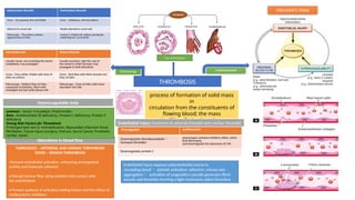
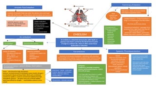
Thrombosis and Embolism
Dr. Dhanya Menon
Assistant Professor
Department of Pathology
THROMBOSIS
process of formation of solid mass
in
circulation from the constituents of
flowing blood; the mass
itself is called a thrombus.
VIRCHOW’S
TRIAD
ETIOPATHOGENESIS
VIRCHOW’S TRIAD
Endothelial injury (common in arterial thrombi and cardiac thrombi)
Procoagulant Antifibrinolytic
Downregulates thrombomodulin 
increases thrombin
plasminogen activator inhibitors (PAIs), which
limit fibrinolysis,
and downregulate the expression of t-PA
Downregulates protein C
Endothelial injury exposes subendothelial matrix to
circulating blood  platelet activation: adhesion, release and
aggregation activation of coagulation cascade generates fibrin
strands and thrombin forming a tight meshwork called thrombus.
TURBULENCE – ARTHERIAL AND CARDIAC THROMBOSIS
STASIS – VENOUS THROMBOSIS
-Promote endothelial activation- enhancing procoagulant
activity and leukocyte adhesion
• Disrupt laminar flow ,bring platelets into contact with
the endothelium
• Prevent washout of activated clotting factors and the inflow of
clotting factor inhibitors
Alterations in blood flow
common - Factor V mutation ,Prothrombin
Rare - Antithrombin III deficiency ,Protein C deficiency, Protein S
deficiency)
Strong Risk Factors for Thrombosis
Prolonged bed rest or immobilization, Myocardial infarction Atrial
fibrillation ,Tissue injury (surgery, fracture, burn) Cancer, Prosthetic
cardiac valves
Hypercoagulable state
Antemortem thrombi Postmortem thrombi
Gross - Dry, granular, firm and friable Gross - Gelatinous, soft and rubbery
Adherent to vessel wall Weakly attached to vessel wall
Microscopy - The surface contains
apparent lines of Zahn
surface is ‘chicken fat’ yellow covering the
underlying red ‘currant jelly’
Morphology
Arterial thrombi Venous thrombi
Usually mural, not occluding the lumen
completely, may propagate
Usually occlusive, take the cast of
the vessel in which formed, may
propagate in both directions
Gross - Grey-white, friable with lines of
Zahn on surface
Gross - Red-blue with fibrin strands and
lines of Zahn
Microscopy - Distinct lines of Zahn
composed of platelets, fibrin with
entangled red and white blood cells
Microscopy - Lines of Zahn with more
abundant red cells
Fate of thrombus
EMBOLISM
An embolus is a detached intravascular solid, liquid, or
gaseous mass that is carried by the blood from its point
of origin to a distant site, where it often causes tissue
dysfunction or infarction
Pulmonary Embolism
Most
common and
fatal form ,
origin - DVT
Risk factors -stasis of venous blood
and hypercoagulable states.
Saddle Embolus = If the thrombus is
large, it is impacted at the bifurcation
of
the main pulmonary artery
Paradoxical embolism - passage of an
embolus from right heart into the left
heart through atrial or
ventricular septal defect. pulmonary
emboli may reach systemic circulation.
Consequences of
Pulmonary embolism
- Sudden death
- Acute cor
pulmonale
- Pulmonary
infarction
- Pulmonary
hemorrhage
- resolution
Systemic Thromboembolism
ORIGIN
80% from intracardiac mural
Thrombi (left ventricular
wall infarcts ,left atrial
dilation and fibrillation)
- Aortic aneurysms,
atherosclerotic plaques, valvular
vegetations,
or venous thrombi (paradoxical
emboli)
In contrast to venous emboli,
arterial emboli can travel to a
wide variety of sites
Most come to rest in the lower
extremities (75%) or the brain
(10%), but other tissues,
including the intestines, kidneys,
spleen, and upper extremities,
may be involved.
presence of microscopic fat globules—sometimes with associated
hematopoietic bone marrow—in the vasculature after fractures of long
bones (90%) or, rarely, in the setting of soft-tissue trauma and burns.
Fat embolism
pulmonary
insufficiency, neurologic symptoms,
anemia, and thrombocytopenia - fatal in
5% to 15% of cases.
1 to 3 days after injury there is a sudden
onset of tachypnea, dyspnea, and
tachycardia; irritability; and restlessness that
can progress to delirium or coma.
Thrombocytopenia - A diffuse petechial rash
(20% to 50% of cases)
Air embolism
Gas embolism Decompression sickness
Venous air embolism
- Operations of head and
neck , trauma
- OG operations and
trauma
- IV infusion of blood and
fluid
Arterial air embolism
- Cardiothoracic surgery and
tyrauma
- Arteriographical
procedures
Decompression sickness / caisson’s disease, divers’ palsy or
aeroembolism
Divers - who descend to high atmospheric
pressure, increased amount of atmospheric gases (mainly nitrogen;)
are dissolved in blood and tissue fluids. When such an individual
ascends too rapidly i.e. comes to normal level suddenly from high
atmospheric pressure the gases come out as minute bubbles,
which have affinity for nitrogen  small bubbles coalesce to form
emboli
Acute form - bends – acute pain
in joints, ligaments and tendons,
chokes respiratory distress;
cerebral effects
Chronic form – ischemic necrosis
Amniotic Fluid Embolism
contents of amniotic fluid may enter the uterine veins and reach
right side of the heart – during labour and immediate post partum
epithelial squames, vernix
caseosa, lanugo hair,bile from
meconium, and mucus
Sudden respiratory
distress and dyspnoea
ii) Deep cyanosis
iii) Cardiovascular shock
iv) Convulsions
v) Coma
vi) Unexpected death

Thrombosis and embolism concept map.pptx

  • 1.
    Thrombosis and Embolism Dr.Dhanya Menon Assistant Professor Department of Pathology
  • 2.
    THROMBOSIS process of formationof solid mass in circulation from the constituents of flowing blood; the mass itself is called a thrombus. VIRCHOW’S TRIAD ETIOPATHOGENESIS VIRCHOW’S TRIAD Endothelial injury (common in arterial thrombi and cardiac thrombi) Procoagulant Antifibrinolytic Downregulates thrombomodulin  increases thrombin plasminogen activator inhibitors (PAIs), which limit fibrinolysis, and downregulate the expression of t-PA Downregulates protein C Endothelial injury exposes subendothelial matrix to circulating blood  platelet activation: adhesion, release and aggregation activation of coagulation cascade generates fibrin strands and thrombin forming a tight meshwork called thrombus. TURBULENCE – ARTHERIAL AND CARDIAC THROMBOSIS STASIS – VENOUS THROMBOSIS -Promote endothelial activation- enhancing procoagulant activity and leukocyte adhesion • Disrupt laminar flow ,bring platelets into contact with the endothelium • Prevent washout of activated clotting factors and the inflow of clotting factor inhibitors Alterations in blood flow common - Factor V mutation ,Prothrombin Rare - Antithrombin III deficiency ,Protein C deficiency, Protein S deficiency) Strong Risk Factors for Thrombosis Prolonged bed rest or immobilization, Myocardial infarction Atrial fibrillation ,Tissue injury (surgery, fracture, burn) Cancer, Prosthetic cardiac valves Hypercoagulable state Antemortem thrombi Postmortem thrombi Gross - Dry, granular, firm and friable Gross - Gelatinous, soft and rubbery Adherent to vessel wall Weakly attached to vessel wall Microscopy - The surface contains apparent lines of Zahn surface is ‘chicken fat’ yellow covering the underlying red ‘currant jelly’ Morphology Arterial thrombi Venous thrombi Usually mural, not occluding the lumen completely, may propagate Usually occlusive, take the cast of the vessel in which formed, may propagate in both directions Gross - Grey-white, friable with lines of Zahn on surface Gross - Red-blue with fibrin strands and lines of Zahn Microscopy - Distinct lines of Zahn composed of platelets, fibrin with entangled red and white blood cells Microscopy - Lines of Zahn with more abundant red cells Fate of thrombus
  • 3.
    EMBOLISM An embolus isa detached intravascular solid, liquid, or gaseous mass that is carried by the blood from its point of origin to a distant site, where it often causes tissue dysfunction or infarction Pulmonary Embolism Most common and fatal form , origin - DVT Risk factors -stasis of venous blood and hypercoagulable states. Saddle Embolus = If the thrombus is large, it is impacted at the bifurcation of the main pulmonary artery Paradoxical embolism - passage of an embolus from right heart into the left heart through atrial or ventricular septal defect. pulmonary emboli may reach systemic circulation. Consequences of Pulmonary embolism - Sudden death - Acute cor pulmonale - Pulmonary infarction - Pulmonary hemorrhage - resolution Systemic Thromboembolism ORIGIN 80% from intracardiac mural Thrombi (left ventricular wall infarcts ,left atrial dilation and fibrillation) - Aortic aneurysms, atherosclerotic plaques, valvular vegetations, or venous thrombi (paradoxical emboli) In contrast to venous emboli, arterial emboli can travel to a wide variety of sites Most come to rest in the lower extremities (75%) or the brain (10%), but other tissues, including the intestines, kidneys, spleen, and upper extremities, may be involved. presence of microscopic fat globules—sometimes with associated hematopoietic bone marrow—in the vasculature after fractures of long bones (90%) or, rarely, in the setting of soft-tissue trauma and burns. Fat embolism pulmonary insufficiency, neurologic symptoms, anemia, and thrombocytopenia - fatal in 5% to 15% of cases. 1 to 3 days after injury there is a sudden onset of tachypnea, dyspnea, and tachycardia; irritability; and restlessness that can progress to delirium or coma. Thrombocytopenia - A diffuse petechial rash (20% to 50% of cases) Air embolism Gas embolism Decompression sickness Venous air embolism - Operations of head and neck , trauma - OG operations and trauma - IV infusion of blood and fluid Arterial air embolism - Cardiothoracic surgery and tyrauma - Arteriographical procedures Decompression sickness / caisson’s disease, divers’ palsy or aeroembolism Divers - who descend to high atmospheric pressure, increased amount of atmospheric gases (mainly nitrogen;) are dissolved in blood and tissue fluids. When such an individual ascends too rapidly i.e. comes to normal level suddenly from high atmospheric pressure the gases come out as minute bubbles, which have affinity for nitrogen  small bubbles coalesce to form emboli Acute form - bends – acute pain in joints, ligaments and tendons, chokes respiratory distress; cerebral effects Chronic form – ischemic necrosis Amniotic Fluid Embolism contents of amniotic fluid may enter the uterine veins and reach right side of the heart – during labour and immediate post partum epithelial squames, vernix caseosa, lanugo hair,bile from meconium, and mucus Sudden respiratory distress and dyspnoea ii) Deep cyanosis iii) Cardiovascular shock iv) Convulsions v) Coma vi) Unexpected death