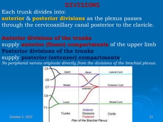
AXILLA
By
ABDULMUJEEB
October 2, 2023 1
Definition
• It is a pyramid shaped space between the
upper part of the arm and the side of the
chest
• Important Nerves, Blood and Lymph
vessels travel through it from root of the
neck to the upper limb
October 2, 2023 2
Axilla is an irregularly shaped pyramidal space with:
Four walls
An inlet (apex)
A floor (base)
October 2, 2023 3
Boundaries of
the Axilla
 Apex:
 Is directed upwards &
medially to the root of
the neck.
 It is called Axillary inlet/
Cervicoaxillary canal.
 It is bounded, by 3
bones:
• Clavicle anteriorly.
• Upper border of the
scapula posteriorly.
• Outer border of the
first rib medially.
C
L
A
V
I
C L E
1
R
I
B
October 2, 2023 4
Base
• Lower end or Base is bounded in front by
the anterior axillary fold formed by
pectoralis major muscle
• Behind by posterior axillary fold formed by
the tendons of latissimus dorsi and teres
major muscles
• Medially by the chest wall
October 2, 2023 5
Anterior wall
1. Pectoralis major
2. Pectoralis minor
3. Subclavius muscles
4. Clavipectoral fascia
October 2, 2023 6
Posterior wall: By the subscapularis, Latissimus
dorsi and teres major muscles
October 2, 2023 7
Walls of The Axilla
• Medial wall:
By the upper 4 or 5 ribs and intercostal
spaces covered by serratus anterior
muscle
• Lateral wall:
By the coracobrachialis and biceps
muscles in the bicipital groove of humerus
October 2, 2023 8
Base
The Base of axilla is formed by the skin
stretching between the anterior and
posterior walls
October 2, 2023 9
Clavipectoral Fascia
• It is a strong sheet of connective tissue
• Split above to enclose the subclavius
muscle and is attached to the clavicle
• Below it splits to enclose the pectoralis
minor muscle
• Then continues downward as the
suspensory ligament of the axilla
• Then joins the fascial floor of armpit
October 2, 2023 10
Contents of The
Axilla
 Cords and branches
of the brachial
plexus
 Axillary artery and
its branches.
 Axillary vein and its
tributaries.
 Axillary lymph nodes.
 Axillary lymphatic
vessels
 Axillary fat.
 Loose connective
tissue.
The neurovascular bundle is enclosed in connective tissue
sheath, called ‘axillary sheath’
Axillary a. & v.
Brachial
plexus
October 2, 2023 11
October 2, 2023 12
Axillary Artery
• Is a continuation of subclavian artery
• Begins at the lateral border of the 1st rib
• Ends at the lower border of teres major
• It continues as the brachial artery
• Closely related to brachial plexus cords
• Enclosed with them in the axillary sheath
• Axillary sheath is continuous with the
prevertebral fascia
• Pectoralis minor divides it into 3 parts
October 2, 2023 13
1st Part of Axillary Artery
• Extends from the lateral border of the 1st
rib to the upper border of pectoralis minor
October 2, 2023 14
October 2, 2023 15
Relation
• Anterior: Pectoralis major, covering fascia,
skin, cephalic vein
• Posterior: Long thoracic nerve
• Lateral: Three cords of brachial plexus
• Medial: Axillary vein
October 2, 2023 16
2nd Part of Axillary Artery
• Lies behind the pectoralis minor muscle
October 2, 2023 17
Relation
• Anterior: Pectoralis minor and major,
covering fascia and skin
• Posterior: Posterior cord of brachial plexus
• Lateral: Lateral cord of brachial plexus
• Medial: medial cord of brachial plexus and
axillary vein
October 2, 2023 18
3rd Part of Axillary Artery
• Extends from lower border of pectoralis
minor to the lower border of teres major
October 2, 2023 19
Relation
• Anterior: Pectoralis major, medial root of
the median nerve
• Posterior: subscapularis, latissimus dorsi
and teres major
• Lateral: Coracobrachialis, biceps, humerus
• Medial: Ulnar nerve, axillary vein, medial
cutaneous nerve of the arm
October 2, 2023 20
Branches
• Branches of axillary artery supply the
thoracic wall and the shoulder region
• 1st Part: Highest thoracic artery
• 2nd Part: Thoracoacromial and lateral
thoracic arteries
• 3rd Part: Subscapular artery, anterior and
posterior circumflex humeral arteries
October 2, 2023 21
October 2, 2023 22
The nerves entering the upper limb
provide the following important functions:
Sensory innervation to skin & deep structures (e.g. joints)
Motor innervation to the muscles
Influence over the diameters of the blood vessels by the
sympathetic vasomotor nerves
Sympathetic secretomotor supply to the sweat glands.
October 2, 2023 23
At the root of the neck, the nerves form
Brachial plexus
Nerve fibers derived from different segments of the spinal
cord arranged and distributed efficiently in various parts of
the upper limb.
October 2, 2023 24
Most nerves in the upper limb arise from the brachial
plexus, a major nerve network supplying the upper limb; it
begins in the neck and extends into the axilla.
Almost
all branches
of the brachial plexus
arise in the axilla
(after the plexus
has crossed the 1st rib).
October 2, 2023 25
The brachial plexus is formed by:
union of the anterior rami of the
last 4 cervical (C5-C8) & first thoracic (T1) nerves
which constitute the roots of the brachial plexus.
Originates in the neck, passes laterally and inferiorly over
rib I, and enters the axilla.
October 2, 2023 26
The parts of the brachial plexus, from medial to lateral, are
roots, trunks, divisions, and cords.
All major nerves that innervate the upper limb originate
from the brachial plexus, mostly from the cords.
October 2, 2023 27
ROOTS
Anterior rami of C5 to C8, and most of T1.
The roots & trunks enter the the neck by passing between
the anterior scalene and middle scalene muscles.
Close to their origin, the roots receive gray rami
communicantes from the sympathetic trunk.
These carry postganglionic sympathetic fibers onto the roots for
distribution to the periphery.
October 2, 2023 28
.
October 2, 2023 29
TRUNKS
In the inferior part of the neck, the roots of the brachial
plexus unite to form three trunks:
Superior trunk formed by union of C5 & C6 roots
Middle trunk a continuation of C7 root
Inferior trunk formed by the union of C8 & T1 roots
The inferior trunk lies on rib I posterior to the subclavian artery; the middle and
superior trunks are more superior in position.
October 2, 2023 30
DIVISIONS
Each trunk divides into:
anterior & posterior divisions as the plexus passes
through the cervicoaxillary canal posterior to the clavicle.
Anterior divisions of the trunks
supply anterior (flexor) compartments of the upper limb
Posterior divisions of the trunks
supply posterior (extensor) compartments.
No peripheral nerves originate directly from the divisions of the brachial plexus.
October 2, 2023 31
October 2, 2023 32
CORDS
The three cords of the brachial plexus originate from the
divisions and are related to the second part of the
axillary artery.
October 2, 2023 34
Proximal posterior to the subclavian artery in the neck
More distal regions surround the axillary artery
The roots of the
plexus usually
pass through
the gap between
anterior &
middle scalene
muscles.
October 2, 2023 35
Lateral cord
Union of anterior divisions of upper & middle trunks
(C5-C7)
Medial cord
Continuation of anterior division of inferior trunk
(C8-T1)
Posterior cord
Union of all three posterior divisions
(C5 to T1)
October 2, 2023 36
Branches
Branches of the roots
Dorsal scapular nerve (C5 root of the brachial plexus)
innervates rhomboid major and minor muscles
Long thoracic nerve (anterior rami of C5 to C7)
innervates serratus anterior muscle
October 2, 2023 37
Branches of the trunks
The only branches from the trunks of the brachial plexus
are 2 nerves ;originate from the superior trunk.
Suprascapular nerve (C5 & C6)
innervates supraspinatus &
infraspinatus muscles
The nerve to subclavius muscle (C5 & C6)
innervates subclavius muscle
October 2, 2023 38
October 2, 2023 39
Branches of the lateral cord
Lateral pectoral nerve pectoralis major muscle
October 2, 2023 40
October 2, 2023 41
Branches of the lateral
cord
Musculocutaneous nerve
Coracobrachialis
Biceps brachii
Brachialis
All 3 flexor muscles @ anterior compartment of the arm
Terminates as lateral cutaneous nerve of forearm.
October 2, 2023 42
Branches of the lateral cord
Lateral root of median nerve
 Largest terminal branch of the lateral cord
 Passes medially to join a similar branch from the
medial cord to form the median nerve.
1. Lateral cord
2. Posterior cord
3. Medial cord
4. Lateral root of median nerve
5. Medial root of median nerve
6. Radial nerve
7. Ulnar nerve
8. Median nerve
October 2, 2023 43
October 2, 2023 44
October 2, 2023 45
Branches of the medial cord
1) Medial pectoral nerve
pectoralis major & minor
2) Medial cutaneous nerve of arm
(medial brachial cutaneous nerve)
medial 1/3 of distal arm
3) Medial cutaneous nerve of forearm
(medial antebrachial cutaneous nerve)
medial surface of the forearm down to the wrist
4) Ulnar nerve
5) Median nerve
October 2, 2023 46
Flexor carpi ulnaris muscle & medial half of the flexor
digitorum profundus muscle
All intrinsic muscles of the hand (except for the three
thenar muscles and the two lateral lumbrical muscles)
Skin over the palmar surface of the little finger, medial
half of the ring finger, and associated palm and wrist,
and the skin over the dorsal surface of the medial part
of the hand
October 2, 2023 47
October 2, 2023 48
Most of the muscles in the anterior compartment of the
forearm
(except for the flexor carpi ulnaris muscle and the medial half of the
flexor digitorum profundus muscle)
In the hand
Three thenar muscles associated with the thumb
Two lateral lumbrical muscles associated with movement
of the index and middle fingers
Skin over the palmar surface of the lateral three and one-
half digits and over the lateral side of the palm and
middle of the wrist.
October 2, 2023 49
October 2, 2023 50
Musculocutaneous nerve, lateral
root of the median nerve,
median nerve, medial root of the
median nerve, ulnar nerve form
an M over the third part of the
axillary artery.
This feature, together with penetration of the
coracobrachialis muscle by the musculocutaneous
nerve, can be used to identify components of the
brachial plexus in the axilla
October 2, 2023 51
Branches of the posterior cord
1) Superior subscapular nerve subscapularis muscle
2) Thoracodorsal nerve latissimus dorsi
3) Inferior subscapular nerve subscapularis & teres major
4) Axillary nerve deltoid and teres minor
5) Radial nerve
All these nerves except the radial nerve innervate muscles
associated with the posterior wall of the axilla; the radial
nerve passes into the arm and forearm.
October 2, 2023 52
October 2, 2023 53
October 2, 2023 54
All muscles
in the posterior compartments of the arm & forearm
Skin on the
Posterior aspect of the arm and forearm
Lower lateral surface of the arm
Dorsal lateral surface of the hand
October 2, 2023 55
October 2, 2023 56

AXILLA 2022.ppt

  • 1.
  • 2.
    Definition • It isa pyramid shaped space between the upper part of the arm and the side of the chest • Important Nerves, Blood and Lymph vessels travel through it from root of the neck to the upper limb October 2, 2023 2
  • 3.
    Axilla is anirregularly shaped pyramidal space with: Four walls An inlet (apex) A floor (base) October 2, 2023 3
  • 4.
    Boundaries of the Axilla Apex:  Is directed upwards & medially to the root of the neck.  It is called Axillary inlet/ Cervicoaxillary canal.  It is bounded, by 3 bones: • Clavicle anteriorly. • Upper border of the scapula posteriorly. • Outer border of the first rib medially. C L A V I C L E 1 R I B October 2, 2023 4
  • 5.
    Base • Lower endor Base is bounded in front by the anterior axillary fold formed by pectoralis major muscle • Behind by posterior axillary fold formed by the tendons of latissimus dorsi and teres major muscles • Medially by the chest wall October 2, 2023 5
  • 6.
    Anterior wall 1. Pectoralismajor 2. Pectoralis minor 3. Subclavius muscles 4. Clavipectoral fascia October 2, 2023 6
  • 7.
    Posterior wall: Bythe subscapularis, Latissimus dorsi and teres major muscles October 2, 2023 7
  • 8.
    Walls of TheAxilla • Medial wall: By the upper 4 or 5 ribs and intercostal spaces covered by serratus anterior muscle • Lateral wall: By the coracobrachialis and biceps muscles in the bicipital groove of humerus October 2, 2023 8
  • 9.
    Base The Base ofaxilla is formed by the skin stretching between the anterior and posterior walls October 2, 2023 9
  • 10.
    Clavipectoral Fascia • Itis a strong sheet of connective tissue • Split above to enclose the subclavius muscle and is attached to the clavicle • Below it splits to enclose the pectoralis minor muscle • Then continues downward as the suspensory ligament of the axilla • Then joins the fascial floor of armpit October 2, 2023 10
  • 11.
    Contents of The Axilla Cords and branches of the brachial plexus  Axillary artery and its branches.  Axillary vein and its tributaries.  Axillary lymph nodes.  Axillary lymphatic vessels  Axillary fat.  Loose connective tissue. The neurovascular bundle is enclosed in connective tissue sheath, called ‘axillary sheath’ Axillary a. & v. Brachial plexus October 2, 2023 11
  • 12.
  • 13.
    Axillary Artery • Isa continuation of subclavian artery • Begins at the lateral border of the 1st rib • Ends at the lower border of teres major • It continues as the brachial artery • Closely related to brachial plexus cords • Enclosed with them in the axillary sheath • Axillary sheath is continuous with the prevertebral fascia • Pectoralis minor divides it into 3 parts October 2, 2023 13
  • 14.
    1st Part ofAxillary Artery • Extends from the lateral border of the 1st rib to the upper border of pectoralis minor October 2, 2023 14
  • 15.
  • 16.
    Relation • Anterior: Pectoralismajor, covering fascia, skin, cephalic vein • Posterior: Long thoracic nerve • Lateral: Three cords of brachial plexus • Medial: Axillary vein October 2, 2023 16
  • 17.
    2nd Part ofAxillary Artery • Lies behind the pectoralis minor muscle October 2, 2023 17
  • 18.
    Relation • Anterior: Pectoralisminor and major, covering fascia and skin • Posterior: Posterior cord of brachial plexus • Lateral: Lateral cord of brachial plexus • Medial: medial cord of brachial plexus and axillary vein October 2, 2023 18
  • 19.
    3rd Part ofAxillary Artery • Extends from lower border of pectoralis minor to the lower border of teres major October 2, 2023 19
  • 20.
    Relation • Anterior: Pectoralismajor, medial root of the median nerve • Posterior: subscapularis, latissimus dorsi and teres major • Lateral: Coracobrachialis, biceps, humerus • Medial: Ulnar nerve, axillary vein, medial cutaneous nerve of the arm October 2, 2023 20
  • 21.
    Branches • Branches ofaxillary artery supply the thoracic wall and the shoulder region • 1st Part: Highest thoracic artery • 2nd Part: Thoracoacromial and lateral thoracic arteries • 3rd Part: Subscapular artery, anterior and posterior circumflex humeral arteries October 2, 2023 21
  • 22.
  • 23.
    The nerves enteringthe upper limb provide the following important functions: Sensory innervation to skin & deep structures (e.g. joints) Motor innervation to the muscles Influence over the diameters of the blood vessels by the sympathetic vasomotor nerves Sympathetic secretomotor supply to the sweat glands. October 2, 2023 23
  • 24.
    At the rootof the neck, the nerves form Brachial plexus Nerve fibers derived from different segments of the spinal cord arranged and distributed efficiently in various parts of the upper limb. October 2, 2023 24
  • 25.
    Most nerves inthe upper limb arise from the brachial plexus, a major nerve network supplying the upper limb; it begins in the neck and extends into the axilla. Almost all branches of the brachial plexus arise in the axilla (after the plexus has crossed the 1st rib). October 2, 2023 25
  • 26.
    The brachial plexusis formed by: union of the anterior rami of the last 4 cervical (C5-C8) & first thoracic (T1) nerves which constitute the roots of the brachial plexus. Originates in the neck, passes laterally and inferiorly over rib I, and enters the axilla. October 2, 2023 26
  • 27.
    The parts ofthe brachial plexus, from medial to lateral, are roots, trunks, divisions, and cords. All major nerves that innervate the upper limb originate from the brachial plexus, mostly from the cords. October 2, 2023 27
  • 28.
    ROOTS Anterior rami ofC5 to C8, and most of T1. The roots & trunks enter the the neck by passing between the anterior scalene and middle scalene muscles. Close to their origin, the roots receive gray rami communicantes from the sympathetic trunk. These carry postganglionic sympathetic fibers onto the roots for distribution to the periphery. October 2, 2023 28
  • 29.
  • 30.
    TRUNKS In the inferiorpart of the neck, the roots of the brachial plexus unite to form three trunks: Superior trunk formed by union of C5 & C6 roots Middle trunk a continuation of C7 root Inferior trunk formed by the union of C8 & T1 roots The inferior trunk lies on rib I posterior to the subclavian artery; the middle and superior trunks are more superior in position. October 2, 2023 30
  • 31.
    DIVISIONS Each trunk dividesinto: anterior & posterior divisions as the plexus passes through the cervicoaxillary canal posterior to the clavicle. Anterior divisions of the trunks supply anterior (flexor) compartments of the upper limb Posterior divisions of the trunks supply posterior (extensor) compartments. No peripheral nerves originate directly from the divisions of the brachial plexus. October 2, 2023 31
  • 32.
  • 34.
    CORDS The three cordsof the brachial plexus originate from the divisions and are related to the second part of the axillary artery. October 2, 2023 34
  • 35.
    Proximal posterior tothe subclavian artery in the neck More distal regions surround the axillary artery The roots of the plexus usually pass through the gap between anterior & middle scalene muscles. October 2, 2023 35
  • 36.
    Lateral cord Union ofanterior divisions of upper & middle trunks (C5-C7) Medial cord Continuation of anterior division of inferior trunk (C8-T1) Posterior cord Union of all three posterior divisions (C5 to T1) October 2, 2023 36
  • 37.
    Branches Branches of theroots Dorsal scapular nerve (C5 root of the brachial plexus) innervates rhomboid major and minor muscles Long thoracic nerve (anterior rami of C5 to C7) innervates serratus anterior muscle October 2, 2023 37
  • 38.
    Branches of thetrunks The only branches from the trunks of the brachial plexus are 2 nerves ;originate from the superior trunk. Suprascapular nerve (C5 & C6) innervates supraspinatus & infraspinatus muscles The nerve to subclavius muscle (C5 & C6) innervates subclavius muscle October 2, 2023 38
  • 39.
  • 40.
    Branches of thelateral cord Lateral pectoral nerve pectoralis major muscle October 2, 2023 40
  • 41.
  • 42.
    Branches of thelateral cord Musculocutaneous nerve Coracobrachialis Biceps brachii Brachialis All 3 flexor muscles @ anterior compartment of the arm Terminates as lateral cutaneous nerve of forearm. October 2, 2023 42
  • 43.
    Branches of thelateral cord Lateral root of median nerve  Largest terminal branch of the lateral cord  Passes medially to join a similar branch from the medial cord to form the median nerve. 1. Lateral cord 2. Posterior cord 3. Medial cord 4. Lateral root of median nerve 5. Medial root of median nerve 6. Radial nerve 7. Ulnar nerve 8. Median nerve October 2, 2023 43
  • 44.
  • 45.
  • 46.
    Branches of themedial cord 1) Medial pectoral nerve pectoralis major & minor 2) Medial cutaneous nerve of arm (medial brachial cutaneous nerve) medial 1/3 of distal arm 3) Medial cutaneous nerve of forearm (medial antebrachial cutaneous nerve) medial surface of the forearm down to the wrist 4) Ulnar nerve 5) Median nerve October 2, 2023 46
  • 47.
    Flexor carpi ulnarismuscle & medial half of the flexor digitorum profundus muscle All intrinsic muscles of the hand (except for the three thenar muscles and the two lateral lumbrical muscles) Skin over the palmar surface of the little finger, medial half of the ring finger, and associated palm and wrist, and the skin over the dorsal surface of the medial part of the hand October 2, 2023 47
  • 48.
  • 49.
    Most of themuscles in the anterior compartment of the forearm (except for the flexor carpi ulnaris muscle and the medial half of the flexor digitorum profundus muscle) In the hand Three thenar muscles associated with the thumb Two lateral lumbrical muscles associated with movement of the index and middle fingers Skin over the palmar surface of the lateral three and one- half digits and over the lateral side of the palm and middle of the wrist. October 2, 2023 49
  • 50.
  • 51.
    Musculocutaneous nerve, lateral rootof the median nerve, median nerve, medial root of the median nerve, ulnar nerve form an M over the third part of the axillary artery. This feature, together with penetration of the coracobrachialis muscle by the musculocutaneous nerve, can be used to identify components of the brachial plexus in the axilla October 2, 2023 51
  • 52.
    Branches of theposterior cord 1) Superior subscapular nerve subscapularis muscle 2) Thoracodorsal nerve latissimus dorsi 3) Inferior subscapular nerve subscapularis & teres major 4) Axillary nerve deltoid and teres minor 5) Radial nerve All these nerves except the radial nerve innervate muscles associated with the posterior wall of the axilla; the radial nerve passes into the arm and forearm. October 2, 2023 52
  • 53.
  • 54.
  • 55.
    All muscles in theposterior compartments of the arm & forearm Skin on the Posterior aspect of the arm and forearm Lower lateral surface of the arm Dorsal lateral surface of the hand October 2, 2023 55
  • 56.