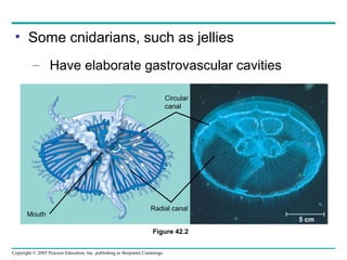
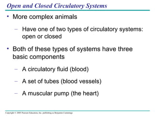
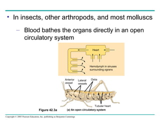
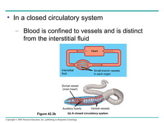
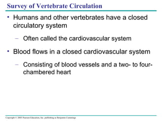
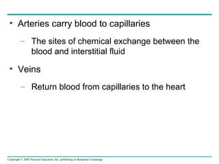
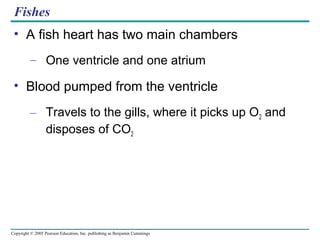
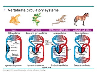
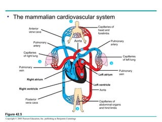
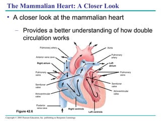
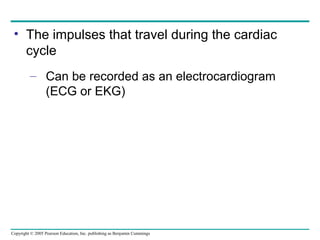
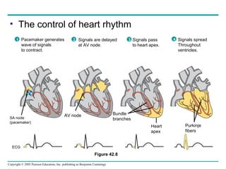
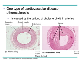
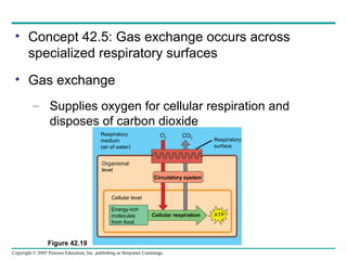
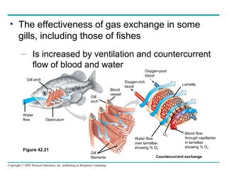
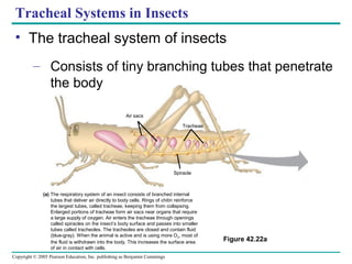
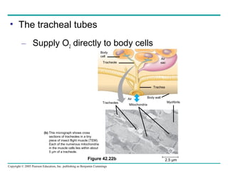
Copyright © 2005 Pearson Education, Inc. publishing as Benjamin Cummings
PowerPoint Lectures for
Biology, Seventh Edition
Neil Campbell and Jane Reece
Lectures by Chris Romero
Chapter 42
Circulation and Gas
Exchange
Copyright © 2005 Pearson Education, Inc. publishing as Benjamin Cummings
• Overview: Trading with the Environment
• Every organism must exchange materials with
its environment
– And this exchange ultimately occurs at the
cellular level
Copyright © 2005 Pearson Education, Inc. publishing as Benjamin Cummings
• In unicellular organisms
– These exchanges occur directly with the
environment
• For most of the cells making up multicellular
organisms
– Direct exchange with the environment is not
possible
Copyright © 2005 Pearson Education, Inc. publishing as Benjamin Cummings
• The feathery gills projecting from a salmon
– Are an example of a specialized exchange
system found in animals
Figure 42.1
Copyright © 2005 Pearson Education, Inc. publishing as Benjamin Cummings
• Concept 42.1: Circulatory systems reflect
phylogeny
• Transport systems
– Functionally connect the organs of exchange
with the body cells
Copyright © 2005 Pearson Education, Inc. publishing as Benjamin Cummings
• Most complex animals have internal transport
systems
– That circulate fluid, providing a lifeline between
the aqueous environment of living cells and the
exchange organs, such as lungs, that exchange
chemicals with the outside environment
Copyright © 2005 Pearson Education, Inc. publishing as Benjamin Cummings
Invertebrate Circulation
• The wide range of invertebrate body size and
form
– Is paralleled by a great diversity in circulatory
systems
Copyright © 2005 Pearson Education, Inc. publishing as Benjamin Cummings
Gastrovascular Cavities
• Simple animals, such as cnidarians
– Have a body wall only two cells thick that
encloses a gastrovascular cavity
• The gastrovascular cavity
– Functions in both digestion and distribution of
substances throughout the body
Copyright © 2005 Pearson Education, Inc. publishing as Benjamin Cummings
• Some cnidarians, such as jellies
– Have elaborate gastrovascular cavities
Figure 42.2
Circular
canal
Radial canal
5 cm
Mouth
Copyright © 2005 Pearson Education, Inc. publishing as Benjamin Cummings
Open and Closed Circulatory Systems
• More complex animals
– Have one of two types of circulatory systems:
open or closed
• Both of these types of systems have three
basic components
– A circulatory fluid (blood)
– A set of tubes (blood vessels)
– A muscular pump (the heart)
Copyright © 2005 Pearson Education, Inc. publishing as Benjamin Cummings
• In insects, other arthropods, and most molluscs
– Blood bathes the organs directly in an open
circulatory system
Heart
Hemolymph in sinuses
surrounding ograns
Anterior
vessel
Tubular heart
Lateral
vessels
Ostia
(a) An open circulatory systemFigure 42.3a
Copyright © 2005 Pearson Education, Inc. publishing as Benjamin Cummings
• In a closed circulatory system
– Blood is confined to vessels and is distinct
from the interstitial fluid
Figure 42.3b
Interstitial
fluid
Heart
Small branch vessels
in each organ
Dorsal vessel
(main heart)
Ventral vesselsAuxiliary hearts
(b) A closed circulatory system
Copyright © 2005 Pearson Education, Inc. publishing as Benjamin Cummings
• Closed systems
– Are more efficient at transporting circulatory
fluids to tissues and cells
Copyright © 2005 Pearson Education, Inc. publishing as Benjamin Cummings
Survey of Vertebrate Circulation
• Humans and other vertebrates have a closed
circulatory system
– Often called the cardiovascular system
• Blood flows in a closed cardiovascular system
– Consisting of blood vessels and a two- to four-
chambered heart
Copyright © 2005 Pearson Education, Inc. publishing as Benjamin Cummings
• Arteries carry blood to capillaries
– The sites of chemical exchange between the
blood and interstitial fluid
• Veins
– Return blood from capillaries to the heart
Copyright © 2005 Pearson Education, Inc. publishing as Benjamin Cummings
Fishes
• A fish heart has two main chambers
– One ventricle and one atrium
• Blood pumped from the ventricle
– Travels to the gills, where it picks up O2 and
disposes of CO2
Copyright © 2005 Pearson Education, Inc. publishing as Benjamin Cummings
Amphibians
• Frogs and other amphibians
– Have a three-chambered heart, with two atria
and one ventricle
• The ventricle pumps blood into a forked artery
– That splits the ventricle’s output into the
pulmocutaneous circuit and the systemic
circuit
Copyright © 2005 Pearson Education, Inc. publishing as Benjamin Cummings
Reptiles (Except Birds)
• Reptiles have double circulation
– With a pulmonary circuit (lungs) and a
systemic circuit
• Turtles, snakes, and lizards
– Have a three-chambered heart
Copyright © 2005 Pearson Education, Inc. publishing as Benjamin Cummings
Mammals and Birds
• In all mammals and birds
– The ventricle is completely divided into
separate right and left chambers
• The left side of the heart pumps and receives
only oxygen-rich blood
– While the right side receives and pumps only
oxygen-poor blood
Copyright © 2005 Pearson Education, Inc. publishing as Benjamin Cummings
• A powerful four-chambered heart
– Was an essential adaptation of the
endothermic way of life characteristic of
mammals and birds
Copyright © 2005 Pearson Education, Inc. publishing as Benjamin Cummings
FISHES AMPHIBIANS REPTILES (EXCEPT BIRDS) MAMMALS AND BIRDS
Systemic capillaries Systemic capillaries Systemic capillaries Systemic capillaries
Lung capillaries Lung capillariesLung and skin capillariesGill capillaries
Right Left Right Left Right Left
Systemic
circuit
Systemic
circuit
Pulmocutaneous
circuit
Pulmonary
circuit
Pulmonary
circuit
Systemic
circulationVein
Atrium (A)
Heart:
ventricle (V)
Artery Gill
circulation
A
V VV VV
A A A AA
Left
Systemic
aorta
Right
systemic
aorta
Figure 42.4
• Vertebrate circulatory systems
Copyright © 2005 Pearson Education, Inc. publishing as Benjamin Cummings
• Concept 42.2: Double circulation in mammals
depends on the anatomy and pumping cycle of
the heart
• The structure and function of the human
circulatory system
– Can serve as a model for exploring
mammalian circulation in general
Copyright © 2005 Pearson Education, Inc. publishing as Benjamin Cummings
Mammalian Circulation: The Pathway
• Heart valves
– Dictate a one-way flow of blood through the
heart
Copyright © 2005 Pearson Education, Inc. publishing as Benjamin Cummings
• Blood begins its flow
– With the right ventricle pumping blood to the
lungs
• In the lungs
– The blood loads O2 and unloads CO2
Copyright © 2005 Pearson Education, Inc. publishing as Benjamin Cummings
• Oxygen-rich blood from the lungs
– Enters the heart at the left atrium and is
pumped to the body tissues by the left ventricle
• Blood returns to the heart
– Through the right atrium
Copyright © 2005 Pearson Education, Inc. publishing as Benjamin Cummings
• The mammalian cardiovascular system
Pulmonary
vein
Right atrium
Right ventricle
Posterior
vena cava Capillaries of
abdominal organs
and hind limbs
Aorta
Left ventricle
Left atrium
Pulmonary
vein
Pulmonary
artery
Capillaries
of left lung
Capillaries of
head and
forelimbs
Anterior
vena cava
Pulmonary
artery
Capillaries
of right lung
Aorta
Figure 42.5
1
10
11
5
4
6
2
9
3
3
7
8
Copyright © 2005 Pearson Education, Inc. publishing as Benjamin Cummings
The Mammalian Heart: A Closer Look
• A closer look at the mammalian heart
– Provides a better understanding of how double
circulation works
Figure 42.6
Aorta
Pulmonary
veins
Semilunar
valve
Atrioventricular
valve
Left ventricle
Right ventricle
Anterior vena cava
Pulmonary artery
Semilunar
valve
Atrioventricular
valve
Posterior
vena cava
Pulmonary
veins
Right atrium
Pulmonary
artery
Left
atrium
Copyright © 2005 Pearson Education, Inc. publishing as Benjamin Cummings
• The heart contracts and relaxes
– In a rhythmic cycle called the cardiac cycle
• The contraction, or pumping, phase of the
cycle
– Is called systole
• The relaxation, or filling, phase of the cycle
– Is called diastole
Copyright © 2005 Pearson Education, Inc. publishing as Benjamin Cummings
• The cardiac cycle
Figure 42.7
Semilunar
valves
closed
AV valves
open
AV valves
closed
Semilunar
valves
open
Atrial and
ventricular
diastole
1
Atrial systole;
ventricular
diastole
2
Ventricular systole;
atrial diastole
3
0.1 sec
0.3 sec
0.4 sec
Copyright © 2005 Pearson Education, Inc. publishing as Benjamin Cummings
• The heart rate, also called the pulse
– Is the number of beats per minute
• The cardiac output
– Is the volume of blood pumped into the
systemic circulation per minute
Copyright © 2005 Pearson Education, Inc. publishing as Benjamin Cummings
Maintaining the Heart’s Rhythmic Beat
• Some cardiac muscle cells are self-excitable
– Meaning they contract without any signal from
the nervous system
Copyright © 2005 Pearson Education, Inc. publishing as Benjamin Cummings
• A region of the heart called the sinoatrial (SA)
node, or pacemaker
– Sets the rate and timing at which all cardiac
muscle cells contract
• Impulses from the SA node
– Travel to the atrioventricular (AV) node
• At the AV node, the impulses are delayed
– And then travel to the Purkinje fibers that make
the ventricles contract
Copyright © 2005 Pearson Education, Inc. publishing as Benjamin Cummings
• The impulses that travel during the cardiac
cycle
– Can be recorded as an electrocardiogram
(ECG or EKG)
Copyright © 2005 Pearson Education, Inc. publishing as Benjamin Cummings
• The control of heart rhythm
Figure 42.8
SA node
(pacemaker)
AV node Bundle
branches
Heart
apex
Purkinje
fibers
2 Signals are delayed
at AV node.
1 Pacemaker generates
wave of signals
to contract.
3 Signals pass
to heart apex.
4 Signals spread
Throughout
ventricles.
ECG
Copyright © 2005 Pearson Education, Inc. publishing as Benjamin Cummings
• The pacemaker is influenced by
– Nerves, hormones, body temperature, and
exercise
Copyright © 2005 Pearson Education, Inc. publishing as Benjamin Cummings
• Concept 42.3: Physical principles govern blood
circulation
• The same physical principles that govern the
movement of water in plumbing systems
– Also influence the functioning of animal
circulatory systems
Copyright © 2005 Pearson Education, Inc. publishing as Benjamin Cummings
Blood Vessel Structure and Function
• The “infrastructure” of the circulatory system
– Is its network of blood vessels
Copyright © 2005 Pearson Education, Inc. publishing as Benjamin Cummings
• All blood vessels
– Are built of similar tissues
– Have three similar layers
Figure 42.9
Artery Vein
100 µm
Artery Vein
Arteriole
Venule
Connective
tissue
Smooth
muscle
Endothelium
Connective
tissue
Smooth
muscle
Endothelium
Valve
Endothelium
Basement
membrane
Capillary
Copyright © 2005 Pearson Education, Inc. publishing as Benjamin Cummings
• Structural differences in arteries, veins, and
capillaries
– Correlate with their different functions
• Arteries have thicker walls
– To accommodate the high pressure of blood
pumped from the heart
Copyright © 2005 Pearson Education, Inc. publishing as Benjamin Cummings
• In the thinner-walled veins
– Blood flows back to the heart mainly as a
result of muscle action
Figure 42.10
Direction of blood flow
in vein (toward heart)
Valve (open)
Skeletal muscle
Valve (closed)
Copyright © 2005 Pearson Education, Inc. publishing as Benjamin Cummings
Blood Flow Velocity
• Physical laws governing the movement of fluids
through pipes
– Influence blood flow and blood pressure
Copyright © 2005 Pearson Education, Inc. publishing as Benjamin Cummings
• The velocity of blood flow varies in the
circulatory system
– And is slowest in the capillary beds as a result of the
high resistance and large total cross-sectional area
Figure 42.11
5,000
4,000
3,000
2,000
1,000
0
Aorta
Arteries
Arterioles
Capillaries
Venules
Veins
Venaecavae
Pressure(mmHg)Velocity(cm/sec)Area(cm2
)
Systolic
pressure
Diastolic
pressure
50
40
30
20
10
0
120
100
80
60
40
20
0
Copyright © 2005 Pearson Education, Inc. publishing as Benjamin Cummings
Blood Pressure
• Blood pressure
– Is the hydrostatic pressure that blood exerts
against the wall of a vessel
Copyright © 2005 Pearson Education, Inc. publishing as Benjamin Cummings
• Systolic pressure
– Is the pressure in the arteries during
ventricular systole
– Is the highest pressure in the arteries
• Diastolic pressure
– Is the pressure in the arteries during diastole
– Is lower than systolic pressure
Copyright © 2005 Pearson Education, Inc. publishing as Benjamin Cummings
• Blood pressure
– Can be easily measured in humans
Figure 42.12
Artery
Rubber cuff
inflated
with air
Artery
closed
120 120
Pressure
in cuff
above 120
Pressure
in cuff
below 120
Pressure
in cuff
below 70
Sounds
audible in
stethoscope
Sounds
stop
Blood pressure
reading: 120/70
A typical blood pressure reading for a 20-year-old
is 120/70. The units for these numbers are mm of
mercury (Hg); a blood pressure of 120 is a force that
can support a column of mercury 120 mm high.
1
A sphygmomanometer, an inflatable cuff attached to a
pressure gauge, measures blood pressure in an artery.
The cuff is wrapped around the upper arm and inflated
until the pressure closes the artery, so that no blood
flows past the cuff. When this occurs, the pressure
exerted by the cuff exceeds the pressure in the artery.
2 A stethoscope is used to listen for sounds of blood flow
below the cuff. If the artery is closed, there is no pulse
below the cuff. The cuff is gradually deflated until blood
begins to flow into the forearm, and sounds from blood
pulsing into the artery below the cuff can be heard with
the stethoscope. This occurs when the blood pressure
is greater than the pressure exerted by the cuff. The
pressure at this point is the systolic pressure.
3
The cuff is loosened further until the blood flows freely
through the artery and the sounds below the cuff
disappear. The pressure at this point is the diastolic
pressure remaining in the artery when the heart is relaxed.
4
70
Copyright © 2005 Pearson Education, Inc. publishing as Benjamin Cummings
• Blood pressure is determined partly by cardiac
output
– And partly by peripheral resistance due to
variable constriction of the arterioles
Copyright © 2005 Pearson Education, Inc. publishing as Benjamin Cummings
Capillary Function
• Capillaries in major organs are usually filled to
capacity
– But in many other sites, the blood supply
varies
Copyright © 2005 Pearson Education, Inc. publishing as Benjamin Cummings
• Two mechanisms
– Regulate the distribution of blood in capillary
beds
• In one mechanism
– Contraction of the smooth muscle layer in the
wall of an arteriole constricts the vessel
Copyright © 2005 Pearson Education, Inc. publishing as Benjamin Cummings
• In a second mechanism
– Precapillary sphincters control the flow of
blood between arterioles and venules
Figure 42.13 a–c
Precapillary sphincters Thoroughfare
channel
Arteriole
Capillaries
Venule
(a) Sphincters relaxed
(b) Sphincters contracted
VenuleArteriole
(c) Capillaries and larger vessels (SEM)
20 µm
Copyright © 2005 Pearson Education, Inc. publishing as Benjamin Cummings
• The critical exchange of substances between
the blood and interstitial fluid
– Takes place across the thin endothelial walls
of the capillaries
Copyright © 2005 Pearson Education, Inc. publishing as Benjamin Cummings
• The difference between blood pressure and
osmotic pressure
– Drives fluids out of capillaries at the arteriole
end and into capillaries at the venule end
At the arterial end of a
capillary, blood pressure is
greater than osmotic pressure,
and fluid flows out of the
capillary into the interstitial fluid.
Capillary Red
blood
cell
15 µm
Tissue cell INTERSTITIAL FLUID
Capillary
Net fluid
movement out
Net fluid
movement in
Direction of
blood flow
Blood pressure
Osmotic pressure
Inward flow
Outward flow
Pressure
Arterial end of capillary Venule end
At the venule end of a capillary,
blood pressure is less than
osmotic pressure, and fluid flows
from the interstitial fluid into the
capillary.
Figure 42.14
Copyright © 2005 Pearson Education, Inc. publishing as Benjamin Cummings
Fluid Return by the Lymphatic System
• The lymphatic system
– Returns fluid to the body from the capillary
beds
– Aids in body defense
Copyright © 2005 Pearson Education, Inc. publishing as Benjamin Cummings
• Fluid reenters the circulation
– Directly at the venous end of the capillary bed
and indirectly through the lymphatic system
Copyright © 2005 Pearson Education, Inc. publishing as Benjamin Cummings
• Concept 42.4: Blood is a connective tissue with
cells suspended in plasma
• Blood in the circulatory systems of vertebrates
– Is a specialized connective tissue
Copyright © 2005 Pearson Education, Inc. publishing as Benjamin Cummings
Blood Composition and Function
• Blood consists of several kinds of cells
– Suspended in a liquid matrix called plasma
• The cellular elements
– Occupy about 45% of the volume of blood
Copyright © 2005 Pearson Education, Inc. publishing as Benjamin Cummings
Plasma
• Blood plasma is about 90% water
• Among its many solutes are
– Inorganic salts in the form of dissolved ions,
sometimes referred to as electrolytes
Copyright © 2005 Pearson Education, Inc. publishing as Benjamin Cummings
• The composition of mammalian plasma
Plasma 55%
Constituent Major functions
Water Solvent for
carrying other
substances
Sodium
Potassium
Calcium
Magnesium
Chloride
Bicarbonat
e
Osmotic balance
pH buffering, and
regulation of
membrane
permeability
Albumin
Fibringen
Immunoglobulins
(antibodies)
Plasma proteins
Icons (blood electrolytes
Osmotic balance,
pH buffering
Substances transported by blood
Nutrients (such as glucose, fatty acids, vitamins)
Waste products of metabolism
Respiratory gases (O2 and CO2)
Hormones
Defense
Figure 42.15
Separated
blood
elements
Clotting
Copyright © 2005 Pearson Education, Inc. publishing as Benjamin Cummings
• Another important class of solutes is the
plasma proteins
– Which influence blood pH, osmotic pressure,
and viscosity
• Various types of plasma proteins
– Function in lipid transport, immunity, and blood
clotting
Copyright © 2005 Pearson Education, Inc. publishing as Benjamin Cummings
Cellular Elements
• Suspended in blood plasma are two classes of
cells
– Red blood cells, which transport oxygen
– White blood cells, which function in defense
• A third cellular element, platelets
– Are fragments of cells that are involved in
clotting
Copyright © 2005 Pearson Education, Inc. publishing as Benjamin Cummings
Figure 42.15
Cellular elements 45%
Cell type Number
per µL (mm3
) of blood
Functions
Erythrocytes
(red blood cells) 5–6 million Transport oxygen
and help transport
carbon dioxide
Leukocytes
(white blood cells)
5,000–10,000 Defense and
immunity
Eosinophil
Basophil
Platelets
Neutrophil
Monocyte
Lymphocyte
250,000−
400,000
Blood clotting
• The cellular elements of mammalian blood
Separated
blood
elements
Copyright © 2005 Pearson Education, Inc. publishing as Benjamin Cummings
Erythrocytes
• Red blood cells, or erythrocytes
– Are by far the most numerous blood cells
– Transport oxygen throughout the body
Copyright © 2005 Pearson Education, Inc. publishing as Benjamin Cummings
Leukocytes
• The blood contains five major types of white
blood cells, or leukocytes
– Monocytes, neutrophils, basophils,
eosinophils, and lymphocytes, which function
in defense by phagocytizing bacteria and
debris or by producing antibodies
Copyright © 2005 Pearson Education, Inc. publishing as Benjamin Cummings
Platelets
• Platelets function in blood clotting
Copyright © 2005 Pearson Education, Inc. publishing as Benjamin Cummings
Stem Cells and the Replacement of Cellular Elements
• The cellular elements of blood wear out
– And are replaced constantly throughout a
person’s life
Copyright © 2005 Pearson Education, Inc. publishing as Benjamin Cummings
• Erythrocytes, leukocytes, and platelets all
develop from a common source
– A single population of cells called pluripotent
stem cells in the red marrow of bones
B cells T cells
Lymphoid
stem cells
Pluripotent stem cells
(in bone marrow)
Myeloid
stem cells
Erythrocytes
Platelets Monocytes
Neutrophils
Eosinophils
Basophils
Lymphocytes
Figure 42.16
Copyright © 2005 Pearson Education, Inc. publishing as Benjamin Cummings
Blood Clotting
• When the endothelium of a blood vessel is
damaged
– The clotting mechanism begins
Copyright © 2005 Pearson Education, Inc. publishing as Benjamin Cummings
• A cascade of complex reactions
– Converts fibrinogen to fibrin, forming a clot
Platelet
plug
Collagen fibers
Platelet releases chemicals
that make nearby platelets sticky
Clotting factors from:
Platelets
Damaged cells
Plasma (factors include calcium, vitamin K)
Prothrombin Thrombin
Fibrinogen Fibrin
5 µm
Fibrin clot
Red blood cell
The clotting process begins
when the endothelium of a
vessel is damaged, exposing
connective tissue in the
vessel wall to blood. Platelets
adhere to collagen fibers in
the connective tissue and
release a substance that
makes nearby platelets sticky.
1 The platelets form a
plug that provides
emergency protection
against blood loss.
2 This seal is reinforced by a clot of fibrin when
vessel damage is severe. Fibrin is formed via a
multistep process: Clotting factors released from
the clumped platelets or damaged cells mix with
clotting factors in the plasma, forming an
activation cascade that converts a plasma protein
called prothrombin to its active form, thrombin.
Thrombin itself is an enzyme that catalyzes the
final step of the clotting process, the conversion of
fibrinogen to fibrin. The threads of fibrin become
interwoven into a patch (see colorized SEM).
3
Figure 42.17
Copyright © 2005 Pearson Education, Inc. publishing as Benjamin Cummings
Cardiovascular Disease
• Cardiovascular diseases
– Are disorders of the heart and the blood
vessels
– Account for more than half the deaths in the
United States
Copyright © 2005 Pearson Education, Inc. publishing as Benjamin Cummings
• One type of cardiovascular disease,
atherosclerosis
– Is caused by the buildup of cholesterol within arteries
Figure 42.18a, b
(a) Normal artery (b) Partly clogged artery
50 µm 250 µm
Smooth muscleConnective
tissue Endothelium Plaque
Copyright © 2005 Pearson Education, Inc. publishing as Benjamin Cummings
• Hypertension, or high blood pressure
– Promotes atherosclerosis and increases the
risk of heart attack and stroke
• A heart attack
– Is the death of cardiac muscle tissue resulting
from blockage of one or more coronary arteries
• A stroke
– Is the death of nervous tissue in the brain,
usually resulting from rupture or blockage of
arteries in the head
Copyright © 2005 Pearson Education, Inc. publishing as Benjamin Cummings
• Concept 42.5: Gas exchange occurs across
specialized respiratory surfaces
• Gas exchange
– Supplies oxygen for cellular respiration and
disposes of carbon dioxide
Figure 42.19
Organismal
level
Cellular level
Circulatory system
Cellular respiration ATP
Energy-rich
molecules
from food
Respiratory
surface
Respiratory
medium
(air of water)
O2 CO2
Copyright © 2005 Pearson Education, Inc. publishing as Benjamin Cummings
• Animals require large, moist respiratory
surfaces for the adequate diffusion of
respiratory gases
– Between their cells and the respiratory
medium, either air or water
Copyright © 2005 Pearson Education, Inc. publishing as Benjamin Cummings
Gills in Aquatic Animals
• Gills are outfoldings of the body surface
– Specialized for gas exchange
Copyright © 2005 Pearson Education, Inc. publishing as Benjamin Cummings
• In some invertebrates
– The gills have a simple shape and are
distributed over much of the body
(a) Sea star. The gills of a sea
star are simple tubular
projections of the skin.
The hollow core of each gill
is an extension of the coelom
(body cavity). Gas exchange
occurs by diffusion across the
gill surfaces, and fluid in the
coelom circulates in and out of
the gills, aiding gas transport.
The surfaces of a sea star’s
tube feet also function in
gas exchange.
Gills
Tube foot
Coelom
Figure 42.20a
Copyright © 2005 Pearson Education, Inc. publishing as Benjamin Cummings
• Many segmented worms have flaplike gills
– That extend from each segment of their body
Figure 42.20b
(b) Marine worm. Many
polychaetes (marine
worms of the phylum
Annelida) have a pair
of flattened appendages
called parapodia on
each body segment. The
parapodia serve as gills
and also function in
crawling and swimming.
Gill
Parapodia
Copyright © 2005 Pearson Education, Inc. publishing as Benjamin Cummings
• The gills of clams, crayfish, and many other
animals
– Are restricted to a local body region
Figure 42.20c, d
(d) Crayfish. Crayfish and
other crustaceans
have long, feathery
gills covered by the
exoskeleton. Specialized
body appendages
drive water over
the gill surfaces.
(c) Scallop. The gills of a
scallop are long,
flattened plates
that project from the
main body mass
inside the hard shell.
Cilia on the gills
circulate water around
the gill surfaces.
Gills
Gills
Copyright © 2005 Pearson Education, Inc. publishing as Benjamin Cummings
• The effectiveness of gas exchange in some
gills, including those of fishes
– Is increased by ventilation and countercurrent
flow of blood and water
Countercurrent exchange
Figure 42.21
Gill arch
Water
flow Operculum
Gill
arch
Blood
vessel
Gill
filaments
Oxygen-poor
blood
Oxygen-rich
blood
Water flow
over lamellae
showing % O2
Blood flow
through capillaries
in lamellae
showing % O2
Lamella
100%
40%
70%
15%
90%
60%
30%
5%
O2
Copyright © 2005 Pearson Education, Inc. publishing as Benjamin Cummings
Figure 42.22a
Tracheae
Air sacs
Spiracle
(a) The respiratory system of an insect consists of branched internal
tubes that deliver air directly to body cells. Rings of chitin reinforce
the largest tubes, called tracheae, keeping them from collapsing.
Enlarged portions of tracheae form air sacs near organs that require
a large supply of oxygen. Air enters the tracheae through openings
called spiracles on the insect’s body surface and passes into smaller
tubes called tracheoles. The tracheoles are closed and contain fluid
(blue-gray). When the animal is active and is using more O2, most of
the fluid is withdrawn into the body. This increases the surface area
of air in contact with cells.
Tracheal Systems in Insects
• The tracheal system of insects
– Consists of tiny branching tubes that penetrate
the body
Copyright © 2005 Pearson Education, Inc. publishing as Benjamin Cummings
• The tracheal tubes
– Supply O2 directly to body cells
Air
sac
Body
cell
Trachea
Tracheole
Tracheoles
Mitochondria
Myofibrils
Body wall
(b) This micrograph shows cross
sections of tracheoles in a tiny
piece of insect flight muscle (TEM).
Each of the numerous mitochondria
in the muscle cells lies within about
5 µm of a tracheole.
Figure 42.22b 2.5 µm
Air
Copyright © 2005 Pearson Education, Inc. publishing as Benjamin Cummings
Lungs
• Spiders, land snails, and most terrestrial
vertebrates
– Have internal lungs
Copyright © 2005 Pearson Education, Inc. publishing as Benjamin Cummings
Mammalian Respiratory Systems: A Closer Look
• A system of branching ducts
– Conveys air to the lungs
Branch
from the
pulmonary
vein
(oxygen-rich
blood)
Terminal
bronchiole
Branch
from the
pulmonary
artery
(oxygen-poor
blood)
Alveoli
Colorized SEMSEM
50µm
50µm
Heart
Left
lung
Nasal
cavity
Pharynx
Larynx
Diaphragm
Bronchiole
Bronchus
Right lung
Trachea
Esophagus
Figure 42.23
Copyright © 2005 Pearson Education, Inc. publishing as Benjamin Cummings
• In mammals, air inhaled through the nostrils
– Passes through the pharynx into the trachea,
bronchi, bronchioles, and dead-end alveoli,
where gas exchange occurs
Copyright © 2005 Pearson Education, Inc. publishing as Benjamin Cummings
• Concept 42.6: Breathing ventilates the lungs
• The process that ventilates the lungs is
breathing
– The alternate inhalation and exhalation of air
Copyright © 2005 Pearson Education, Inc. publishing as Benjamin Cummings
How an Amphibian Breathes
• An amphibian such as a frog
– Ventilates its lungs by positive pressure
breathing, which forces air down the trachea
Copyright © 2005 Pearson Education, Inc. publishing as Benjamin Cummings
How a Mammal Breathes
• Mammals ventilate their lungs
– By negative pressure breathing, which pulls air
into the lungs
Air inhaled Air exhaled
INHALATION
Diaphragm contracts
(moves down)
EXHALATION
Diaphragm relaxes
(moves up)
Diaphragm
Lung
Rib cage
expands as
rib muscles
contract
Rib cage gets
smaller as
rib muscles
relax
Figure 42.24
Copyright © 2005 Pearson Education, Inc. publishing as Benjamin Cummings
• Lung volume increases
– As the rib muscles and diaphragm contract
Copyright © 2005 Pearson Education, Inc. publishing as Benjamin Cummings
How a Bird Breathes
• Besides lungs, bird have eight or nine air sacs
– That function as bellows that keep air flowing
through the lungs
INHALATION
Air sacs fill
EXHALATION
Air sacs empty; lungs fill
Anterior
air sacs
Trachea
Lungs LungsPosterior
air sacs
Air Air
1 mm
Air tubes
(parabronchi)
in lung
Figure 42.25
Copyright © 2005 Pearson Education, Inc. publishing as Benjamin Cummings
• Air passes through the lungs
– In one direction only
• Every exhalation
– Completely renews the air in the lungs
Copyright © 2005 Pearson Education, Inc. publishing as Benjamin Cummings
Control of Breathing in Humans
• The main breathing control centers
– Are located in two regions of the brain, the
medulla oblongata and the pons
Figure 42.26
Pons
Breathing
control
centers Medulla
oblongata
Diaphragm
Carotid
arteries
Aorta
Cerebrospinal
fluid
Rib muscles
In a person at rest, these
nerve impulses result in
about 10 to 14 inhalations
per minute. Between
inhalations, the muscles
relax and the person exhales.
The medulla’s control center
also helps regulate blood CO2 level.
Sensors in the medulla detect
changes
in the pH (reflecting CO2
concentration)
of the blood and cerebrospinal fluid
bathing the surface of the brain.Nerve impulses relay changes in
CO2 and O2 concentrations. Other
sensors in the walls of the aorta
and carotid arteries in the neck
detect changes in blood pH and
send nerve impulses to the medulla.
In response, the medulla’s breathing
control center alters the rate and
depth of breathing, increasing both
to dispose of excess CO2 or decreasing
both if CO2 levels are depressed.
The control center in the
medulla sets the basic
rhythm, and a control center
in the pons moderates it,
smoothing out the
transitions between
inhalations and exhalations.
1
Nerve impulses trigger
muscle contraction. Nerves
from a breathing control center
in the medulla oblongata of the
brain send impulses to the
diaphragm and rib muscles,
stimulating them to contract
and causing inhalation.
2
The sensors in the aorta and
carotid arteries also detect changes
in O2 levels in the blood and signal
the medulla to increase the breathing
rate when levels become very low.
6
5
3
4
Copyright © 2005 Pearson Education, Inc. publishing as Benjamin Cummings
• The centers in the medulla
– Regulate the rate and depth of breathing in
response to pH changes in the cerebrospinal
fluid
• The medulla adjusts breathing rate and depth
– To match metabolic demands
Copyright © 2005 Pearson Education, Inc. publishing as Benjamin Cummings
• Sensors in the aorta and carotid arteries
– Monitor O2 and CO2 concentrations in the blood
– Exert secondary control over breathing
Copyright © 2005 Pearson Education, Inc. publishing as Benjamin Cummings
• Concept 42.7: Respiratory pigments bind and
transport gases
• The metabolic demands of many organisms
– Require that the blood transport large
quantities of O2 and CO2
Copyright © 2005 Pearson Education, Inc. publishing as Benjamin Cummings
The Role of Partial Pressure Gradients
• Gases diffuse down pressure gradients
– In the lungs and other organs
• Diffusion of a gas
– Depends on differences in a quantity called
partial pressure
Copyright © 2005 Pearson Education, Inc. publishing as Benjamin Cummings
• A gas always diffuses from a region of higher
partial pressure
– To a region of lower partial pressure
Copyright © 2005 Pearson Education, Inc. publishing as Benjamin Cummings
• In the lungs and in the tissues
– O2 and CO2 diffuse from where their partial
pressures are higher to where they are lower
Copyright © 2005 Pearson Education, Inc. publishing as Benjamin Cummings
Inhaled air Exhaled air
160 0.2
O2 CO2
O2 CO2
O2 CO2
O2 CO2
O2 CO2
O2 CO2
O2 CO2
O2 CO2
40 45
40 45
100 40
104 40
104 40
120 27
CO2
O2
Alveolar
epithelial
cells
Pulmonary
arteries
Blood
entering
alveolar
capillaries
Blood
leaving
tissue
capillaries
Blood
entering
tissue
capillaries
Blood
leaving
alveolar
capillaries
CO2
O2
Tissue
capillaries
Heart
Alveolar
capillaries
of lung
<40 >45
Tissue
cells
Pulmonary
veins
Systemic
arteriesSystemic
veins
O2
CO2
O2
CO
2
Alveolar spaces
1
2
4
3
Figure 42.27
Copyright © 2005 Pearson Education, Inc. publishing as Benjamin Cummings
Respiratory Pigments
• Respiratory pigments
– Are proteins that transport oxygen
– Greatly increase the amount of oxygen that
blood can carry
Copyright © 2005 Pearson Education, Inc. publishing as Benjamin Cummings
Oxygen Transport
• The respiratory pigment of almost all
vertebrates
– Is the protein hemoglobin, contained in the
erythrocytes
Copyright © 2005 Pearson Education, Inc. publishing as Benjamin Cummings
• Like all respiratory pigments
– Hemoglobin must reversibly bind O2, loading O2
in the lungs and unloading it in other parts of
the body
Heme group Iron atom
O2 loaded
in lungs
O2 unloaded
In tissues
Polypeptide chain
O2
O2
Figure 42.28
Copyright © 2005 Pearson Education, Inc. publishing as Benjamin Cummings
• Loading and unloading of O2
– Depend on cooperation between the subunits
of the hemoglobin molecule
• The binding of O2 to one subunit induces the
other subunits to bind O2 with more affinity
Copyright © 2005 Pearson Education, Inc. publishing as Benjamin Cummings
• Cooperative O2 binding and release
– Is evident in the dissociation curve for
hemoglobin
• A drop in pH
– Lowers the affinity of hemoglobin for O2
Copyright © 2005 Pearson Education, Inc. publishing as Benjamin Cummings
O2 unloaded from
hemoglobin
during normal
metabolism
O2 reserve that can
be unloaded from
hemoglobin to
tissues with high
metabolism
Tissues during
exercise
Tissues
at rest
100
80
60
40
20
0
100
80
60
40
20
0
100806040200
100806040200
Lungs
PO2
(mm Hg)
PO2
(mm Hg)
O2saturationofhemoglobin(%)O2saturationofhemoglobin(%)
Bohr shift:
Additional O2
released from
hemoglobin at
lower pH
(higher CO2
concentration)
pH 7.4
pH 7.2
(a) PO2
and Hemoglobin Dissociation at 37°C and pH 7.4
(b) pH and Hemoglobin Dissociation
Figure 42.29a, b
Copyright © 2005 Pearson Education, Inc. publishing as Benjamin Cummings
Carbon Dioxide Transport
• Hemoglobin also helps transport CO2
– And assists in buffering
Copyright © 2005 Pearson Education, Inc. publishing as Benjamin Cummings
• Carbon from respiring cells
– Diffuses into the blood plasma and then into
erythrocytes and is ultimately released in the
lungs
Copyright © 2005 Pearson Education, Inc. publishing as Benjamin Cummings
Figure 42.30
Tissue cell
CO2
Interstitial
fluid
CO2 produced
CO2 transport
from tissues
CO2
CO2
Blood plasma
within capillary
Capillary
wall
H2O
Red
blood
cell
HbCarbonic acid
H2CO3
HCO3
–
H+
+
Bicarbonate
HCO3
–
Hemoglobin
picks up
CO2 and H+
HCO3
–
HCO3
– H+
+
H2CO3
Hb
Hemoglobin
releases
CO2 and H+
CO2 transport
to lungs
H2O
CO2
CO2
CO2
CO2
Alveolar space in lung
2
1
3
4
5
6
7
8
9
10
11
To lungs
Carbon dioxide produced by
body tissues diffuses into the
interstitial fluid and the plasma.
Over 90% of the CO2 diffuses
into red blood cells, leaving only 7%
in the plasma as dissolved CO2.
Some CO2 is picked up and
transported by hemoglobin.
However, most CO2 reacts with water
in red blood cells, forming carbonic
acid (H2CO3), a reaction catalyzed by
carbonic anhydrase contained. Within
red blood cells.
Carbonic acid dissociates into a
biocarbonate ion (HCO3
–
) and a
hydrogen ion (H+
).
Hemoglobin binds most of the
H+
from H2CO3 preventing the H+
from acidifying the blood and thus
preventing the Bohr shift.
CO2 diffuses into the alveolar
space, from which it is expelled
during exhalation. The reduction
of CO2 concentration in the plasma
drives the breakdown of H2CO3
Into CO2 and water in the red blood
cells (see step 9), a reversal of the
reaction that occurs in the tissues
(see step 4).
Most of the HCO3
–
diffuse
into the plasma where it is
carried in the bloodstream to
the lungs.
In the HCO3
–
diffuse
from the plasma red blood cells,
combining with H+
released from
hemoglobin and forming H2CO3.
Carbonic acid is converted back
into CO2 and water.
CO2 formed from H2CO3 is unloaded
from hemoglobin and diffuses into the
interstitial fluid.
1
2
3
4
5
6
7
8
9
10
11
Copyright © 2005 Pearson Education, Inc. publishing as Benjamin Cummings
Elite Animal Athletes
• Migratory and diving mammals
– Have evolutionary adaptations that allow them
to perform extraordinary feats
Copyright © 2005 Pearson Education, Inc. publishing as Benjamin Cummings
The Ultimate Endurance Runner
• The extreme O2 consumption of the antelope-
like pronghorn
– Underlies its ability to run at high speed over
long distances
Figure 42.31
Copyright © 2005 Pearson Education, Inc. publishing as Benjamin Cummings
Diving Mammals
• Deep-diving air breathers
– Stockpile O2 and deplete it slowly

42-circulation-text-and gas exchange

  • 1.
    Copyright © 2005Pearson Education, Inc. publishing as Benjamin Cummings PowerPoint Lectures for Biology, Seventh Edition Neil Campbell and Jane Reece Lectures by Chris Romero Chapter 42 Circulation and Gas Exchange
  • 2.
    Copyright © 2005Pearson Education, Inc. publishing as Benjamin Cummings • Overview: Trading with the Environment • Every organism must exchange materials with its environment – And this exchange ultimately occurs at the cellular level
  • 3.
    Copyright © 2005Pearson Education, Inc. publishing as Benjamin Cummings • In unicellular organisms – These exchanges occur directly with the environment • For most of the cells making up multicellular organisms – Direct exchange with the environment is not possible
  • 4.
    Copyright © 2005Pearson Education, Inc. publishing as Benjamin Cummings • The feathery gills projecting from a salmon – Are an example of a specialized exchange system found in animals Figure 42.1
  • 5.
    Copyright © 2005Pearson Education, Inc. publishing as Benjamin Cummings • Concept 42.1: Circulatory systems reflect phylogeny • Transport systems – Functionally connect the organs of exchange with the body cells
  • 6.
    Copyright © 2005Pearson Education, Inc. publishing as Benjamin Cummings • Most complex animals have internal transport systems – That circulate fluid, providing a lifeline between the aqueous environment of living cells and the exchange organs, such as lungs, that exchange chemicals with the outside environment
  • 7.
    Copyright © 2005Pearson Education, Inc. publishing as Benjamin Cummings Invertebrate Circulation • The wide range of invertebrate body size and form – Is paralleled by a great diversity in circulatory systems
  • 8.
    Copyright © 2005Pearson Education, Inc. publishing as Benjamin Cummings Gastrovascular Cavities • Simple animals, such as cnidarians – Have a body wall only two cells thick that encloses a gastrovascular cavity • The gastrovascular cavity – Functions in both digestion and distribution of substances throughout the body
  • 9.
    Copyright © 2005Pearson Education, Inc. publishing as Benjamin Cummings • Some cnidarians, such as jellies – Have elaborate gastrovascular cavities Figure 42.2 Circular canal Radial canal 5 cm Mouth
  • 10.
    Copyright © 2005Pearson Education, Inc. publishing as Benjamin Cummings Open and Closed Circulatory Systems • More complex animals – Have one of two types of circulatory systems: open or closed • Both of these types of systems have three basic components – A circulatory fluid (blood) – A set of tubes (blood vessels) – A muscular pump (the heart)
  • 11.
    Copyright © 2005Pearson Education, Inc. publishing as Benjamin Cummings • In insects, other arthropods, and most molluscs – Blood bathes the organs directly in an open circulatory system Heart Hemolymph in sinuses surrounding ograns Anterior vessel Tubular heart Lateral vessels Ostia (a) An open circulatory systemFigure 42.3a
  • 12.
    Copyright © 2005Pearson Education, Inc. publishing as Benjamin Cummings • In a closed circulatory system – Blood is confined to vessels and is distinct from the interstitial fluid Figure 42.3b Interstitial fluid Heart Small branch vessels in each organ Dorsal vessel (main heart) Ventral vesselsAuxiliary hearts (b) A closed circulatory system
  • 13.
    Copyright © 2005Pearson Education, Inc. publishing as Benjamin Cummings • Closed systems – Are more efficient at transporting circulatory fluids to tissues and cells
  • 14.
    Copyright © 2005Pearson Education, Inc. publishing as Benjamin Cummings Survey of Vertebrate Circulation • Humans and other vertebrates have a closed circulatory system – Often called the cardiovascular system • Blood flows in a closed cardiovascular system – Consisting of blood vessels and a two- to four- chambered heart
  • 15.
    Copyright © 2005Pearson Education, Inc. publishing as Benjamin Cummings • Arteries carry blood to capillaries – The sites of chemical exchange between the blood and interstitial fluid • Veins – Return blood from capillaries to the heart
  • 16.
    Copyright © 2005Pearson Education, Inc. publishing as Benjamin Cummings Fishes • A fish heart has two main chambers – One ventricle and one atrium • Blood pumped from the ventricle – Travels to the gills, where it picks up O2 and disposes of CO2
  • 17.
    Copyright © 2005Pearson Education, Inc. publishing as Benjamin Cummings Amphibians • Frogs and other amphibians – Have a three-chambered heart, with two atria and one ventricle • The ventricle pumps blood into a forked artery – That splits the ventricle’s output into the pulmocutaneous circuit and the systemic circuit
  • 18.
    Copyright © 2005Pearson Education, Inc. publishing as Benjamin Cummings Reptiles (Except Birds) • Reptiles have double circulation – With a pulmonary circuit (lungs) and a systemic circuit • Turtles, snakes, and lizards – Have a three-chambered heart
  • 19.
    Copyright © 2005Pearson Education, Inc. publishing as Benjamin Cummings Mammals and Birds • In all mammals and birds – The ventricle is completely divided into separate right and left chambers • The left side of the heart pumps and receives only oxygen-rich blood – While the right side receives and pumps only oxygen-poor blood
  • 20.
    Copyright © 2005Pearson Education, Inc. publishing as Benjamin Cummings • A powerful four-chambered heart – Was an essential adaptation of the endothermic way of life characteristic of mammals and birds
  • 21.
    Copyright © 2005Pearson Education, Inc. publishing as Benjamin Cummings FISHES AMPHIBIANS REPTILES (EXCEPT BIRDS) MAMMALS AND BIRDS Systemic capillaries Systemic capillaries Systemic capillaries Systemic capillaries Lung capillaries Lung capillariesLung and skin capillariesGill capillaries Right Left Right Left Right Left Systemic circuit Systemic circuit Pulmocutaneous circuit Pulmonary circuit Pulmonary circuit Systemic circulationVein Atrium (A) Heart: ventricle (V) Artery Gill circulation A V VV VV A A A AA Left Systemic aorta Right systemic aorta Figure 42.4 • Vertebrate circulatory systems
  • 22.
    Copyright © 2005Pearson Education, Inc. publishing as Benjamin Cummings • Concept 42.2: Double circulation in mammals depends on the anatomy and pumping cycle of the heart • The structure and function of the human circulatory system – Can serve as a model for exploring mammalian circulation in general
  • 23.
    Copyright © 2005Pearson Education, Inc. publishing as Benjamin Cummings Mammalian Circulation: The Pathway • Heart valves – Dictate a one-way flow of blood through the heart
  • 24.
    Copyright © 2005Pearson Education, Inc. publishing as Benjamin Cummings • Blood begins its flow – With the right ventricle pumping blood to the lungs • In the lungs – The blood loads O2 and unloads CO2
  • 25.
    Copyright © 2005Pearson Education, Inc. publishing as Benjamin Cummings • Oxygen-rich blood from the lungs – Enters the heart at the left atrium and is pumped to the body tissues by the left ventricle • Blood returns to the heart – Through the right atrium
  • 26.
    Copyright © 2005Pearson Education, Inc. publishing as Benjamin Cummings • The mammalian cardiovascular system Pulmonary vein Right atrium Right ventricle Posterior vena cava Capillaries of abdominal organs and hind limbs Aorta Left ventricle Left atrium Pulmonary vein Pulmonary artery Capillaries of left lung Capillaries of head and forelimbs Anterior vena cava Pulmonary artery Capillaries of right lung Aorta Figure 42.5 1 10 11 5 4 6 2 9 3 3 7 8
  • 27.
    Copyright © 2005Pearson Education, Inc. publishing as Benjamin Cummings The Mammalian Heart: A Closer Look • A closer look at the mammalian heart – Provides a better understanding of how double circulation works Figure 42.6 Aorta Pulmonary veins Semilunar valve Atrioventricular valve Left ventricle Right ventricle Anterior vena cava Pulmonary artery Semilunar valve Atrioventricular valve Posterior vena cava Pulmonary veins Right atrium Pulmonary artery Left atrium
  • 28.
    Copyright © 2005Pearson Education, Inc. publishing as Benjamin Cummings • The heart contracts and relaxes – In a rhythmic cycle called the cardiac cycle • The contraction, or pumping, phase of the cycle – Is called systole • The relaxation, or filling, phase of the cycle – Is called diastole
  • 29.
    Copyright © 2005Pearson Education, Inc. publishing as Benjamin Cummings • The cardiac cycle Figure 42.7 Semilunar valves closed AV valves open AV valves closed Semilunar valves open Atrial and ventricular diastole 1 Atrial systole; ventricular diastole 2 Ventricular systole; atrial diastole 3 0.1 sec 0.3 sec 0.4 sec
  • 30.
    Copyright © 2005Pearson Education, Inc. publishing as Benjamin Cummings • The heart rate, also called the pulse – Is the number of beats per minute • The cardiac output – Is the volume of blood pumped into the systemic circulation per minute
  • 31.
    Copyright © 2005Pearson Education, Inc. publishing as Benjamin Cummings Maintaining the Heart’s Rhythmic Beat • Some cardiac muscle cells are self-excitable – Meaning they contract without any signal from the nervous system
  • 32.
    Copyright © 2005Pearson Education, Inc. publishing as Benjamin Cummings • A region of the heart called the sinoatrial (SA) node, or pacemaker – Sets the rate and timing at which all cardiac muscle cells contract • Impulses from the SA node – Travel to the atrioventricular (AV) node • At the AV node, the impulses are delayed – And then travel to the Purkinje fibers that make the ventricles contract
  • 33.
    Copyright © 2005Pearson Education, Inc. publishing as Benjamin Cummings • The impulses that travel during the cardiac cycle – Can be recorded as an electrocardiogram (ECG or EKG)
  • 34.
    Copyright © 2005Pearson Education, Inc. publishing as Benjamin Cummings • The control of heart rhythm Figure 42.8 SA node (pacemaker) AV node Bundle branches Heart apex Purkinje fibers 2 Signals are delayed at AV node. 1 Pacemaker generates wave of signals to contract. 3 Signals pass to heart apex. 4 Signals spread Throughout ventricles. ECG
  • 35.
    Copyright © 2005Pearson Education, Inc. publishing as Benjamin Cummings • The pacemaker is influenced by – Nerves, hormones, body temperature, and exercise
  • 36.
    Copyright © 2005Pearson Education, Inc. publishing as Benjamin Cummings • Concept 42.3: Physical principles govern blood circulation • The same physical principles that govern the movement of water in plumbing systems – Also influence the functioning of animal circulatory systems
  • 37.
    Copyright © 2005Pearson Education, Inc. publishing as Benjamin Cummings Blood Vessel Structure and Function • The “infrastructure” of the circulatory system – Is its network of blood vessels
  • 38.
    Copyright © 2005Pearson Education, Inc. publishing as Benjamin Cummings • All blood vessels – Are built of similar tissues – Have three similar layers Figure 42.9 Artery Vein 100 µm Artery Vein Arteriole Venule Connective tissue Smooth muscle Endothelium Connective tissue Smooth muscle Endothelium Valve Endothelium Basement membrane Capillary
  • 39.
    Copyright © 2005Pearson Education, Inc. publishing as Benjamin Cummings • Structural differences in arteries, veins, and capillaries – Correlate with their different functions • Arteries have thicker walls – To accommodate the high pressure of blood pumped from the heart
  • 40.
    Copyright © 2005Pearson Education, Inc. publishing as Benjamin Cummings • In the thinner-walled veins – Blood flows back to the heart mainly as a result of muscle action Figure 42.10 Direction of blood flow in vein (toward heart) Valve (open) Skeletal muscle Valve (closed)
  • 41.
    Copyright © 2005Pearson Education, Inc. publishing as Benjamin Cummings Blood Flow Velocity • Physical laws governing the movement of fluids through pipes – Influence blood flow and blood pressure
  • 42.
    Copyright © 2005Pearson Education, Inc. publishing as Benjamin Cummings • The velocity of blood flow varies in the circulatory system – And is slowest in the capillary beds as a result of the high resistance and large total cross-sectional area Figure 42.11 5,000 4,000 3,000 2,000 1,000 0 Aorta Arteries Arterioles Capillaries Venules Veins Venaecavae Pressure(mmHg)Velocity(cm/sec)Area(cm2 ) Systolic pressure Diastolic pressure 50 40 30 20 10 0 120 100 80 60 40 20 0
  • 43.
    Copyright © 2005Pearson Education, Inc. publishing as Benjamin Cummings Blood Pressure • Blood pressure – Is the hydrostatic pressure that blood exerts against the wall of a vessel
  • 44.
    Copyright © 2005Pearson Education, Inc. publishing as Benjamin Cummings • Systolic pressure – Is the pressure in the arteries during ventricular systole – Is the highest pressure in the arteries • Diastolic pressure – Is the pressure in the arteries during diastole – Is lower than systolic pressure
  • 45.
    Copyright © 2005Pearson Education, Inc. publishing as Benjamin Cummings • Blood pressure – Can be easily measured in humans Figure 42.12 Artery Rubber cuff inflated with air Artery closed 120 120 Pressure in cuff above 120 Pressure in cuff below 120 Pressure in cuff below 70 Sounds audible in stethoscope Sounds stop Blood pressure reading: 120/70 A typical blood pressure reading for a 20-year-old is 120/70. The units for these numbers are mm of mercury (Hg); a blood pressure of 120 is a force that can support a column of mercury 120 mm high. 1 A sphygmomanometer, an inflatable cuff attached to a pressure gauge, measures blood pressure in an artery. The cuff is wrapped around the upper arm and inflated until the pressure closes the artery, so that no blood flows past the cuff. When this occurs, the pressure exerted by the cuff exceeds the pressure in the artery. 2 A stethoscope is used to listen for sounds of blood flow below the cuff. If the artery is closed, there is no pulse below the cuff. The cuff is gradually deflated until blood begins to flow into the forearm, and sounds from blood pulsing into the artery below the cuff can be heard with the stethoscope. This occurs when the blood pressure is greater than the pressure exerted by the cuff. The pressure at this point is the systolic pressure. 3 The cuff is loosened further until the blood flows freely through the artery and the sounds below the cuff disappear. The pressure at this point is the diastolic pressure remaining in the artery when the heart is relaxed. 4 70
  • 46.
    Copyright © 2005Pearson Education, Inc. publishing as Benjamin Cummings • Blood pressure is determined partly by cardiac output – And partly by peripheral resistance due to variable constriction of the arterioles
  • 47.
    Copyright © 2005Pearson Education, Inc. publishing as Benjamin Cummings Capillary Function • Capillaries in major organs are usually filled to capacity – But in many other sites, the blood supply varies
  • 48.
    Copyright © 2005Pearson Education, Inc. publishing as Benjamin Cummings • Two mechanisms – Regulate the distribution of blood in capillary beds • In one mechanism – Contraction of the smooth muscle layer in the wall of an arteriole constricts the vessel
  • 49.
    Copyright © 2005Pearson Education, Inc. publishing as Benjamin Cummings • In a second mechanism – Precapillary sphincters control the flow of blood between arterioles and venules Figure 42.13 a–c Precapillary sphincters Thoroughfare channel Arteriole Capillaries Venule (a) Sphincters relaxed (b) Sphincters contracted VenuleArteriole (c) Capillaries and larger vessels (SEM) 20 µm
  • 50.
    Copyright © 2005Pearson Education, Inc. publishing as Benjamin Cummings • The critical exchange of substances between the blood and interstitial fluid – Takes place across the thin endothelial walls of the capillaries
  • 51.
    Copyright © 2005Pearson Education, Inc. publishing as Benjamin Cummings • The difference between blood pressure and osmotic pressure – Drives fluids out of capillaries at the arteriole end and into capillaries at the venule end At the arterial end of a capillary, blood pressure is greater than osmotic pressure, and fluid flows out of the capillary into the interstitial fluid. Capillary Red blood cell 15 µm Tissue cell INTERSTITIAL FLUID Capillary Net fluid movement out Net fluid movement in Direction of blood flow Blood pressure Osmotic pressure Inward flow Outward flow Pressure Arterial end of capillary Venule end At the venule end of a capillary, blood pressure is less than osmotic pressure, and fluid flows from the interstitial fluid into the capillary. Figure 42.14
  • 52.
    Copyright © 2005Pearson Education, Inc. publishing as Benjamin Cummings Fluid Return by the Lymphatic System • The lymphatic system – Returns fluid to the body from the capillary beds – Aids in body defense
  • 53.
    Copyright © 2005Pearson Education, Inc. publishing as Benjamin Cummings • Fluid reenters the circulation – Directly at the venous end of the capillary bed and indirectly through the lymphatic system
  • 54.
    Copyright © 2005Pearson Education, Inc. publishing as Benjamin Cummings • Concept 42.4: Blood is a connective tissue with cells suspended in plasma • Blood in the circulatory systems of vertebrates – Is a specialized connective tissue
  • 55.
    Copyright © 2005Pearson Education, Inc. publishing as Benjamin Cummings Blood Composition and Function • Blood consists of several kinds of cells – Suspended in a liquid matrix called plasma • The cellular elements – Occupy about 45% of the volume of blood
  • 56.
    Copyright © 2005Pearson Education, Inc. publishing as Benjamin Cummings Plasma • Blood plasma is about 90% water • Among its many solutes are – Inorganic salts in the form of dissolved ions, sometimes referred to as electrolytes
  • 57.
    Copyright © 2005Pearson Education, Inc. publishing as Benjamin Cummings • The composition of mammalian plasma Plasma 55% Constituent Major functions Water Solvent for carrying other substances Sodium Potassium Calcium Magnesium Chloride Bicarbonat e Osmotic balance pH buffering, and regulation of membrane permeability Albumin Fibringen Immunoglobulins (antibodies) Plasma proteins Icons (blood electrolytes Osmotic balance, pH buffering Substances transported by blood Nutrients (such as glucose, fatty acids, vitamins) Waste products of metabolism Respiratory gases (O2 and CO2) Hormones Defense Figure 42.15 Separated blood elements Clotting
  • 58.
    Copyright © 2005Pearson Education, Inc. publishing as Benjamin Cummings • Another important class of solutes is the plasma proteins – Which influence blood pH, osmotic pressure, and viscosity • Various types of plasma proteins – Function in lipid transport, immunity, and blood clotting
  • 59.
    Copyright © 2005Pearson Education, Inc. publishing as Benjamin Cummings Cellular Elements • Suspended in blood plasma are two classes of cells – Red blood cells, which transport oxygen – White blood cells, which function in defense • A third cellular element, platelets – Are fragments of cells that are involved in clotting
  • 60.
    Copyright © 2005Pearson Education, Inc. publishing as Benjamin Cummings Figure 42.15 Cellular elements 45% Cell type Number per µL (mm3 ) of blood Functions Erythrocytes (red blood cells) 5–6 million Transport oxygen and help transport carbon dioxide Leukocytes (white blood cells) 5,000–10,000 Defense and immunity Eosinophil Basophil Platelets Neutrophil Monocyte Lymphocyte 250,000− 400,000 Blood clotting • The cellular elements of mammalian blood Separated blood elements
  • 61.
    Copyright © 2005Pearson Education, Inc. publishing as Benjamin Cummings Erythrocytes • Red blood cells, or erythrocytes – Are by far the most numerous blood cells – Transport oxygen throughout the body
  • 62.
    Copyright © 2005Pearson Education, Inc. publishing as Benjamin Cummings Leukocytes • The blood contains five major types of white blood cells, or leukocytes – Monocytes, neutrophils, basophils, eosinophils, and lymphocytes, which function in defense by phagocytizing bacteria and debris or by producing antibodies
  • 63.
    Copyright © 2005Pearson Education, Inc. publishing as Benjamin Cummings Platelets • Platelets function in blood clotting
  • 64.
    Copyright © 2005Pearson Education, Inc. publishing as Benjamin Cummings Stem Cells and the Replacement of Cellular Elements • The cellular elements of blood wear out – And are replaced constantly throughout a person’s life
  • 65.
    Copyright © 2005Pearson Education, Inc. publishing as Benjamin Cummings • Erythrocytes, leukocytes, and platelets all develop from a common source – A single population of cells called pluripotent stem cells in the red marrow of bones B cells T cells Lymphoid stem cells Pluripotent stem cells (in bone marrow) Myeloid stem cells Erythrocytes Platelets Monocytes Neutrophils Eosinophils Basophils Lymphocytes Figure 42.16
  • 66.
    Copyright © 2005Pearson Education, Inc. publishing as Benjamin Cummings Blood Clotting • When the endothelium of a blood vessel is damaged – The clotting mechanism begins
  • 67.
    Copyright © 2005Pearson Education, Inc. publishing as Benjamin Cummings • A cascade of complex reactions – Converts fibrinogen to fibrin, forming a clot Platelet plug Collagen fibers Platelet releases chemicals that make nearby platelets sticky Clotting factors from: Platelets Damaged cells Plasma (factors include calcium, vitamin K) Prothrombin Thrombin Fibrinogen Fibrin 5 µm Fibrin clot Red blood cell The clotting process begins when the endothelium of a vessel is damaged, exposing connective tissue in the vessel wall to blood. Platelets adhere to collagen fibers in the connective tissue and release a substance that makes nearby platelets sticky. 1 The platelets form a plug that provides emergency protection against blood loss. 2 This seal is reinforced by a clot of fibrin when vessel damage is severe. Fibrin is formed via a multistep process: Clotting factors released from the clumped platelets or damaged cells mix with clotting factors in the plasma, forming an activation cascade that converts a plasma protein called prothrombin to its active form, thrombin. Thrombin itself is an enzyme that catalyzes the final step of the clotting process, the conversion of fibrinogen to fibrin. The threads of fibrin become interwoven into a patch (see colorized SEM). 3 Figure 42.17
  • 68.
    Copyright © 2005Pearson Education, Inc. publishing as Benjamin Cummings Cardiovascular Disease • Cardiovascular diseases – Are disorders of the heart and the blood vessels – Account for more than half the deaths in the United States
  • 69.
    Copyright © 2005Pearson Education, Inc. publishing as Benjamin Cummings • One type of cardiovascular disease, atherosclerosis – Is caused by the buildup of cholesterol within arteries Figure 42.18a, b (a) Normal artery (b) Partly clogged artery 50 µm 250 µm Smooth muscleConnective tissue Endothelium Plaque
  • 70.
    Copyright © 2005Pearson Education, Inc. publishing as Benjamin Cummings • Hypertension, or high blood pressure – Promotes atherosclerosis and increases the risk of heart attack and stroke • A heart attack – Is the death of cardiac muscle tissue resulting from blockage of one or more coronary arteries • A stroke – Is the death of nervous tissue in the brain, usually resulting from rupture or blockage of arteries in the head
  • 71.
    Copyright © 2005Pearson Education, Inc. publishing as Benjamin Cummings • Concept 42.5: Gas exchange occurs across specialized respiratory surfaces • Gas exchange – Supplies oxygen for cellular respiration and disposes of carbon dioxide Figure 42.19 Organismal level Cellular level Circulatory system Cellular respiration ATP Energy-rich molecules from food Respiratory surface Respiratory medium (air of water) O2 CO2
  • 72.
    Copyright © 2005Pearson Education, Inc. publishing as Benjamin Cummings • Animals require large, moist respiratory surfaces for the adequate diffusion of respiratory gases – Between their cells and the respiratory medium, either air or water
  • 73.
    Copyright © 2005Pearson Education, Inc. publishing as Benjamin Cummings Gills in Aquatic Animals • Gills are outfoldings of the body surface – Specialized for gas exchange
  • 74.
    Copyright © 2005Pearson Education, Inc. publishing as Benjamin Cummings • In some invertebrates – The gills have a simple shape and are distributed over much of the body (a) Sea star. The gills of a sea star are simple tubular projections of the skin. The hollow core of each gill is an extension of the coelom (body cavity). Gas exchange occurs by diffusion across the gill surfaces, and fluid in the coelom circulates in and out of the gills, aiding gas transport. The surfaces of a sea star’s tube feet also function in gas exchange. Gills Tube foot Coelom Figure 42.20a
  • 75.
    Copyright © 2005Pearson Education, Inc. publishing as Benjamin Cummings • Many segmented worms have flaplike gills – That extend from each segment of their body Figure 42.20b (b) Marine worm. Many polychaetes (marine worms of the phylum Annelida) have a pair of flattened appendages called parapodia on each body segment. The parapodia serve as gills and also function in crawling and swimming. Gill Parapodia
  • 76.
    Copyright © 2005Pearson Education, Inc. publishing as Benjamin Cummings • The gills of clams, crayfish, and many other animals – Are restricted to a local body region Figure 42.20c, d (d) Crayfish. Crayfish and other crustaceans have long, feathery gills covered by the exoskeleton. Specialized body appendages drive water over the gill surfaces. (c) Scallop. The gills of a scallop are long, flattened plates that project from the main body mass inside the hard shell. Cilia on the gills circulate water around the gill surfaces. Gills Gills
  • 77.
    Copyright © 2005Pearson Education, Inc. publishing as Benjamin Cummings • The effectiveness of gas exchange in some gills, including those of fishes – Is increased by ventilation and countercurrent flow of blood and water Countercurrent exchange Figure 42.21 Gill arch Water flow Operculum Gill arch Blood vessel Gill filaments Oxygen-poor blood Oxygen-rich blood Water flow over lamellae showing % O2 Blood flow through capillaries in lamellae showing % O2 Lamella 100% 40% 70% 15% 90% 60% 30% 5% O2
  • 78.
    Copyright © 2005Pearson Education, Inc. publishing as Benjamin Cummings Figure 42.22a Tracheae Air sacs Spiracle (a) The respiratory system of an insect consists of branched internal tubes that deliver air directly to body cells. Rings of chitin reinforce the largest tubes, called tracheae, keeping them from collapsing. Enlarged portions of tracheae form air sacs near organs that require a large supply of oxygen. Air enters the tracheae through openings called spiracles on the insect’s body surface and passes into smaller tubes called tracheoles. The tracheoles are closed and contain fluid (blue-gray). When the animal is active and is using more O2, most of the fluid is withdrawn into the body. This increases the surface area of air in contact with cells. Tracheal Systems in Insects • The tracheal system of insects – Consists of tiny branching tubes that penetrate the body
  • 79.
    Copyright © 2005Pearson Education, Inc. publishing as Benjamin Cummings • The tracheal tubes – Supply O2 directly to body cells Air sac Body cell Trachea Tracheole Tracheoles Mitochondria Myofibrils Body wall (b) This micrograph shows cross sections of tracheoles in a tiny piece of insect flight muscle (TEM). Each of the numerous mitochondria in the muscle cells lies within about 5 µm of a tracheole. Figure 42.22b 2.5 µm Air
  • 80.
    Copyright © 2005Pearson Education, Inc. publishing as Benjamin Cummings Lungs • Spiders, land snails, and most terrestrial vertebrates – Have internal lungs
  • 81.
    Copyright © 2005Pearson Education, Inc. publishing as Benjamin Cummings Mammalian Respiratory Systems: A Closer Look • A system of branching ducts – Conveys air to the lungs Branch from the pulmonary vein (oxygen-rich blood) Terminal bronchiole Branch from the pulmonary artery (oxygen-poor blood) Alveoli Colorized SEMSEM 50µm 50µm Heart Left lung Nasal cavity Pharynx Larynx Diaphragm Bronchiole Bronchus Right lung Trachea Esophagus Figure 42.23
  • 82.
    Copyright © 2005Pearson Education, Inc. publishing as Benjamin Cummings • In mammals, air inhaled through the nostrils – Passes through the pharynx into the trachea, bronchi, bronchioles, and dead-end alveoli, where gas exchange occurs
  • 83.
    Copyright © 2005Pearson Education, Inc. publishing as Benjamin Cummings • Concept 42.6: Breathing ventilates the lungs • The process that ventilates the lungs is breathing – The alternate inhalation and exhalation of air
  • 84.
    Copyright © 2005Pearson Education, Inc. publishing as Benjamin Cummings How an Amphibian Breathes • An amphibian such as a frog – Ventilates its lungs by positive pressure breathing, which forces air down the trachea
  • 85.
    Copyright © 2005Pearson Education, Inc. publishing as Benjamin Cummings How a Mammal Breathes • Mammals ventilate their lungs – By negative pressure breathing, which pulls air into the lungs Air inhaled Air exhaled INHALATION Diaphragm contracts (moves down) EXHALATION Diaphragm relaxes (moves up) Diaphragm Lung Rib cage expands as rib muscles contract Rib cage gets smaller as rib muscles relax Figure 42.24
  • 86.
    Copyright © 2005Pearson Education, Inc. publishing as Benjamin Cummings • Lung volume increases – As the rib muscles and diaphragm contract
  • 87.
    Copyright © 2005Pearson Education, Inc. publishing as Benjamin Cummings How a Bird Breathes • Besides lungs, bird have eight or nine air sacs – That function as bellows that keep air flowing through the lungs INHALATION Air sacs fill EXHALATION Air sacs empty; lungs fill Anterior air sacs Trachea Lungs LungsPosterior air sacs Air Air 1 mm Air tubes (parabronchi) in lung Figure 42.25
  • 88.
    Copyright © 2005Pearson Education, Inc. publishing as Benjamin Cummings • Air passes through the lungs – In one direction only • Every exhalation – Completely renews the air in the lungs
  • 89.
    Copyright © 2005Pearson Education, Inc. publishing as Benjamin Cummings Control of Breathing in Humans • The main breathing control centers – Are located in two regions of the brain, the medulla oblongata and the pons Figure 42.26 Pons Breathing control centers Medulla oblongata Diaphragm Carotid arteries Aorta Cerebrospinal fluid Rib muscles In a person at rest, these nerve impulses result in about 10 to 14 inhalations per minute. Between inhalations, the muscles relax and the person exhales. The medulla’s control center also helps regulate blood CO2 level. Sensors in the medulla detect changes in the pH (reflecting CO2 concentration) of the blood and cerebrospinal fluid bathing the surface of the brain.Nerve impulses relay changes in CO2 and O2 concentrations. Other sensors in the walls of the aorta and carotid arteries in the neck detect changes in blood pH and send nerve impulses to the medulla. In response, the medulla’s breathing control center alters the rate and depth of breathing, increasing both to dispose of excess CO2 or decreasing both if CO2 levels are depressed. The control center in the medulla sets the basic rhythm, and a control center in the pons moderates it, smoothing out the transitions between inhalations and exhalations. 1 Nerve impulses trigger muscle contraction. Nerves from a breathing control center in the medulla oblongata of the brain send impulses to the diaphragm and rib muscles, stimulating them to contract and causing inhalation. 2 The sensors in the aorta and carotid arteries also detect changes in O2 levels in the blood and signal the medulla to increase the breathing rate when levels become very low. 6 5 3 4
  • 90.
    Copyright © 2005Pearson Education, Inc. publishing as Benjamin Cummings • The centers in the medulla – Regulate the rate and depth of breathing in response to pH changes in the cerebrospinal fluid • The medulla adjusts breathing rate and depth – To match metabolic demands
  • 91.
    Copyright © 2005Pearson Education, Inc. publishing as Benjamin Cummings • Sensors in the aorta and carotid arteries – Monitor O2 and CO2 concentrations in the blood – Exert secondary control over breathing
  • 92.
    Copyright © 2005Pearson Education, Inc. publishing as Benjamin Cummings • Concept 42.7: Respiratory pigments bind and transport gases • The metabolic demands of many organisms – Require that the blood transport large quantities of O2 and CO2
  • 93.
    Copyright © 2005Pearson Education, Inc. publishing as Benjamin Cummings The Role of Partial Pressure Gradients • Gases diffuse down pressure gradients – In the lungs and other organs • Diffusion of a gas – Depends on differences in a quantity called partial pressure
  • 94.
    Copyright © 2005Pearson Education, Inc. publishing as Benjamin Cummings • A gas always diffuses from a region of higher partial pressure – To a region of lower partial pressure
  • 95.
    Copyright © 2005Pearson Education, Inc. publishing as Benjamin Cummings • In the lungs and in the tissues – O2 and CO2 diffuse from where their partial pressures are higher to where they are lower
  • 96.
    Copyright © 2005Pearson Education, Inc. publishing as Benjamin Cummings Inhaled air Exhaled air 160 0.2 O2 CO2 O2 CO2 O2 CO2 O2 CO2 O2 CO2 O2 CO2 O2 CO2 O2 CO2 40 45 40 45 100 40 104 40 104 40 120 27 CO2 O2 Alveolar epithelial cells Pulmonary arteries Blood entering alveolar capillaries Blood leaving tissue capillaries Blood entering tissue capillaries Blood leaving alveolar capillaries CO2 O2 Tissue capillaries Heart Alveolar capillaries of lung <40 >45 Tissue cells Pulmonary veins Systemic arteriesSystemic veins O2 CO2 O2 CO 2 Alveolar spaces 1 2 4 3 Figure 42.27
  • 97.
    Copyright © 2005Pearson Education, Inc. publishing as Benjamin Cummings Respiratory Pigments • Respiratory pigments – Are proteins that transport oxygen – Greatly increase the amount of oxygen that blood can carry
  • 98.
    Copyright © 2005Pearson Education, Inc. publishing as Benjamin Cummings Oxygen Transport • The respiratory pigment of almost all vertebrates – Is the protein hemoglobin, contained in the erythrocytes
  • 99.
    Copyright © 2005Pearson Education, Inc. publishing as Benjamin Cummings • Like all respiratory pigments – Hemoglobin must reversibly bind O2, loading O2 in the lungs and unloading it in other parts of the body Heme group Iron atom O2 loaded in lungs O2 unloaded In tissues Polypeptide chain O2 O2 Figure 42.28
  • 100.
    Copyright © 2005Pearson Education, Inc. publishing as Benjamin Cummings • Loading and unloading of O2 – Depend on cooperation between the subunits of the hemoglobin molecule • The binding of O2 to one subunit induces the other subunits to bind O2 with more affinity
  • 101.
    Copyright © 2005Pearson Education, Inc. publishing as Benjamin Cummings • Cooperative O2 binding and release – Is evident in the dissociation curve for hemoglobin • A drop in pH – Lowers the affinity of hemoglobin for O2
  • 102.
    Copyright © 2005Pearson Education, Inc. publishing as Benjamin Cummings O2 unloaded from hemoglobin during normal metabolism O2 reserve that can be unloaded from hemoglobin to tissues with high metabolism Tissues during exercise Tissues at rest 100 80 60 40 20 0 100 80 60 40 20 0 100806040200 100806040200 Lungs PO2 (mm Hg) PO2 (mm Hg) O2saturationofhemoglobin(%)O2saturationofhemoglobin(%) Bohr shift: Additional O2 released from hemoglobin at lower pH (higher CO2 concentration) pH 7.4 pH 7.2 (a) PO2 and Hemoglobin Dissociation at 37°C and pH 7.4 (b) pH and Hemoglobin Dissociation Figure 42.29a, b
  • 103.
    Copyright © 2005Pearson Education, Inc. publishing as Benjamin Cummings Carbon Dioxide Transport • Hemoglobin also helps transport CO2 – And assists in buffering
  • 104.
    Copyright © 2005Pearson Education, Inc. publishing as Benjamin Cummings • Carbon from respiring cells – Diffuses into the blood plasma and then into erythrocytes and is ultimately released in the lungs
  • 105.
    Copyright © 2005Pearson Education, Inc. publishing as Benjamin Cummings Figure 42.30 Tissue cell CO2 Interstitial fluid CO2 produced CO2 transport from tissues CO2 CO2 Blood plasma within capillary Capillary wall H2O Red blood cell HbCarbonic acid H2CO3 HCO3 – H+ + Bicarbonate HCO3 – Hemoglobin picks up CO2 and H+ HCO3 – HCO3 – H+ + H2CO3 Hb Hemoglobin releases CO2 and H+ CO2 transport to lungs H2O CO2 CO2 CO2 CO2 Alveolar space in lung 2 1 3 4 5 6 7 8 9 10 11 To lungs Carbon dioxide produced by body tissues diffuses into the interstitial fluid and the plasma. Over 90% of the CO2 diffuses into red blood cells, leaving only 7% in the plasma as dissolved CO2. Some CO2 is picked up and transported by hemoglobin. However, most CO2 reacts with water in red blood cells, forming carbonic acid (H2CO3), a reaction catalyzed by carbonic anhydrase contained. Within red blood cells. Carbonic acid dissociates into a biocarbonate ion (HCO3 – ) and a hydrogen ion (H+ ). Hemoglobin binds most of the H+ from H2CO3 preventing the H+ from acidifying the blood and thus preventing the Bohr shift. CO2 diffuses into the alveolar space, from which it is expelled during exhalation. The reduction of CO2 concentration in the plasma drives the breakdown of H2CO3 Into CO2 and water in the red blood cells (see step 9), a reversal of the reaction that occurs in the tissues (see step 4). Most of the HCO3 – diffuse into the plasma where it is carried in the bloodstream to the lungs. In the HCO3 – diffuse from the plasma red blood cells, combining with H+ released from hemoglobin and forming H2CO3. Carbonic acid is converted back into CO2 and water. CO2 formed from H2CO3 is unloaded from hemoglobin and diffuses into the interstitial fluid. 1 2 3 4 5 6 7 8 9 10 11
  • 106.
    Copyright © 2005Pearson Education, Inc. publishing as Benjamin Cummings Elite Animal Athletes • Migratory and diving mammals – Have evolutionary adaptations that allow them to perform extraordinary feats
  • 107.
    Copyright © 2005Pearson Education, Inc. publishing as Benjamin Cummings The Ultimate Endurance Runner • The extreme O2 consumption of the antelope- like pronghorn – Underlies its ability to run at high speed over long distances Figure 42.31
  • 108.
    Copyright © 2005Pearson Education, Inc. publishing as Benjamin Cummings Diving Mammals • Deep-diving air breathers – Stockpile O2 and deplete it slowly