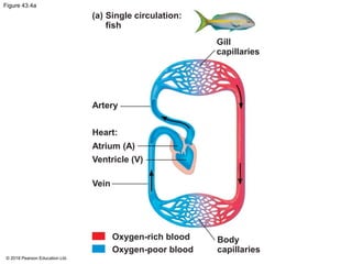
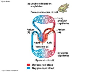
The document outlines the various animal transport systems, emphasizing the importance of circulatory systems in facilitating the exchange of substances at the cellular level, particularly in multicellular organisms. It describes the differences between open and closed circulatory systems, highlights the structure and function of the cardiovascular system in vertebrates, and details the processes of blood circulation and heart function. Additionally, it covers mechanisms of blood pressure regulation and the significance of capillary function in tissue perfusion.


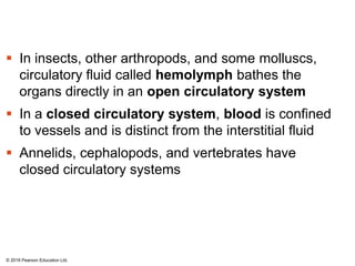
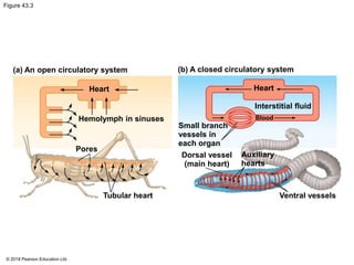
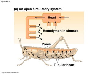
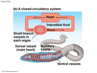


![Polymer [ बहुलक ] Chemistry Notes PDF - Irfanullah Mehar - JJ Sir Chemistry.pdf](https://cdn.slidesharecdn.com/ss_thumbnails/polymerchemistrynotespdf-irfanullahmehar-jjsirchemistry-260210172118-3f9b37f7-thumbnail.jpg?width=640&height=640&fit=bounds)


