En esta presentación se encuentra la explicación sobre la tomografía Axial Computarizada, se habla sobre su historia, partes, operación general y especifica del equipo, tipos de densidades y sus aplicaciones más comunes y las innovadoras.
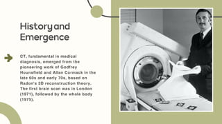
CT, fundamental inmedical
diagnosis, emerged from the
pioneering work of Godfrey
Hounsfield and Allan Cormack in the
late 60s and early 70s, based on
Radon's 3D reconstruction theory.
The first brain scan was in London
(1971), followed by the whole body
(1975).
Historyand
Emergence
3.
KEY PHYSICAL
PRINCIPLES:
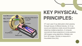
CT scanuses X-ray attenuation when passing
through tissues of different density. An X-ray
source and detectors rotate around the patient,
obtaining multiple projections. A computer
reconstructs these projections in cross-sections
(2D images) using algorithms. Multiple cuts
form 3D images. Density is measured in
Hounsfield Units (HU).
4.
The gantry containsthe X-ray tube and the
detectors. The data acquisition system (DAS)
digitizes the signals. The patient's table moves
through the gantry. The computer controls the
process and reconstructs the images, which are
displayed in the operator's console and stored in
the PACS. Other components are the high voltage
generator and collimators.
MAINPARTS:
5.
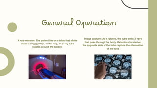
General Operation
X-ray emission:The patient lies on a table that slides
inside a ring (gantry). In this ring, an X-ray tube
rotates around the patient.
Image capture: As it rotates, the tube emits X-rays
that pass through the body. Detectors located on
the opposite side of the tube capture the attenuation
of the rays
6.
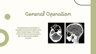
General Operation
Computerized reconstruction:A
computer processes the data to form
sliced
(axial) images of the body.
These can then be reconstructed in
3D or in other planes (sagittal,
coronal).
7.
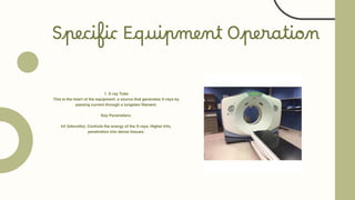
Specific Equipment Operation
1.X-ray Tube
This is the heart of the equipment: a source that generates X-rays by
passing current through a tungsten filament.
Key Parameters:
kV (kilovolts): Controls the energy of the X-rays. Higher kVs,
penetration into dense tissues.
8.
Specific Equipment Operation
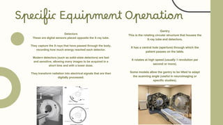
Detectors
Theseare digital sensors placed opposite the X-ray tube.
They capture the X-rays that have passed through the body,
recording how much energy reached each detector.
Modern detectors (such as solid-state detectors) are fast
and sensitive, allowing many images to be acquired in a
short time and with a lower dose.
They transform radiation into electrical signals that are then
digitally processed.
Gantry
This is the rotating circular structure that houses the
X-ray tube and detectors.
It has a central hole (aperture) through which the
patient passes on the table.
It rotates at high speed (usually 1 revolution per
second or more).
Some models allow the gantry to be tilted to adapt
the scanning angle (useful in neuroimaging or
specific studies).
9.
Specific Equipment Operation
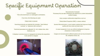
ControlConsole
This is the technician's or radiologist's workstation.
From here, the following are used:
Patient data is entered.
Scanning protocols are selected (for example, "skull without
contrast" or "abdomen with contrast").
Technical parameters are adjusted: kV, mA, rotation time, slice
thickness, etc.
It displays a preview (scout or topogram) and allows the acquisition
to be started or stopped
Reconstruction Computer
Processes the raw data collected by the detectors to create interpretable
medical images.
Uses complex mathematical algorithms, such as:
Filtered Back Projection (FBP): a classic method.
Iterative Reconstruction: a more modern method that improves image
quality with less noise and lower dose.
In newer equipment, it can use artificial intelligence to improve
sharpness or reduce artifacts.
Images are reconstructed in different planes and can be processed in
3D, multiplanar slices, etc.
10.
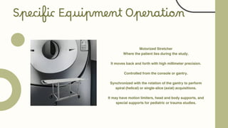
Motorized Stretcher
Where thepatient lies during the study.
It moves back and forth with high millimeter precision.
Controlled from the console or gantry.
Synchronized with the rotation of the gantry to perform
spiral (helical) or single-slice (axial) acquisitions.
It may have motion limiters, head and body supports, and
special supports for pediatric or trauma studies.
Specific Equipment Operation
11.
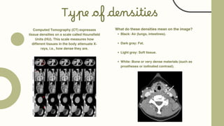
Type of densities
ComputedTomography (CT) expresses
tissue densities on a scale called Hounsfield
Units (HU). This scale measures how
different tissues in the body attenuate X-
rays, i.e., how dense they are.
What do these densities mean on the image?
Black: Air (lungs, intestines).
Dark gray: Fat.
Light gray: Soft tissue.
White: Bone or very dense materials (such as
prostheses or iodinated contrast).
12.
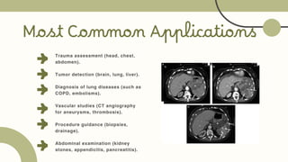
Most Common Applications
Traumaassessment (head, chest,
abdomen).
Tumor detection (brain, lung, liver).
Diagnosis of lung diseases (such as
COPD, embolisms).
Vascular studies (CT angiography
for aneurysms, thrombosis).
Procedure guidance (biopsies,
drainage).
Abdominal examination (kidney
stones, appendicitis, pancreatitis).
13.
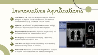
Innovative Applications
Dual-energy CT:Uses two X-ray sources with different
energies to improve tissue differentiation and material
characterization (e.g., urates vs. calcium stones).
Spectral CT: Provides images based on the full energy
spectrum, improving contrast and reducing dose.
AI-powered reconstruction: Improves image quality and
reduces artifacts with lower radiation doses.
4D imaging: Dynamic assessment (e.g., respiratory
motion or real-time cardiac flow).
Low-dose CT: Applications in screening (such as early
detection of lung cancer in smokers).
Radiomics: Advanced quantitative image feature analysis
to support clinical decisions with artificial intelligence.