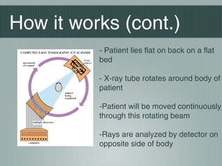
Computed
Tomography
(CT) Scans

Brandon Lau Edward L
4B IB Psychology
Background
information
X-Ray Computed Tomography is a medical imaging procedure
using computer processed x-rays to produce tomographic images
The produced cross-sectional images are used primarily for
diagnostic and therapeutic processes
Greek Foundation of the word: ‘Tomo’ = Slice
Invented by Hounsfield and Cormack in 1972, both won Nobel
prize for the invention
About 6,000 CT scanners in US and about 30,000 worldwide today
Background Info
(cont.)

Usage of CT scans has dramatically increased over the past two
decades
Estimated 72 million scans were performed in the United
States in 2007
Older versions of the scan were known as Computed Axial
Tomography or CAT Scans
Although previously images produced were in the axial plane
(limited to one angle), modern scanners allow data to be
reformatted as volumetric 3D representations of structures
How it works
Since the 1970s, CT scans have become an important tool in
medical imaging to supplement X-rays and medical
ultrasonography
Unlike simple X-ray scans, the combination of computed
technology allows soft tissue to be visible
A x-ray tube and detector are physically rotated behind a circular
shroud
Pixels in an image obtained by CT scanning are displayed in terms
of relative radio-density
Images can be acquired from any angle and at any depth
How it works (cont.)
- Patient lies flat on back on a flat
bed
- X-ray tube rotates around body of
patient
-Patient will be moved continuously
through this rotating beam
-Rays are analyzed by detector on
opposite side of body
Results of CT Scan

A visual representation of the raw data is
referred to as a sinogram (not yet sufficient
for interpretation)
Data must then be processed using a form
of tomographic reconstruction
This process produces a series of crosssectional images
Can show a full body CT scan or be
focused on just the brain/head
Images produced are called Tomograms
Individual scans can be combined to
create 3D images
Strengths
Has many advantages over traditional 2D medical radiography
Eliminates the superimposition of images of structures outside
the area of interests
Differences between tissues that differ in physical density by
less than 1% can be distinguished
Capable of showing soft tissue and structural changes
eg. Brain tumors, Brain damage, Liver, and other organs
Far more useful than an MRI in terms of skull fractures
Cheaper than a MRI but equally as fast
Can be performed on patients with implanted medical devices
Weaknesses
Can only produce structural images
Unable to provide any information on brain
activity
Inferior to MRIs when it comes to soft
tissue contrast
Potentially Dangerous (see next slide)
Dangers
Exposure to radiation

Especially dangerous to young children and pregnant women
Half of all CT scans in the United States involve intravenously
injected radio contrast agents
Mild side affects of these agents include nausea, vomiting, and
rashes
Rare cases of extreme reactions include contrast-induced
nephropathy, occurring in approximate 2-7% of people who receive
these agents
Ionizing radiation in the form of x-rays are energetic enough to
directly damage DNA
Small increased risk of cancer
Citations
Siemens Medical Solutions. (n.d.). Retrieved from
http://www.imaginis.com/ct-scan/brief-history-of-ct
Stoppler, M. C. (n.d.). Retrieved from
http://www.medicinenet.com/cat_scan/article.htm

Images From:
http://www.callusdoc.com/AImages/canstockphoto4116197%20(1).jpg
http://media.web.britannica.com/eb-media/59/62659-004-A8C6FDA4.gif
http://assets.knowledge.allianz.com/img/ct_scan_head_q_1_48138.
http://dz-world-health.blogspot.com/2012_07_01_archive.html

Computed Tomography Scans

  • 1.
  • 2.
    Background information X-Ray Computed Tomographyis a medical imaging procedure using computer processed x-rays to produce tomographic images The produced cross-sectional images are used primarily for diagnostic and therapeutic processes Greek Foundation of the word: ‘Tomo’ = Slice Invented by Hounsfield and Cormack in 1972, both won Nobel prize for the invention About 6,000 CT scanners in US and about 30,000 worldwide today
  • 3.
    Background Info (cont.) Usage ofCT scans has dramatically increased over the past two decades Estimated 72 million scans were performed in the United States in 2007 Older versions of the scan were known as Computed Axial Tomography or CAT Scans Although previously images produced were in the axial plane (limited to one angle), modern scanners allow data to be reformatted as volumetric 3D representations of structures
  • 4.
    How it works Sincethe 1970s, CT scans have become an important tool in medical imaging to supplement X-rays and medical ultrasonography Unlike simple X-ray scans, the combination of computed technology allows soft tissue to be visible A x-ray tube and detector are physically rotated behind a circular shroud Pixels in an image obtained by CT scanning are displayed in terms of relative radio-density Images can be acquired from any angle and at any depth
  • 5.
    How it works(cont.) - Patient lies flat on back on a flat bed - X-ray tube rotates around body of patient -Patient will be moved continuously through this rotating beam -Rays are analyzed by detector on opposite side of body
  • 6.
    Results of CTScan A visual representation of the raw data is referred to as a sinogram (not yet sufficient for interpretation) Data must then be processed using a form of tomographic reconstruction This process produces a series of crosssectional images Can show a full body CT scan or be focused on just the brain/head Images produced are called Tomograms Individual scans can be combined to create 3D images
  • 7.
    Strengths Has many advantagesover traditional 2D medical radiography Eliminates the superimposition of images of structures outside the area of interests Differences between tissues that differ in physical density by less than 1% can be distinguished Capable of showing soft tissue and structural changes eg. Brain tumors, Brain damage, Liver, and other organs Far more useful than an MRI in terms of skull fractures Cheaper than a MRI but equally as fast Can be performed on patients with implanted medical devices
  • 8.
    Weaknesses Can only producestructural images Unable to provide any information on brain activity Inferior to MRIs when it comes to soft tissue contrast Potentially Dangerous (see next slide)
  • 9.
    Dangers Exposure to radiation Especiallydangerous to young children and pregnant women Half of all CT scans in the United States involve intravenously injected radio contrast agents Mild side affects of these agents include nausea, vomiting, and rashes Rare cases of extreme reactions include contrast-induced nephropathy, occurring in approximate 2-7% of people who receive these agents Ionizing radiation in the form of x-rays are energetic enough to directly damage DNA Small increased risk of cancer
  • 10.
    Citations Siemens Medical Solutions.(n.d.). Retrieved from http://www.imaginis.com/ct-scan/brief-history-of-ct Stoppler, M. C. (n.d.). Retrieved from http://www.medicinenet.com/cat_scan/article.htm Images From: http://www.callusdoc.com/AImages/canstockphoto4116197%20(1).jpg http://media.web.britannica.com/eb-media/59/62659-004-A8C6FDA4.gif http://assets.knowledge.allianz.com/img/ct_scan_head_q_1_48138. http://dz-world-health.blogspot.com/2012_07_01_archive.html