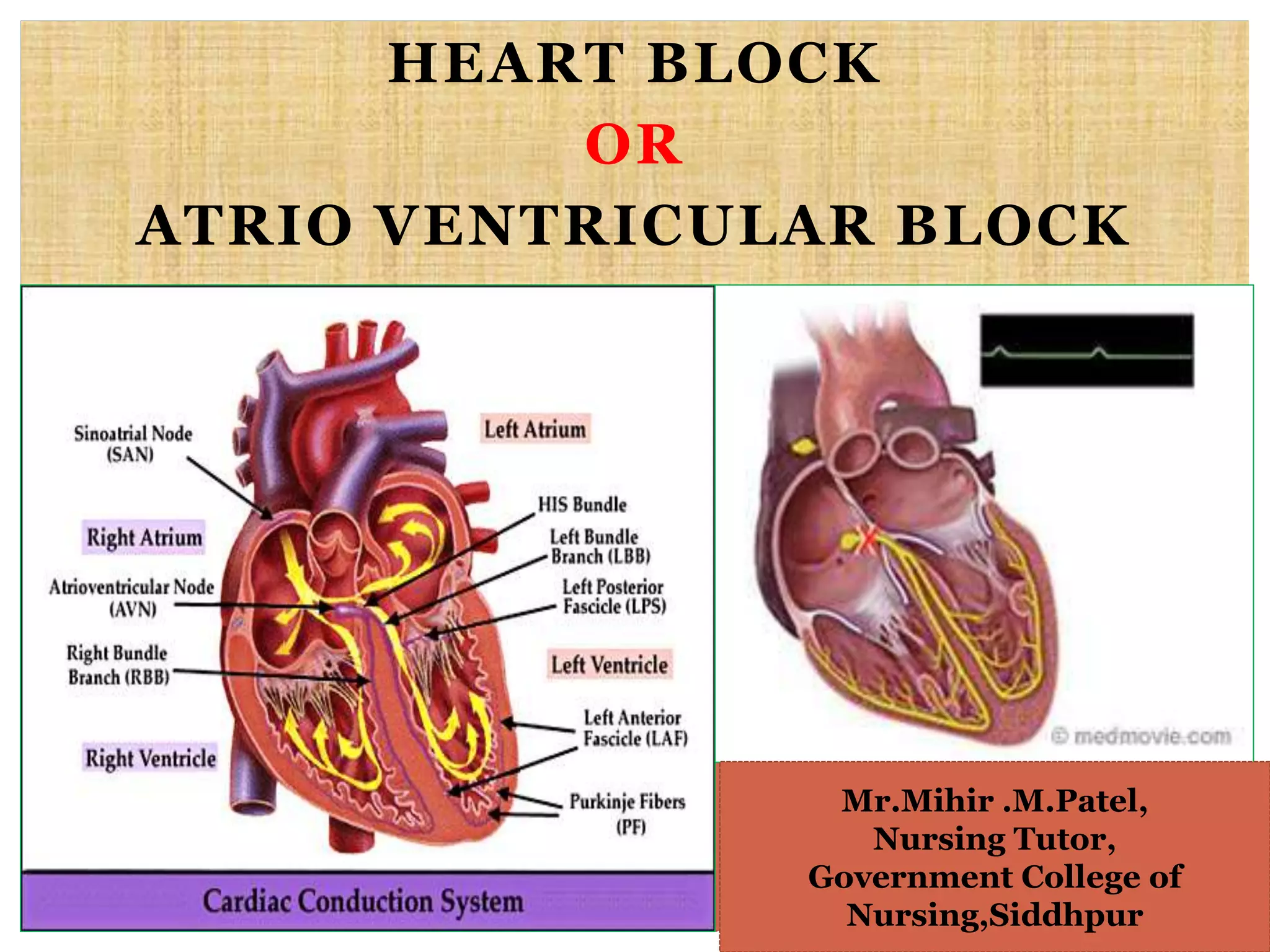
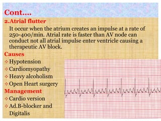
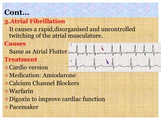
HEART BLOCK
OR
ATRIO VENTRICULAR BLOCK
Mr.Mihir .M.Patel,
Nursing Tutor,
Government College of
Nursing,Siddhpur
Definition
 It occur when the conduction of impulses through the
AV nodal area is decreased or stopped.
Etiology
Medications e.g. Digitalis, calcium channel blocker,
beta blockers
Myocardial ischemia
Myocardial infraction
Increased vagal tone e.g. suctioning, pressure over
eye.
Classification
First Degree Heart Block
It occurs when all the atrial impulses are conducted
through the AV node into the ventricle at a rate slower
than the normal.
ECG Characteristics
Ventricular and artrial rate
QRS Complex: Normal
PR Interval more than 0.20 sec.
P wave : Normal
Cont…
Second Degree Heart Block
It occurs when all but one of the atrial impulses are
conducted through the AV node into the ventricle.
Each impulse takes a longer time than the previous
impulse
ECG characteristics
 Ventricular and atrial rate
 Rhythm:RR interval gradually shorten
 QRS Complex: normal/abnormal
 PR interval: longer with every ECG
 P wave: Normal
 P:QRS-3:2,4:3,5:4.
Cont….
Treatment
Inj.Atropine is administered to increase heart rate
Temporary pacemaker may be used
If patients is asymptomatic monitor the rhythm
slowly.
Third Degree Heart Block
It occurs when no atrial impulse is conducted through
the AV node into the ventricles. Here 2 impulses
stimulate heart, one stimulate the ventricle and other
the atria.
ECG characteristics
Ventricular and atrial rate
Rhythm:PP and RR interval are regular but not equal
PR interval :irregular
P:QRS-P waves are more than QRS complexes.
Clinical Manifestation
 Activity Intolerance
 Weakness
 Hypotension
 Dizziness
 Shortness of Breath
 Dyspnea
 Palpitation
 Altered mental status
 Anxiety
 Respiratory Insufficiency
 Syncope
 Seizures
 Pallor
 Decreased urine output
Diagnostic Evaluation
 History Collection
 Physical Examination
 12 lead ECG
 Serum Electrolyte level
 Exercise stress testing
 Holter monitoring
 Atrial Electrogram (It is similar to an ECG but is used
to monitor atrial rhythm)
Management
 Medical Management
 Defibrillation and Cardioversion
 CPR
 Automatic External Defrillator
 Cardiac Pacing
 Antirhythmic Drugs
Class-1 Sodium channel blocker e.g. disopramide
Class-2 Beta adrenergic blocker e.g. Metoprolol
Class-3 Potassium channel Blocker e.g. Verapamil
 Oxygen Administration
 Implantable cardioverter defibrillator
 Anti coagulation
 Reduce intake of caffeine, low fat and cholesterol
Surgical Management
Left Ventricular Infarctectomy
Myocardial Revascularization
Encircling Ventriculotomy
Nursing Management
Monitor the pt heart rhythm Properly
Monitor CO
Document dysrhythmias in ECG
Monitor the patients lab value
Administer anti dysrhythmias agents
Administer Oxygen
Minimize the anxiety
DYSARRHYTHMIAS
 It is the disorder of the formation or conduction of
electrical impulses within the heart altering the heart
rate ,rhythm or both potentially causing altered blood
flow.
Etiology
Cardiac Condition
Cardiomyopathy
Heart Failure
Myocardial cell degeneration
Myocardial infarction
Valve Disease
Other Condition
Acid Base Imbalance
Alcohol
Caffeine and tobacco intake
Connective tissue disorder
Drugs toxicity
Electric shock
Electrolyte Imbalance
Hypoxia
Shock
Metabolic Disorders
Poisoning
Pathophysiology
Cardiac cycle depends on a balance of basic regulatory
substance (Na,K,Ca and catecholamine)
Imbalance in the regulator can cause disturb pacing
function, conduction or myocardial contractility
Dysarrythmias
Classification
They are classified as the following;
Sinus node
Atrial node
Normal Junctional
Ventricular dysarrhythmias
Sinus Node Arrhythmias
 Sinus Bradycardia
It occures when the sinus node creates an impulse at a
slower than normal rate
Causes
Low metabolic demand
Vagal Stimulation
Increased ICP
Myocardial infarction
Cont…
ECG Characteristics
Ventricular and atrial rate less than 60.
Rhythm: Normal
P wave: Normal and Consistent
P:QRS ratio-1:1
Treatment
Inj.Atropine 0.5-1.0 mg/iv
IV catecholamine
Transcutaneous Pacing
Cont…
Sinus Tachycardia
It occures when the sinus node creates an impulse at a
faster than normal rate.
Causes
 Acute blood loss
 CCF
 Pain
 Hyper metabolic State
 Fever
 Anxiety
Cont…
ECG Characteristics
Ventricular and Atrial rate more than 100.
Rhythm: Normal
P Wave: Normal and consistent
P:QRS ratio-1:1
Cont….
Sinus Arrhythmias
It occures when the sinus node create an impulse at a
irregular rhythm, rate usually increases with
inspiration and decrease with expiration
ECG Characteristics
Ventricular and atrial rate :60-100
Rhythm Irregular
QRS Wave Abnormal
P Wave; Normal
P:QRS ratio-1:1
Atrial Dysarrythmias
1. Pre Mature Atrial Complex
It occurs when an electrical impulse starts in the
atrium before the normal impulse of the sinus node.
Causes
Intake of Caffeine
Alcohol
Nicotine
Anxiety
Infarction
Note: Treatment is not usually required
Cont….
2.Atrial flutter
It occur when the atrium creates an impulse at a rate of
250-400/min. Atrial rate is faster than AV node can
conduct not all atrial impulse enter ventricle causing a
therapeutic AV block.
Causes
Hypotension
Cardiomyopathy
Heavy alcoholism
Open Heart surgery
Management
Cardio version
Ad.B-blocker and
Digitalis
Cont…
3.Atrial Fibrillation
It causes a rapid,disorganised and uncontrolled
twitching of the atrial musculature.
Causes
Same as Atrial Flutter
Treatment
Cardio version
Medication: Amiodarone
Calcium Channel Blockers
Warfarin
Digoxin to improve cardiac function
Pacemaker
Junctional Dysarrythmias
1.Premature Junctional Complexes
It is an impulse that starts in the AV nodal area before
the next normal sinus impulse reaches AV node.
2.Junctional Rhythm
It occurs when the AV node instead of the SA node
becomes the pacemaker of the Heart
Treatment: Emergency Pacing
3.Atrioventricular Nodal reentry tachycardia
It occurs when an impulse is conducted to an area in
the AV node that causes the impulse to be re routed
back into the same area over and over again at a faster
rate.
Ventricular Dysarrhythmias
Premature Ventricular Complex
It is an impulse that start in the ventricle and is
conducted through the ventricle before the next normal
sinus impulse
Ventricular Tachycardia
It is defined as more than 3 PVC in a row. It is treated
with immediate defibrillation
Ventricular Fibrillation
It is rapid but disorganized ventricular rhythm that
causes ineffective waves from ventricle. No atrial
activity seen in ECG.
Cont…
Treatment
CPR and emergency services
Transcutaneous pacing

361711208-Heart-Block-ppt.pptx

  • 1.
    HEART BLOCK OR ATRIO VENTRICULARBLOCK Mr.Mihir .M.Patel, Nursing Tutor, Government College of Nursing,Siddhpur
  • 2.
    Definition  It occurwhen the conduction of impulses through the AV nodal area is decreased or stopped. Etiology Medications e.g. Digitalis, calcium channel blocker, beta blockers Myocardial ischemia Myocardial infraction Increased vagal tone e.g. suctioning, pressure over eye.
  • 3.
    Classification First Degree HeartBlock It occurs when all the atrial impulses are conducted through the AV node into the ventricle at a rate slower than the normal. ECG Characteristics Ventricular and artrial rate QRS Complex: Normal PR Interval more than 0.20 sec. P wave : Normal
  • 4.
    Cont… Second Degree HeartBlock It occurs when all but one of the atrial impulses are conducted through the AV node into the ventricle. Each impulse takes a longer time than the previous impulse ECG characteristics  Ventricular and atrial rate  Rhythm:RR interval gradually shorten  QRS Complex: normal/abnormal  PR interval: longer with every ECG  P wave: Normal  P:QRS-3:2,4:3,5:4.
  • 5.
    Cont…. Treatment Inj.Atropine is administeredto increase heart rate Temporary pacemaker may be used If patients is asymptomatic monitor the rhythm slowly.
  • 6.
    Third Degree HeartBlock It occurs when no atrial impulse is conducted through the AV node into the ventricles. Here 2 impulses stimulate heart, one stimulate the ventricle and other the atria. ECG characteristics Ventricular and atrial rate Rhythm:PP and RR interval are regular but not equal PR interval :irregular P:QRS-P waves are more than QRS complexes.
  • 7.
    Clinical Manifestation  ActivityIntolerance  Weakness  Hypotension  Dizziness  Shortness of Breath  Dyspnea  Palpitation  Altered mental status  Anxiety  Respiratory Insufficiency  Syncope  Seizures  Pallor  Decreased urine output
  • 8.
    Diagnostic Evaluation  HistoryCollection  Physical Examination  12 lead ECG  Serum Electrolyte level  Exercise stress testing  Holter monitoring  Atrial Electrogram (It is similar to an ECG but is used to monitor atrial rhythm)
  • 9.
    Management  Medical Management Defibrillation and Cardioversion  CPR  Automatic External Defrillator  Cardiac Pacing  Antirhythmic Drugs Class-1 Sodium channel blocker e.g. disopramide Class-2 Beta adrenergic blocker e.g. Metoprolol Class-3 Potassium channel Blocker e.g. Verapamil  Oxygen Administration  Implantable cardioverter defibrillator  Anti coagulation  Reduce intake of caffeine, low fat and cholesterol
  • 10.
    Surgical Management Left VentricularInfarctectomy Myocardial Revascularization Encircling Ventriculotomy
  • 11.
    Nursing Management Monitor thept heart rhythm Properly Monitor CO Document dysrhythmias in ECG Monitor the patients lab value Administer anti dysrhythmias agents Administer Oxygen Minimize the anxiety
  • 12.
    DYSARRHYTHMIAS  It isthe disorder of the formation or conduction of electrical impulses within the heart altering the heart rate ,rhythm or both potentially causing altered blood flow.
  • 13.
    Etiology Cardiac Condition Cardiomyopathy Heart Failure Myocardialcell degeneration Myocardial infarction Valve Disease Other Condition Acid Base Imbalance Alcohol Caffeine and tobacco intake Connective tissue disorder Drugs toxicity Electric shock Electrolyte Imbalance Hypoxia Shock Metabolic Disorders Poisoning
  • 14.
    Pathophysiology Cardiac cycle dependson a balance of basic regulatory substance (Na,K,Ca and catecholamine) Imbalance in the regulator can cause disturb pacing function, conduction or myocardial contractility Dysarrythmias
  • 15.
    Classification They are classifiedas the following; Sinus node Atrial node Normal Junctional Ventricular dysarrhythmias
  • 16.
    Sinus Node Arrhythmias Sinus Bradycardia It occures when the sinus node creates an impulse at a slower than normal rate Causes Low metabolic demand Vagal Stimulation Increased ICP Myocardial infarction
  • 17.
    Cont… ECG Characteristics Ventricular andatrial rate less than 60. Rhythm: Normal P wave: Normal and Consistent P:QRS ratio-1:1 Treatment Inj.Atropine 0.5-1.0 mg/iv IV catecholamine Transcutaneous Pacing
  • 18.
    Cont… Sinus Tachycardia It occureswhen the sinus node creates an impulse at a faster than normal rate. Causes  Acute blood loss  CCF  Pain  Hyper metabolic State  Fever  Anxiety
  • 19.
    Cont… ECG Characteristics Ventricular andAtrial rate more than 100. Rhythm: Normal P Wave: Normal and consistent P:QRS ratio-1:1
  • 20.
    Cont…. Sinus Arrhythmias It occureswhen the sinus node create an impulse at a irregular rhythm, rate usually increases with inspiration and decrease with expiration ECG Characteristics Ventricular and atrial rate :60-100 Rhythm Irregular QRS Wave Abnormal P Wave; Normal P:QRS ratio-1:1
  • 21.
    Atrial Dysarrythmias 1. PreMature Atrial Complex It occurs when an electrical impulse starts in the atrium before the normal impulse of the sinus node. Causes Intake of Caffeine Alcohol Nicotine Anxiety Infarction Note: Treatment is not usually required
  • 22.
    Cont…. 2.Atrial flutter It occurwhen the atrium creates an impulse at a rate of 250-400/min. Atrial rate is faster than AV node can conduct not all atrial impulse enter ventricle causing a therapeutic AV block. Causes Hypotension Cardiomyopathy Heavy alcoholism Open Heart surgery Management Cardio version Ad.B-blocker and Digitalis
  • 23.
    Cont… 3.Atrial Fibrillation It causesa rapid,disorganised and uncontrolled twitching of the atrial musculature. Causes Same as Atrial Flutter Treatment Cardio version Medication: Amiodarone Calcium Channel Blockers Warfarin Digoxin to improve cardiac function Pacemaker
  • 24.
    Junctional Dysarrythmias 1.Premature JunctionalComplexes It is an impulse that starts in the AV nodal area before the next normal sinus impulse reaches AV node. 2.Junctional Rhythm It occurs when the AV node instead of the SA node becomes the pacemaker of the Heart Treatment: Emergency Pacing 3.Atrioventricular Nodal reentry tachycardia It occurs when an impulse is conducted to an area in the AV node that causes the impulse to be re routed back into the same area over and over again at a faster rate.
  • 25.
    Ventricular Dysarrhythmias Premature VentricularComplex It is an impulse that start in the ventricle and is conducted through the ventricle before the next normal sinus impulse Ventricular Tachycardia It is defined as more than 3 PVC in a row. It is treated with immediate defibrillation Ventricular Fibrillation It is rapid but disorganized ventricular rhythm that causes ineffective waves from ventricle. No atrial activity seen in ECG.
  • 26.
    Cont… Treatment CPR and emergencyservices Transcutaneous pacing

Editor's Notes