Downloaded 155 times

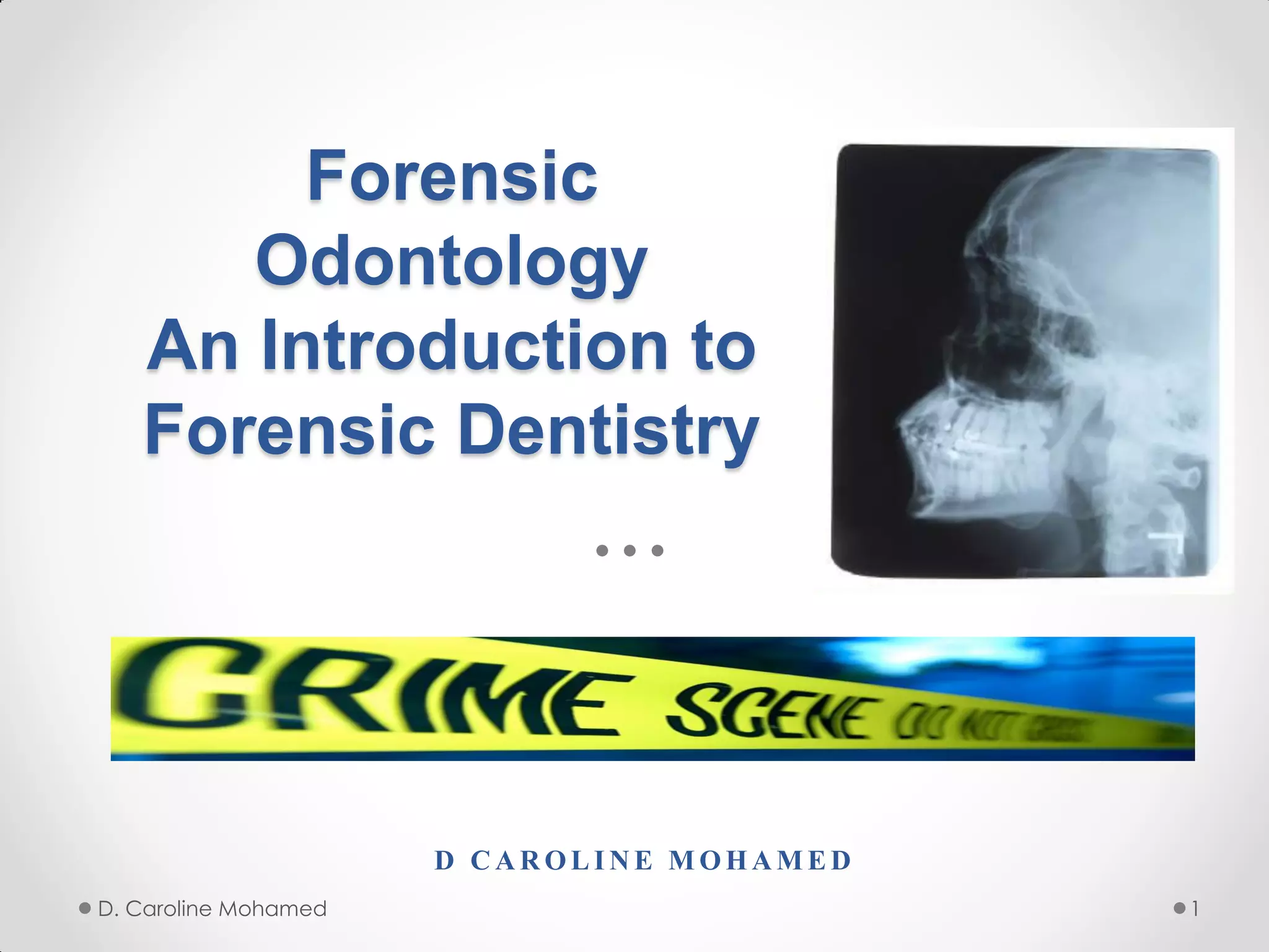
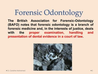
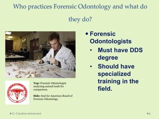
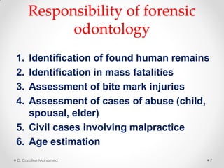
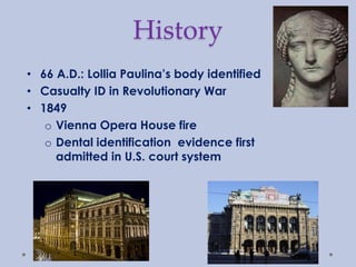

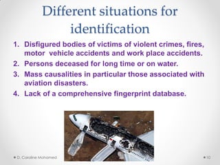
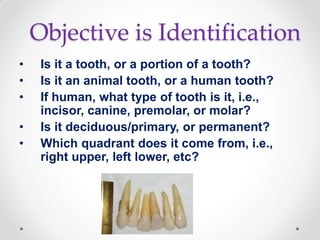

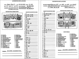
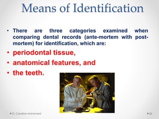
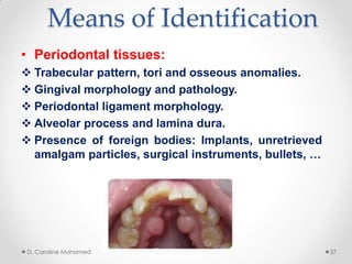
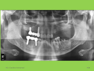
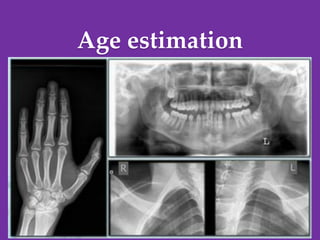

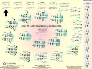
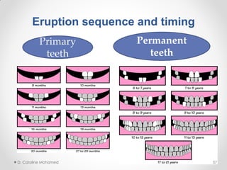
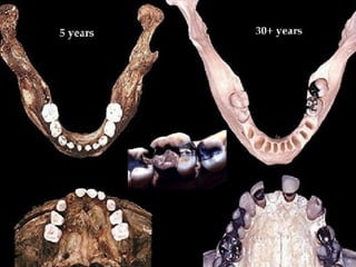

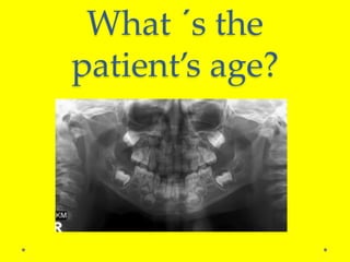
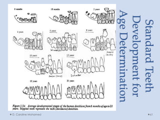
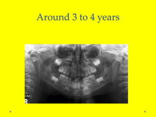
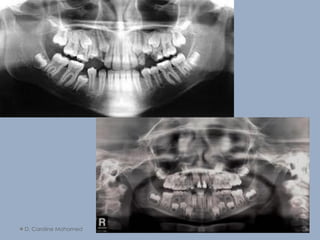
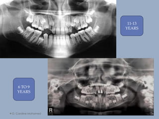


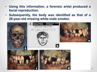
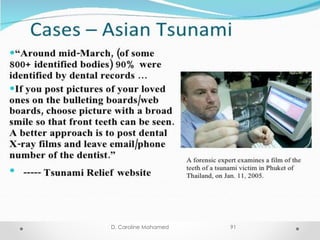
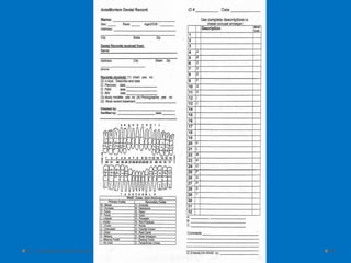

This document provides an overview of forensic odontology. It defines forensic odontology as the application of dental expertise to legal investigations. Forensic odontologists assist with identifications using dental records, bite mark analysis, and age estimations. Identifications can be made by comparing ante-mortem and post-mortem dental records or by developing a dental profile when records are unavailable. Additional areas discussed include the responsibilities of forensic odontologists, their training, the history of the field, and techniques used for analysis.


![CASE_PRESENTATION_ON_subdural_hematoma(SDH)[1 FINAL PPT]-1.pptx](https://cdn.slidesharecdn.com/ss_thumbnails/casepresentationonsubduralhematomasdh1finalppt-1-260129172522-d405d375-thumbnail.jpg?width=640&height=640&fit=bounds)


