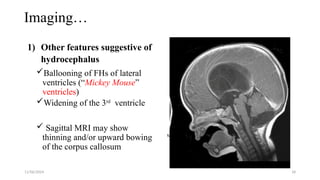
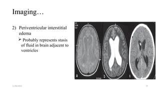
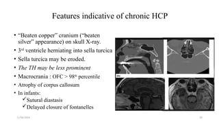
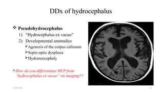
Hydrocephalus and it’s management
Presenter: Dr. Kemal T. (GSR-3)
Moderator: Dr. Yetsedaw. (Assistant
proff. of Neurosurgery)
11/06/2024 2
Outline
• Anatomy of ventricles and CSF physiology
• Definition
• Classification
• Pathophysiology
• Clinical feature
• Diagnosis
• Management
• Reference
11/06/2024 3
Anatomy and physiology
• The ventricular system of the brain consists of:
• The two lateral ventricles (largest),
• midline 3rd and 4th ventricle.
11/06/2024 4
CSF Production and Circulation
• 80% of CSF is produced by the choroid plexuses.
• In the adult, CSF is produced at a rate of about 0.3
ml/min.
• The average total CSF volume in the body is 150 ml
• CSF is absorbed primarily by arachnoid villi
(granulations) that extend into the dural venous
sinuses.
11/06/2024 5
Function of CSF
11/06/2024 6
Hydrocephalus
 Hydrocephalus – Greek word :
Hydro - water &
Kefale - skull  (“water in the head”)
 An abnormal accumulation of CSF within the ventricles
of the brain due to disturbance of CSF absorption, flow or
production
 It is almost always associated with increased intracranial
pressure (ICP)
11/06/2024 7
Epidemiology
Estimated prevalence: 1–1.5%
Incidence :
• Congenital HCP ≈ 0.9–1.8/1000 births (reported range from 0.2 to
3.5/ 1000 births).
• Infant HCP - 3-5/1000 life births
Incidence of acquired hydrocephalus is unknown
In Ethiopia : 2000 - 4000 new cases per year
11/06/2024 8
11/06/2024 9
Pathophysiology
Hydrocephalus
Subnormal CSF
reabsorption.
Non-communicating
Masses
Communicating
Infection and
post SAH
CSF
overproduction
choroid plexus
papillomas
Special types
NPH
Entrapped fourth
ventricle
Arrested
hydrocephalus
11/06/2024 10
Specific etiologies of hydrocephalus
1. Congenital
Chiari Type 2 malformation
and/or MMC
Chiari Type 1 malformation
Primary aqueductal stenosis
Secondary aqueductal gliosis
Dandy Walker malformation
X-linked inherited disorder
2. Acquired
A. Infectious
B. Post-hemorrhagic
C. Secondary to masses
D. Post-op
A. 20% of pediatric patients develop
permanent hydrocephalus
following p-fossa tumour removal
11/06/2024 11
Clinical Manifestation
• Clinical features of hydrocephalus are influenced by:
Age
Cause
Site of obstruction
Duration
Rapidity of onset
11/06/2024 12
In Infants and young children
• Cranium enlarges at rate > facial
growth
• Irritability, poor head control,
Nausea
• Fontanelle full and bulging
• Frontal bossing
• Engorgement of scalp Veins
• “Macewen's sign”
• CN-3 and CN-6 palsy
• “Setting sun sign”
(upward gaze palsy)
• Hyperactive reflexes
• Irregular respirations
with apneic spells
11/06/2024 13
In older children & adults
Slowly enlarging ventricles may initially be asymptomatic.
Symptoms of increased ICP
 Headache
 Nausea/Vomiting
 Gait changes
 Upgaze and/or abducens palsy
 Papilledema.
11/06/2024 14
Diagnosis
History and physical examination
Laboratory Studies
• No specific blood tests in the workup for hydrocephalus.
• Genetic testing recommended when X-linked HCP is suspected.
Imaging
Ultrasound
 In a newborn, preferred technique for the initial
examination
15
Imaging…
Head CT scan
• Fast, reliable, and does not interfere
with implanted medical devices.
• Usually can be accomplished
without sedation.
• Disadvantages - radiation exposure
MRI
• Modality of choice in patients with
unexplained hydrocephalus
• Superior visualization of
pathological processes in the CSF
pathway, including CSF flow
dynamics.
11/06/2024 16
CT/MRI criteria for hydrocephalus
• HCP is suggested when
either:-
1. Both TH is ≥ 2 mm in
width + sylvian &
interhemispheric fissures
and cerebral sulci are not
visible OR
2. Both TH are ≥ 2 mm and
the ratio FH/ ID > 0.5
11/06/2024 17
Imaging…
• Evans ratio or index:
Ratio of FH to maximal BPD
at same slice > 0.3 suggests
hydrocephalus.
11/06/2024 18
Imaging…
1) Other features suggestive of
hydrocephalus
Ballooning of FHs of lateral
ventricles (“Mickey Mouse”
ventricles)
Widening of the 3rd
ventricle
 Sagittal MRI may show
thinning and/or upward bowing
of the corpus callosum
11/06/2024 19
Imaging…
2) Periventricular interstitial
edema
 Probably represents stasis
of fluid in brain adjacent to
ventricles
11/06/2024 20
Features indicative of chronic HCP
• “Beaten copper” cranium (“beaten
silver” appearance) on skull X-ray.
• 3rd
ventricle herniating into sella turcica
• Sella turcica may be eroded.
• The TH may be less prominent
• Macrocrania : OFC > 98th
percentile
• Atrophy of corpus callosum
• In infants:
Sutural diastasis
Delayed closure of fontanelles
11/06/2024 21
Workup of HCP….
Lumbar Puncture
• Indicated if an infection causes are suspected.
• Valuable test in evaluating NPH, but should be performed
only after CT or MRI of the head.
• Predict response rate of CSF shunting in pts. with NPH.
11/06/2024 22
DDx of hydrocephalus
 Pseudohydrocephalus
1) “Hydrocephalus ex vacuo”
2) Developmental anomalies
Agenesis of the corpus callosum
Septo-optic dysplasia
Hydranencephaly
How do you differentiate HCP from
“hydrocephalus ex vacuo” on imaging??
11/06/2024 23
Treatment of Hydrocephalus
• Options of management
 Medical therapy
Diuretics,
Fibrinolysis, and
Serial lumbar punctures
 Surgical therapy
Shunt
Third ventriculostomy
Choroid plexectomy
Embolization of the
malformation
11/06/2024 24
Treatment….
Medical treatment of hydrocephalus
I. Diuretic therapy
Acetazolamide (a carbonic anhydrase inhibitor)
o25 mg/kg/day PO divided TID × 1 day, increase 25 mg/kg/day each
day until 100 mg/kg/day is reached
Simultaneously start furosemide:
o1 mg/kg/day PO divided TID
11/06/2024 25
Medical mgt…
Maintain therapy for a 6 month trial, then taper dosage over
2–4 weeks.
Resume 3–4 months of treatment if progressive HCP occurs.
FOLLOW UP
Watch for electrolyte imbalance
Weekly U/S or CT scan
 Insert ventricular shunt if progressive ventriculomegaly occurs
11/06/2024 26
Treatment of Hydrocephalus Cont...
II. Spinal taps
HCP after IVH and SAH may be transient.
Ventricular tap or LP may temporize until resorption resumes
If reabsorption does not resume when the protein content of
the CSF is <100 mg/dl, a shunt will usually be indicated.
11/06/2024 27
Treatment of Hydrocephalus Cont...
Surgical
The definitive management of hydrocephalus at
present
Goals of therapy:-
Optimum brain function
Good cosmotic result.
11/06/2024 28
Shunts
 Only 25 % of patients with HCP are successfully treated
without shunt placement.
 The principle of shunt is to create a communication between
CSF and a drainage cavity or systemic circulation
 Surgical goal:
Redirect CSF to another area of the body to normalize ICP
11/06/2024 29
Shunts….cont.
Types of shunts Others
1. Torkildsen shunt
 shunts ventricle to cisternal
space
 rarely used
2. Ventriculopleural shunt
3. Gall bladder
4. Ureter or bladder
11/06/2024 30
Components of Shunt Systems
 Ventricular catheter
 Valve and
 Distal catheter
11/06/2024 31
Surgical Technique
General Principles
GA and endotracheal intubation
Supine with head turned to opposite side & neck extended.
Infection Avoidance (“shunt protocols”)
• Restrict personnel trafficking
• Prophylactic IV antibiotic
• Hair is clipped rather than shaved
• Antibiotics impregnated shunt system & suture materials
• Changing glove before shunt touching implants
11/06/2024 32
Steps for VP shunt insertion
1. Cranial and abdominal exposure
2. Abdominal access
3. Subcutaneous tunnelling
4. Ventricular access
5. Shunt assembly and testing
6. Closure
11/06/2024 33
Ventricular Access
Frontal (Coronal) Approach
Kocher’s point.
Occipitoparietal Approach
Frazier point
Keen’s point
Dandy’s point
34
Abdominal incision and tunneling
11/06/2024 35
Ventricular Access, Shunt Assembly & Closure
Connect peritoneal catheter to shunt valve & tightly secure
with 2.0 silk suture
Puncture the ventricle (Ventricular Access)
Free hand (blindly)
Intraoperative ultrasound
Frameless stereotactic neuronavigation
Connect ventricular catheter to shunt valve & secure with 2.0
silk suture
11/06/2024 36
Cont…
 Distal catheter is inserted into the peritoneum directly
 Check for functionality of the shunt
 Irrigate all wounds with saline, antibiotic solutions??
 Close the incision in layer
11/06/2024 37
Complications of various shunts
11/06/2024 38
Other complications…
Bleeding
Hardware erosion through skin
Seizures
Conduit for metastasis
Silcon allergy
Disconnection at any point
Peritoneal catheter migration
Abdominal CSF pseudocyst
11/06/2024 39
Endoscopic third ventriculostomy (ETV)
Indications
• Obstructive HCP
• In shunt infection
• In SDH after shunting
• In slit ventricle syndrome
Contraindications
• Communicating HCP.
• If predicted success rate is low.
• Overall success rate is = 56%
11/06/2024 40
(ETV)….Success rate for different etiologies
• Acquired aqueductal stenosis
• Tumor obstructing CSF flow
High success rate (>75
%)
• Previously shunted MMC
• Congenital aqueductal stenosis
• Arachnoid cyst
• Recurrent shunt infections/malfunction
Intermediate success
rates
(50-75 %)
• Post hemorrhagic HCP
• Post infectious HCP
• MMC
Low success rates
(<50 %)
41
Treatment of Hydrocephalus Cont...
ETV Complications:-
• Hypothalamic injury
• Injury to pituitary stalk or gland
• Transient 3rd and 6th nerve palsies
• Injury to basilar artery, p-comm, or PCA
• Uncontrollable bleeding
• Cardiac arrest
• Traumatic basilar artery aneurysm
11/06/2024 42
43
Post-operative follow-up
Need lifelong follow up
• Thorough history esp. Dev’tal history
• Monitoring of vital signs
• Serial head circumference measurements, and
• Serial Neurologic examination and
• Advice family how to know shunt infection and malfunction.
• Psychosocial support
11/06/2024 44
Special forms of hydrocephalus
1. Arrested hydrocephalus
Situation where there is no progression or harmful
sequelae due to HCP that would require the presence of a
CSF shunt
Arrested HCP Dx criteria (in the absence of a CSF shunt):
a) Near normal ventricular size
b) Normal head growth curve
c) Continued psychomotor development
11/06/2024 45
2. Entrapped (isolated) fourth ventricle
4th
ventricle that neither communicates with the 3rd
ventricle
nor with the basal cisterns
Usually seen with chronic shunting of the lateral ventricles
Occurs in 2–3% of shunted patients
May also occur in Dandy Walker malformation if the aqueduct
is also obstructed
11/06/2024 46
Entrapped fourth ventricle….
• Presentation
Headache, nausea/vomiting
Lower cranial nerve palsies: swallowing difficulties
Bilateral abducens palsy, Ataxia
 Reduced level of consciousness
• Treatment
Shunting the ventricle either with a separate VP shunt, or by linking
into an existing shunt
11/06/2024 47
3. Normal Pressure Hydrocephalus (NPH)
 Is a form of communicating HCP with normal or slightly
elevated CSF pressure
Is characterized by Triad of:-
• Abnormal gait, Urinary incontinence & Dementia
Features on CT and MRI
Ventriculomegaly in the absence or out of proportion to sulcal
enlargement
Treatment
VP Shunt is the procedure of choice
Lumbar-peritoneal shunt
48
Prevention
 Hydrocephalus isn't a preventable condition, but there are ways
to reduce the risk.
 Regular ANC follow up.
 Prevent infection
 Prevent head injury
11/06/2024 49
Summary
• Knowing anatomy of ventricles and CSF physiology is very
important for management of HCP.
• If not treated early hydrocephalus can cause significant
neurological impairment with acute and long standing deficits.
• HCP can be managed medically and surgically, with surgical
management being the standard of treatment.
• Patients should be followed after treatment of HCP for any
complications related to treatment and neurologic improvement.
11/06/2024 50
References
• Greenberg handbook of Neurosurgery, 10th Edition
• Youmans & Winn Neurological Surgery, 7th ed. 2017
• Schmidek & Sweet Operative Neurosurgical Techniques, 7th
ed.
• Up to date 21.6
11/06/2024 51
THANK YOU

Hydrocephalus Kemalbedrumohammedumerrbest.pptx

  • 1.
    Hydrocephalus and it’smanagement Presenter: Dr. Kemal T. (GSR-3) Moderator: Dr. Yetsedaw. (Assistant proff. of Neurosurgery)
  • 2.
    11/06/2024 2 Outline • Anatomyof ventricles and CSF physiology • Definition • Classification • Pathophysiology • Clinical feature • Diagnosis • Management • Reference
  • 3.
    11/06/2024 3 Anatomy andphysiology • The ventricular system of the brain consists of: • The two lateral ventricles (largest), • midline 3rd and 4th ventricle.
  • 4.
    11/06/2024 4 CSF Productionand Circulation • 80% of CSF is produced by the choroid plexuses. • In the adult, CSF is produced at a rate of about 0.3 ml/min. • The average total CSF volume in the body is 150 ml • CSF is absorbed primarily by arachnoid villi (granulations) that extend into the dural venous sinuses.
  • 5.
  • 6.
    11/06/2024 6 Hydrocephalus  Hydrocephalus– Greek word : Hydro - water & Kefale - skull  (“water in the head”)  An abnormal accumulation of CSF within the ventricles of the brain due to disturbance of CSF absorption, flow or production  It is almost always associated with increased intracranial pressure (ICP)
  • 7.
    11/06/2024 7 Epidemiology Estimated prevalence:1–1.5% Incidence : • Congenital HCP ≈ 0.9–1.8/1000 births (reported range from 0.2 to 3.5/ 1000 births). • Infant HCP - 3-5/1000 life births Incidence of acquired hydrocephalus is unknown In Ethiopia : 2000 - 4000 new cases per year
  • 8.
  • 9.
    11/06/2024 9 Pathophysiology Hydrocephalus Subnormal CSF reabsorption. Non-communicating Masses Communicating Infectionand post SAH CSF overproduction choroid plexus papillomas Special types NPH Entrapped fourth ventricle Arrested hydrocephalus
  • 10.
    11/06/2024 10 Specific etiologiesof hydrocephalus 1. Congenital Chiari Type 2 malformation and/or MMC Chiari Type 1 malformation Primary aqueductal stenosis Secondary aqueductal gliosis Dandy Walker malformation X-linked inherited disorder 2. Acquired A. Infectious B. Post-hemorrhagic C. Secondary to masses D. Post-op A. 20% of pediatric patients develop permanent hydrocephalus following p-fossa tumour removal
  • 11.
    11/06/2024 11 Clinical Manifestation •Clinical features of hydrocephalus are influenced by: Age Cause Site of obstruction Duration Rapidity of onset
  • 12.
    11/06/2024 12 In Infantsand young children • Cranium enlarges at rate > facial growth • Irritability, poor head control, Nausea • Fontanelle full and bulging • Frontal bossing • Engorgement of scalp Veins • “Macewen's sign” • CN-3 and CN-6 palsy • “Setting sun sign” (upward gaze palsy) • Hyperactive reflexes • Irregular respirations with apneic spells
  • 13.
    11/06/2024 13 In olderchildren & adults Slowly enlarging ventricles may initially be asymptomatic. Symptoms of increased ICP  Headache  Nausea/Vomiting  Gait changes  Upgaze and/or abducens palsy  Papilledema.
  • 14.
    11/06/2024 14 Diagnosis History andphysical examination Laboratory Studies • No specific blood tests in the workup for hydrocephalus. • Genetic testing recommended when X-linked HCP is suspected. Imaging Ultrasound  In a newborn, preferred technique for the initial examination
  • 15.
    15 Imaging… Head CT scan •Fast, reliable, and does not interfere with implanted medical devices. • Usually can be accomplished without sedation. • Disadvantages - radiation exposure MRI • Modality of choice in patients with unexplained hydrocephalus • Superior visualization of pathological processes in the CSF pathway, including CSF flow dynamics.
  • 16.
    11/06/2024 16 CT/MRI criteriafor hydrocephalus • HCP is suggested when either:- 1. Both TH is ≥ 2 mm in width + sylvian & interhemispheric fissures and cerebral sulci are not visible OR 2. Both TH are ≥ 2 mm and the ratio FH/ ID > 0.5
  • 17.
    11/06/2024 17 Imaging… • Evansratio or index: Ratio of FH to maximal BPD at same slice > 0.3 suggests hydrocephalus.
  • 18.
    11/06/2024 18 Imaging… 1) Otherfeatures suggestive of hydrocephalus Ballooning of FHs of lateral ventricles (“Mickey Mouse” ventricles) Widening of the 3rd ventricle  Sagittal MRI may show thinning and/or upward bowing of the corpus callosum
  • 19.
    11/06/2024 19 Imaging… 2) Periventricularinterstitial edema  Probably represents stasis of fluid in brain adjacent to ventricles
  • 20.
    11/06/2024 20 Features indicativeof chronic HCP • “Beaten copper” cranium (“beaten silver” appearance) on skull X-ray. • 3rd ventricle herniating into sella turcica • Sella turcica may be eroded. • The TH may be less prominent • Macrocrania : OFC > 98th percentile • Atrophy of corpus callosum • In infants: Sutural diastasis Delayed closure of fontanelles
  • 21.
    11/06/2024 21 Workup ofHCP…. Lumbar Puncture • Indicated if an infection causes are suspected. • Valuable test in evaluating NPH, but should be performed only after CT or MRI of the head. • Predict response rate of CSF shunting in pts. with NPH.
  • 22.
    11/06/2024 22 DDx ofhydrocephalus  Pseudohydrocephalus 1) “Hydrocephalus ex vacuo” 2) Developmental anomalies Agenesis of the corpus callosum Septo-optic dysplasia Hydranencephaly How do you differentiate HCP from “hydrocephalus ex vacuo” on imaging??
  • 23.
    11/06/2024 23 Treatment ofHydrocephalus • Options of management  Medical therapy Diuretics, Fibrinolysis, and Serial lumbar punctures  Surgical therapy Shunt Third ventriculostomy Choroid plexectomy Embolization of the malformation
  • 24.
    11/06/2024 24 Treatment…. Medical treatmentof hydrocephalus I. Diuretic therapy Acetazolamide (a carbonic anhydrase inhibitor) o25 mg/kg/day PO divided TID × 1 day, increase 25 mg/kg/day each day until 100 mg/kg/day is reached Simultaneously start furosemide: o1 mg/kg/day PO divided TID
  • 25.
    11/06/2024 25 Medical mgt… Maintaintherapy for a 6 month trial, then taper dosage over 2–4 weeks. Resume 3–4 months of treatment if progressive HCP occurs. FOLLOW UP Watch for electrolyte imbalance Weekly U/S or CT scan  Insert ventricular shunt if progressive ventriculomegaly occurs
  • 26.
    11/06/2024 26 Treatment ofHydrocephalus Cont... II. Spinal taps HCP after IVH and SAH may be transient. Ventricular tap or LP may temporize until resorption resumes If reabsorption does not resume when the protein content of the CSF is <100 mg/dl, a shunt will usually be indicated.
  • 27.
    11/06/2024 27 Treatment ofHydrocephalus Cont... Surgical The definitive management of hydrocephalus at present Goals of therapy:- Optimum brain function Good cosmotic result.
  • 28.
    11/06/2024 28 Shunts  Only25 % of patients with HCP are successfully treated without shunt placement.  The principle of shunt is to create a communication between CSF and a drainage cavity or systemic circulation  Surgical goal: Redirect CSF to another area of the body to normalize ICP
  • 29.
    11/06/2024 29 Shunts….cont. Types ofshunts Others 1. Torkildsen shunt  shunts ventricle to cisternal space  rarely used 2. Ventriculopleural shunt 3. Gall bladder 4. Ureter or bladder
  • 30.
    11/06/2024 30 Components ofShunt Systems  Ventricular catheter  Valve and  Distal catheter
  • 31.
    11/06/2024 31 Surgical Technique GeneralPrinciples GA and endotracheal intubation Supine with head turned to opposite side & neck extended. Infection Avoidance (“shunt protocols”) • Restrict personnel trafficking • Prophylactic IV antibiotic • Hair is clipped rather than shaved • Antibiotics impregnated shunt system & suture materials • Changing glove before shunt touching implants
  • 32.
    11/06/2024 32 Steps forVP shunt insertion 1. Cranial and abdominal exposure 2. Abdominal access 3. Subcutaneous tunnelling 4. Ventricular access 5. Shunt assembly and testing 6. Closure
  • 33.
    11/06/2024 33 Ventricular Access Frontal(Coronal) Approach Kocher’s point. Occipitoparietal Approach Frazier point Keen’s point Dandy’s point
  • 34.
  • 35.
    11/06/2024 35 Ventricular Access,Shunt Assembly & Closure Connect peritoneal catheter to shunt valve & tightly secure with 2.0 silk suture Puncture the ventricle (Ventricular Access) Free hand (blindly) Intraoperative ultrasound Frameless stereotactic neuronavigation Connect ventricular catheter to shunt valve & secure with 2.0 silk suture
  • 36.
    11/06/2024 36 Cont…  Distalcatheter is inserted into the peritoneum directly  Check for functionality of the shunt  Irrigate all wounds with saline, antibiotic solutions??  Close the incision in layer
  • 37.
  • 38.
    11/06/2024 38 Other complications… Bleeding Hardwareerosion through skin Seizures Conduit for metastasis Silcon allergy Disconnection at any point Peritoneal catheter migration Abdominal CSF pseudocyst
  • 39.
    11/06/2024 39 Endoscopic thirdventriculostomy (ETV) Indications • Obstructive HCP • In shunt infection • In SDH after shunting • In slit ventricle syndrome Contraindications • Communicating HCP. • If predicted success rate is low. • Overall success rate is = 56%
  • 40.
    11/06/2024 40 (ETV)….Success ratefor different etiologies • Acquired aqueductal stenosis • Tumor obstructing CSF flow High success rate (>75 %) • Previously shunted MMC • Congenital aqueductal stenosis • Arachnoid cyst • Recurrent shunt infections/malfunction Intermediate success rates (50-75 %) • Post hemorrhagic HCP • Post infectious HCP • MMC Low success rates (<50 %)
  • 41.
    41 Treatment of HydrocephalusCont... ETV Complications:- • Hypothalamic injury • Injury to pituitary stalk or gland • Transient 3rd and 6th nerve palsies • Injury to basilar artery, p-comm, or PCA • Uncontrollable bleeding • Cardiac arrest • Traumatic basilar artery aneurysm
  • 42.
  • 43.
    43 Post-operative follow-up Need lifelongfollow up • Thorough history esp. Dev’tal history • Monitoring of vital signs • Serial head circumference measurements, and • Serial Neurologic examination and • Advice family how to know shunt infection and malfunction. • Psychosocial support
  • 44.
    11/06/2024 44 Special formsof hydrocephalus 1. Arrested hydrocephalus Situation where there is no progression or harmful sequelae due to HCP that would require the presence of a CSF shunt Arrested HCP Dx criteria (in the absence of a CSF shunt): a) Near normal ventricular size b) Normal head growth curve c) Continued psychomotor development
  • 45.
    11/06/2024 45 2. Entrapped(isolated) fourth ventricle 4th ventricle that neither communicates with the 3rd ventricle nor with the basal cisterns Usually seen with chronic shunting of the lateral ventricles Occurs in 2–3% of shunted patients May also occur in Dandy Walker malformation if the aqueduct is also obstructed
  • 46.
    11/06/2024 46 Entrapped fourthventricle…. • Presentation Headache, nausea/vomiting Lower cranial nerve palsies: swallowing difficulties Bilateral abducens palsy, Ataxia  Reduced level of consciousness • Treatment Shunting the ventricle either with a separate VP shunt, or by linking into an existing shunt
  • 47.
    11/06/2024 47 3. NormalPressure Hydrocephalus (NPH)  Is a form of communicating HCP with normal or slightly elevated CSF pressure Is characterized by Triad of:- • Abnormal gait, Urinary incontinence & Dementia Features on CT and MRI Ventriculomegaly in the absence or out of proportion to sulcal enlargement Treatment VP Shunt is the procedure of choice Lumbar-peritoneal shunt
  • 48.
    48 Prevention  Hydrocephalus isn'ta preventable condition, but there are ways to reduce the risk.  Regular ANC follow up.  Prevent infection  Prevent head injury
  • 49.
    11/06/2024 49 Summary • Knowinganatomy of ventricles and CSF physiology is very important for management of HCP. • If not treated early hydrocephalus can cause significant neurological impairment with acute and long standing deficits. • HCP can be managed medically and surgically, with surgical management being the standard of treatment. • Patients should be followed after treatment of HCP for any complications related to treatment and neurologic improvement.
  • 50.
    11/06/2024 50 References • Greenberghandbook of Neurosurgery, 10th Edition • Youmans & Winn Neurological Surgery, 7th ed. 2017 • Schmidek & Sweet Operative Neurosurgical Techniques, 7th ed. • Up to date 21.6
  • 51.

Editor's Notes

  • #3 The ventricles are four fluid-filled cavities located within the brain; these are the two lateral ventricles, the third ventricle, and the fourth ventricle. The two lateral ventricles communicate through the lnterventrlcular foramlna (of Monro) with the third ventricle. The third ventricle is connected to the fourth ventricle by the narrow cerebral aqueduct (aqueduct of Sylvlua). The fourth ventricle, in turn, is continuous with the narrow central canal of the spinal cord and, through the three forarntna In its roof, with the subarachnoid space. CSF from fourth ventricle can exit to subarachnoid space through Two lateral foramina of Luschka & Single, median foramen of Magendie The central canal in the spinal cord has a small dllatatlon at its Inferior end, referred to as the terminal ventricle. The ventricles are lined throughout with ependyma and are filled with CSF. The ventricles are developmentally derived from the cavity of the neural tube.
  • #4 CSF Is formed mainly in the choroid plexuses of the lateral, third, and fourth ventricles; some originates from the ependymal cells llnlng the ventricles and from the brain substance through the perlvascular spaces. The CSF Is found in the ventricles of the brain and in the subarachnoid space around the brain and spinal cord. The choroid plems projects Into the ventricle on Its medial aspect and is a vascular fringe composed of pia mater covered with the ependymal lining of the ventricular cavity It is a highly organized tissue that lines all the ventricles of the brain except the frontal/occipital horn of the lateral ventricles and the cerebral aqueduct. 80% of CSF is produced by the choroid plexuses in both lateral ventricles and in the 4th ventricle. The rest by:- Interstitial space. ependymal lining of the ventricles, and in the spine, in the dura of the nerve root sleeves. CSF is secreted 400–500 mL/day or 0.3 ml/min in adult and turned over” ≈ 3 times every day. Rate is independent of the intracranial pressure. CSF is absorbed primarily by arachnoid villi (granulations) that extend into the dural venous sinuses. Other sites of absorption include the choroid plexuses and glymphatics. Importantly, CSP production is not pressure regulated (as in the case of blood pressure) and it continues to be produced even if the reabsorption mechanisms are obstructed. The main sites for CSF absorption are the arachnoid villi that project into the dural venous sinuses, especially the mperlorsaglttalslnua Structurally, each arachnoid villus ls a divertlculum. of the subarachnold space that pierces the dura mater. CSF absorption into the venous sinuses occurs when CSF pressure exceeds the venous pressure in the sinus. Electron-microscopic studies of the arachnoid villi indicate that fine tubules lined with endothellwn permit a direct flow of fluid from the subarachnoid space into the lwnen of the venous sinuses. Should the venous pressure rise and exceed CSF pressure, compression of the tips of the villi closes the tubules and prevents the reflux of blood into the subarachnold space. The arachnoid villi thus serve as valves. Some CSF probably is absorbed directly into the veins in the subarachnoid space and some possibly escapes through the perineural lymph vessels of the cranial and spinal nerves. Because CSF production from the choroid plexuses is constant, the rate of CSF absorption through the arachnoid vlll1 controls CSF pressw-e. The arachnoid villi consist of a cluster of cells that project from the subarachnoid space to the sinus lumen; these are covered by a layer of endothelium with tight junctions that are continuous with the inner layer of the sinuses. This assembly acts as a one-way valve, allowing passive absorption of CSF down a pressure gradient; if the CSF pressure is less than the venous pressure, the arachnoid villi close and do not allow blood to pass into the ventricular system. (upto date)
  • #5 CSF, which bathes the external and internal surfaces of the brain and spinal cord, serves as a cushion between the central nervous system (CNS) and the surrounding bones, thus protecting lt against mechanical trauma. The choroid plexuses actively eecrete CSF and this creates a small pressure gradient. At the same time, they actively transport nervous system metabolites from the CSF lnto the blood. Because the CSF is an ideal physiologic substrate, it probably plays an active part In the nourishment of the nervous tissue
  • #7 Although no reliable incidence estimate exists for HCP in most developing countries, it is likely higher due to nutritional deficiencies, low infant birth weight, greater incidence of perinatal and neonatal infections, and delayed antenatal diagnosis. In Ethiopia: 2000-4000 new cases per year In most cases in adults, hydrocephalus is acquired; common causes include infection, hemorrhage (subarachnoid or intraventricular), trauma, obstruction by mass lesions, and postoperative causes, particularly after procedures at the skull base, the posterior fossa, and within the cerebral ventricles.
  • #8 a cross-sectional facility-based study design over a two-time period, i.e. a 2-year retrospective and 1 yr a prospective shows prevalence of HCP 22% (223/1000 live birth) etiology being NTD, Aqueductal stenosis and post infectious In descending order.
  • #9 -In non-comm.g HCP there is gross anatomic lesion obstructing flow of CSF -In Commu. HCP – the problem is with CSF reabsorption (increased ICP w/o anatomic lesion obstructing flow). Occur in SAH, Extensive meningeal disease, NPH Subnormal CSF reabsorption:- Two main functional subdivisions:- obstructive hydrocephalus:- Block proximal to the arachnoid granulations (AG). On CT or MRI: enlargement of ventricles proximal to block (e.g. obstruction of aqueduct of Sylvius: lateral and 3rd ventricular enlargement out of proportion to the 4th ventricle. Referred to as triventricular hydrocephalus) Communicating hydrocephalus:- defect in CSF reabsorption by the AG CSF overproduction:- rare. E.g. choroid plexus papillomas; even here, reabsorption is probably defective in some. obstructive hydrocephalus (AKA non-communicating): block proximal to the arachnoid granulations (AG). On CT or MRI: enlargement of ventricles proximal to block (e.g. obstruction of aqueduct of Sylvius → lateral and 3rd ventricular enlargement out of proportion to the 4th ventricle, sometimes referred to as tri-ventricular hydrocephalus) CSF overproduction: rare. As with some choroid plexus papillomas; even here, reabsorption is probably defective in some as normal individuals could probably tolerate the slightly elevated CSF production rate of these tumors. Obstruction of the aqueduct of Sylvius (aqueductal stenosis) causes dilation of the lateral and third ventricles, while the size of the fourth ventricle remains relatively normal. This is a very common cause of hydrocephalus in infants and children.
  • #10 Congenital:- Chiari Type 2 malformation and/or myelomeningocele (MM) (usually occur together) Chiari Type 1 malformation: HCP may occur with 4th ventricle outlet obstruction primary aqueductal stenosis (usually presents in infancy, rarely in adulthood) secondary aqueductal gliosis: due to intrauterine infection or germinal matrix hemorrhage3 Dandy Walker malformation:- atresia of foramina of Luschka & Magendie. The incidence of this in patients with HCP is 2.4% X-linked inherited disorder:- rare X-linked hydrocephalus  — The most common genetic form of congenital hydrocephalus is X-linked hydrocephalus with stenosis of the aqueduct of Sylvius (aqueductal stenosis), which accounts for about 5 percent of cases of congenital hydrocephalus. Approximately 50 percent of affected boys have adducted thumbs, which is helpful in making the diagnosis. Acquired a) infectious (the most common cause of communicating HCP) ● post-meningitis; especially purulent and basal, including TB, cryptococcus (p.390) ● cysticercosis b) post-hemorrhagic (2nd most common cause of communicating HCP) ● post-SAH ● post-intraventricular hemorrhage (IVH): many will develop transient HCP. 20–50% of patients with large IVH develop permanent HCP, requiring a shunt Secondary to masses:- Non neoplastic: e.g. vascular malformation Neoplastic: most produce obstructive HCP, Tumors around aqueduct, e.g. medulloblastoma. A colloid cyst at foramen of Monro. Pituitary tumor Acquired Infectious (the most common cause of communicating HCP) Post-meningitis; especially purulent and basal, including TB, cryptococcus Cysticercosis Post-hemorrhagic (2nd most common cause of communicating HCP) post-SAH post-intraventricular hemorrhage (IVH): many will develop transient HCP. 20–50% of patients with large IVH develop permanent HCP requiring a shunt Secondary to masses:- Non neoplastic: e.g. vascular malformation Neoplastic: most produce obstructive HCP by blocking CSF pathways, especially tumors around aqueduct, e.g. medulloblastoma. A colloid cyst can block CSF flow at the foramen of Monro. Pituitary tumor: suprasellar extension of tumor or expansion from pituitary apoplexy Post-op:- 20% of pediatric patients develop permanent hydrocephalus (requiring shunt) following p-fossa tumor removal. Neurosarcoidosis “constitutional ventriculomegaly”: asymptomatic. Needs no treatment. Associated with spinal tumors:- ? due to ↑ protein?, ↑ venous pressure?, previous hemorrhage in some? Dandy-Walker malformation is a less common but important cause of infantile hydrocephalus. It is an abnormality of cerebellar development resulting in an extremely large fourth ventricle, elevation of the tentorium, and, in some cases, supratentorial hydrocephalus. Germinal matrix hemorrhages, also known as periventricular-intraventricular hemorrhages (PVIH), are the commonest type of intracranial hemorrhage in neonates and are related to perinatal stress affecting the highly vascularized subependymal germinal matrix. The majority of cases occur in premature births within the first week of life. They are a cause of significant morbidity and mortality in this population.
  • #11 The signs and symptoms of hydrocephalus result from increased intracranial pressure (ICP) and dilatation of the ventricles. The time of presentation depends upon the acuity of the process. If accumulation of excessive cerebrospinal fluid (CSF) is slow, allowing adjustments to occur, the patient may have a long period without symptoms. Rapid progression of ventricular dilatation typically results in early development of symptoms. The pathophysiology of hydrocephalus depends upon the underlying cause, upon how quickly the condition develops, and upon the presence of compensatory mechanisms: Hydrocephalus that begins in infancy before fusion of the cranial sutures, if untreated, typically results in marked enlargement of the head and in less destruction of brain tissue, compared with hydrocephalus that develops acutely. This is because the skull expands, partially relieving the intracranial pressure. In addition, the force of the intracranial pressure is distributed over the greater surface area of an enlarged ventricular system, so there is less pressure on the brain parenchyma compared with hydrocephalus that develops in a ventricular system that is not previously enlarged. If hydrocephalus occurs acutely or occurs after fusion of the cranial sutures, the head does not enlarge. This results in significantly increased intracranial pressure and in more rapid destruction of brain tissue. The progression of ventricular dilatation is usually uneven. The frontal and occipital horns typically enlarge first and to the greatest extent. Their progressive enlargement disrupts the ependymal lining of the ventricles, allowing the cerebrospinal fluid (CSF) to move directly into the brain tissue. This reduces CSF pressure but also leads to edema of the subependymal areas and to progressive involvement of the white matter. As the hydrocephalus progresses, edema and ischemia develop in the periventricular brain tissue, leading to atrophy of the white matter. The gyri become flattened, and the sulci become compressed against the cranium, obliterating the subarachnoid space over the hemispheres. The width of the cerebral mantle may be substantially reduced; gray matter is better preserved than white matter, even in advanced stages. The vascular system is compressed, and the venous pressure in the dural sinuses increases. Blindness from hydrocephalus Blindness is a rare complication of hydrocephalus and/or shunt malfunction. Possible causes include: 1. Occlusion of PCA caused by downward transtentorial herniation 2. Chronic papilledema causing injury to optic nerve at the optic disc 3. Dilatation of the 3rd ventricle with compression of optic chiasm Physical Physical findings in infants include the following: Head enlargement: Head circumference is at or above the 98th percentile for age. or an increase rapidly across percentiles on the head growth curve. Dysjunction of sutures: This can be seen or palpated. Dilated scalp veins: The scalp is thin and shiny with easily visible veins. Tense fontanelle: The anterior fontanelle in infants who are held erect and are not crying may be excessively tense. Setting-sun sign: In infants, it is characteristic of increased intracranial pressure (ICP). Ocular globes are deviated downward, the upper lids are retracted, and the white sclerae may be visible above the iris. Increased limb tone: Spasticity preferentially affects the lower limbs. The cause is stretching of the periventricular pyramidal tract fibers by hydrocephalus. Physical findings in children include the following: Papilledema: If the raised ICP is not treated, this can lead to optic atrophy and vision loss. The absence of papilledema does not rule out increased intracranial pressure, since it does not develop acutely. Failure of upward gaze: This is due to pressure on the tectal plate through the suprapineal recess. The limitation of upward gaze is of supranuclear origin. When the pressure is severe, other elements of the dorsal midbrain syndrome (ie, Parinaud syndrome) may be observed, such as light-near dissociation, convergence-retraction nystagmus, and eyelid retraction (Collier sign). Macewen sign: A "cracked pot" sound is noted on percussion of the head. Unsteady gait: This is related to spasticity in the lower extremities. Large head: Sutures are closed, but chronic increased ICP will lead to progressive macrocephaly. Unilateral or bilateral sixth nerve palsy is secondary to increased ICP. Children with ventriculoperitoneal (VP) shunts may be more likely to have congenital esotropia. [11] Physical findings in adults include the following: The following are physical findings found in NPH: Muscle strength is usually normal. No sensory loss is noted. Reflexes may be increased, and the Babinski response may be found in one or both feet. These findings should prompt search for vascular risk factors (causing associated brain microangiopathy or vascular Parkinsonism), which are common in NPH patients. Difficulty in walking varies from mild imbalance to inability to walk or to stand. The classic gait impairment consists of short steps, wide base, externally rotated feet, and lack of festination (hastening of cadence with progressively shortening stride length, a hallmark of the gait impairment of Parkinson disease). These abnormalities may progress to the point of apraxia. Patients may not know how to take steps despite preservation of other learned motor tasks. Frontal release signs such as sucking and grasping reflexes appear in late stages. Whether ICP is high or normal often dictates how the hydrocephalus manifests. High ICP causes headache, nausea, and vomiting, as well as papilledema and abducens nerve palsy. Common symptoms when ICP is normal are cognitive impairment, gait disturbance, and urinary incontinence It must be remembered that ICP may gradually fall over time; many cases of untreated high-ICP hydrocephalus can resolve if left untreated. These cases can be considered secondary normal-pressure hydrocephalus (NPH). Whether a patient's hydrocephalus is communicating or noncommunicating may influence symptoms and treatment.
  • #12 Hydrocephalus is an important cause of macrocephaly in infants. Excessive head growth may be noted on serial measurements of head circumference plotted on growth curves. However, significant dilatation of the ventricles can occur before head growth becomes abnormal. The anterior fontanelle may become full or distended. The sutures feel more widely split due to an enlarging head circumference. There is an abnormal percussion note to the head when the sutures are spread (the “cracked pot” sound or Macewen’s sign). frontal bossing (protuberance of the frontal bone manifesting as a prominent forehead) Cranial nerves Compression of the third or sixth cranial nerve may result in extraocular muscle pareses leading to diplopia. Pressure on the midbrain may result in impairment of upward gaze. This is known as the setting-sun sign because of the appearance of the sclera visible above the iris ( picture 1 ), and it may be part of a larger constellation of neuro-ophthalmologic signs known as Parinaud syndrome Fundus — Funduscopic examination may reveal papilledema. Motor function — Stretching of the fibers from the motor cortex around the dilated ventricles may result in spasticity of the extremities, especially the legs. Physical findings in infants include the following: Head enlargement: Head circumference is at or above the 98th percentile for age. or an increase rapidly across percentiles on the head growth curve. Dysjunction of sutures: This can be seen or palpated. Dilated scalp veins: The scalp is thin and shiny with easily visible veins. Tense fontanelle: The anterior fontanelle in infants who are held erect and are not crying may be excessively tense Enlar. and eng’’t of scalp veins: due to reversal of flow from intracerebral sinuses, due to increased ICP ● Macewen’s sign: cracked pot sound on percussing over-dilated ventricles ● “setting sun sign” (upward gaze palsy); upper eye lid retracted & eyes turn down, due to pressure wave transmitted to mid brain w/c contain vertical gaze centre Macewen's sign:-cracked pot sound on percussing over dilated ventricles 6th cranial nerve palsy Setting sun sign (upward gaze palsy):- from pressure on region of suprapineal recess. Splaying of cranial sutures (on plain skull x-ray)
  • #13 Symptoms of hydrocephalus are nonspecific and independent of the etiology [ 17 ]. Headache is a prominent symptom. It is caused by distortion of the meninges and blood vessels. The pain often varies in intensity and location and may be intermittent or persistent. Headaches due to increased ICP tend to occur in the early morning and may be associated with nausea and vomiting. They tend to occur in the morning because venous pressure is higher in the recumbent position; this reduces CSF absorption and increases ICP. Gait change = is caused by expansion of the lateral ventricles to impinge on the corticospinal tract motor fibers 6th nerve (abducens) palsy: the long intracranial course is postulated to render this nerve very sensitive to pressure “setting sun sign” (upward gaze palsy); upper eye lid retracted & eyes turn down, due to pressure wave transmitted to mid brain w/c contain vertical gaze centre
  • #14 Hydrocephalus should be suspected in an infant whose head circumference is enlarged at birth or in whom serial measurements cross percentiles in standard growth curves, indicating excessive head growth In some cases, the diagnosis is made by antenatal ultrasonography. Hydrocephalus should be considered in children with severe headache and other features suggesting increased intracranial pressure (ICP). CSF Analysis in post hemorrhagic and post meningitis hydrocephalus Imaging  — The diagnosis of hydrocephalus is confirmed by neuroimaging. Ultrasonography In a newborn, ultrasonography is the preferred technique for the initial examination because it is portable and avoids ionizing radiation. Good for imaging the lateral ventricles but does not assess the posterior fossa have no role in older infants and children the diagnostic accuracy of ultrasound also depends upon the expertise of the user. As the anterior fontanelle closes, the ultrasound is no longer a useful diagnostic modality.
  • #15 In older infants and children with suspected hydrocephalus, computerized tomography (CT) or magnetic resonance imaging (MRI) should be performed. These imaging studies will also detect associated central nervous system (CNS) malformations or tumors. CT is fast, is reliable, and does not interfere with implanted medical devices. Head CT scanning usually can be accomplished without sedation. Disadvantages of CT scanning include radiation exposure. MRI is generally the imaging modality of choice in patients with unexplained hydrocephalus, if it is readily available. MRI provides superior visualization of pathological processes in the cerebrospinal fluid (CSF) pathway, including CSF flow dynamics. There are numerous sequences, but few will provide useful information regarding hydrocephalus. T2-weighted imaging provides information regarding the CSF spaces and cisterns. Specific sequences such as Turbo-spin echo (TSE), three-dimensional constructive interference in the steady state (3D-CISS), and cine phase contrast (cine PC) have gained wide acceptance in evaluating CSF flow and anatomy MRI cine is an MRI technique to measure CSF stroke volume (SV) in the cerebral aqueduct. Cine phase-contrast MRI measurements of SV in the cerebral aqueduct does not appear to be useful in predicting response to shunting. Brain imaging can help to distinguish obstructive (non-communicating) from absorptive (communicating) hydrocephalus. This distinction informs treatment decisions about shunting versus third ventriculostomy. The radiographic hallmark of communicating hydrocephalus is dilation of the entire ventricular system, including the fourth ventricle Gadolinium-enhanced images are added when tumor is suspected. To demonstrate detailed anatomy of obstructing membranes and CSF pathways, heavily T2-weighted images provide exquisite detail Beyond ventricular size and etiology, MRI techniques have an expanding role in hydrocephalus.
  • #16 Temporal horns - On axial CT, the TH are often best visualized on the highest cut through the petrous temporal bone. In the absence of HCP, the TH should be barely visible. HCP is suggested when either : The size of both TH is ≥2 mm, sylvian & interhemispheric fissures and sulci are not visible. both TH are ≥ 2 mm, and the ratio FH/ID > 0.5 (where FH is the largest width of the frontal horns, and ID is the internal diameter from inner-table to inner-table at this level ) ( TH is the most sensitive marker of HCP) Other features of hydrocephalus Ballooning of frontal horns of lateral ventricles and third ventricle (ie, "Mickey mouse" ventricles) aqueductal obstruction. Periventricular low density on CT, or periventricular high intensity signal on T2WI on MRI represents stasis of fluid in brain adjacent to ventricles. Evans ratio is FH/BPD measured in the same CT slice: >0.3 suggests hydrocephalus. Upward bowing of the corpus callosum on sagittal MRI suggests acute hydrocephalus. The ratio of frontal horn to internal diameter measured at the same level > 50 % suggest HCP
  • #17 Evans ratio or index (originally described for ventriculography): ratio of FH to maximal biparietal diameter (BPD) measured in the same CT slice: > 0.3 suggests hydrocephalus. Problems with the Evans ratio as adapted to axial imaging: 1. it varies with the angle of the slice 2. the maximal BPD may not be on the same slice as the maximal FH (the original ventriculographic description divides the width of the frontal horns by the maximal BPD) 3. measurements that rely on the frontal horn diameter tend to underestimate hydrocephalus in pediatrics possibly because of disproportionate dilatation of the occipital horns in pediatrics. 4. in reality, the variability of the ratio may be greater than the value of FH
  • #18 1.ballooning of frontal horns of lateral ventricles (“Mickey Mouse” ventricles) widening of the 3rd ventricle (the 3rd ventricle should normally be slit-like) Sagittal MRI may show thinning of the corpus callosum (generally present with chronic HCP) and/or upward bowing of the corpus callosum
  • #19 2. periventricular low density on CT, or periventricular high intensity signal on T2WI on MRI suggesting transependymal absorption of CSF (note: a misnomer: CSF does not actually penetrate the ependymal lining, proven with CSF labeling studies; probably represents stasis of fluid in brain adjacent to ventricles)
  • #20 beaten copper cranium (some refer to beaten silver appearance) on plain skull X-ray.36 By itself, does not correlate with increased ICP, however when associated with #3 and #4 below, does suggest ↑ ICP. May be seen in craniosynostosis There is no specific time frame to define chronic HCP May be due to pressure effect (exert on cranium) May be seen in craniosynostosis; 7. In infants Sutural diastasis Delayed closure of fontanelles Failure to thrive or developmental delay
  • #22 conditions that may mimic HCP but are not due to inadequate CSF absorption are occasionally referred to as “pseudohydrocephalus” and include: Hydrocephalus ex vacuo Enlargement of the ventricles due cerebral atrophy, usually as a function of normal aging, but accelerated by certain disease processes e.g. Alzheimer’s disease Traumatic brain injury Does not represent altered CSF hydrodynamics, but rather the loss of brain tissue Hydrocephalus ex vacuo, also known as compensatory enlargement of ventricle ( to accommodate the space created by cerebral atrophy) Posttraumatic hydrocephalus was associated with worse outcome Developmental anomalies where the ventricles or portions of the ventricles appear enlarged Hydrocephalus versus atrophy  — It may be difficult to differentiate hydrocephalus from ventriculomegaly due to cerebral atrophy (“hydrocephalus ex-vacuo”). The following characteristics are suggestive of hydrocephalus, rather than ventriculomegaly secondary to atrophic brain: Enlargement of the recesses of the third ventricle Dilation of the temporal horns of the lateral ventricle Interstitial edema of the periventricular tissues (seen on T2-weighted or FLAIR [fluid-attenuated inversion recovery] MRI sequences) Effacement of the cortical sulci Agenesis of the corpus callosum A failure of commissuration occurring ≈ 2 weeks after conception. Results in expansion of the third ventricle and separation of the lateral ventricles (which develop dilated occipital horns and atria, and concave medial borders). Hydranencephaly Total or near-total absence of the cerebrum, most commonly due to bilateral ICA infarcts. It is critical to differentiate this from severe (“maximal”) hydrocephalus (HCP) since shunting for true HCP may produce some re-expansion of the cortical mantle; see means to differentiate Hydranencephaly is a condition in which the brain's cerebral hemispheres are absent to a great degree and the remaining cranial cavity is filled with cerebrospinal fluid. Hydranencephaly is a type of cephalic disorder. hydranencephaly : Total or near-total absence of the cerebrum, most commonly due to bilateral ICA infarcts ( a post-neurulation defect. )
  • #23 Most cases of hydrocephalus are progressive, meaning that neurological deterioration will occur if the hydrocephalus is not effectively and continuously treated. Medical therapy  — Nonsurgical treatment for hydrocephalus includes the use of diuretics, fibrinolysis, and serial lumbar punctures. These procedures have significant complications and are less effective than surgical treatment. The most effective treatment is surgical drainage, using a shunt or third ventriculostomy. Shunting can be effective for hydrocephalus caused either by obstruction or by impaired cerebrospinal fluid (CSF) absorption (communicating hydrocephalus). By contrast, third ventriculostomy is only effective for obstructive hydrocephalus; it may be the optimal procedure for obstructive hydrocephalus including aqueductal stenosis [ 26 ]. However, many types of hydrocephalus have both obstructive and absorptive components, so the selection of procedure is not always clear
  • #24 The diuretics furosemide and acetazolamide decrease CSF production. They have been used for short periods in slowly progressive hydrocephalus in patients too unstable for surgery. Satisfactory control of HCP was reported in ≈ 50% of patients of age < 1 year who had stable vital signs, normal renal function and no symptoms of elevated ICP (apnea, lethargy, vomiting) using the following Medical treatment in hydrocephalus is used to delay surgical intervention. It may be tried in premature infants with posthemorrhagic hydrocephalus (in the absence of acute hydrocephalus). Normal CSF absorption may resume spontaneously during this interim period. Medications affect CSF dynamics by the following mechanisms: Decreasing CSF secretion by the choroid plexus - Acetazolamide and furosemide Increasing CSF reabsorption - Isosorbide (effectiveness is questionable) Patients on acetazolamide (ACZ) or furosemide (FUR) should be followed for possible electrolyte imbalance and metabolic acidosis. Clinical signs that should prompt attention are lethargy, tachypnea, or diarrhea HCP remains a surgically treated condition. Acetazolamide may be helpful for temporizing Acetazolamide is one of the most widely used drugs for lowering intracranial pressure (ICP) Mechanism of action is ependymal cell are dependent on active transport system that use enzyme carbonic anhydrase to produce CSF , SO Acetazolamide inhibit this enzyme & reduce CSF secretion to counteract acidosis, use tricitrate (Polycitra®): a) start 4 ml/kg/day divided QID (each ml is equivalent to 2 mEq of bicarbonate, and contains 1 mEq K+ and 1 mEq Na+) b) measure serial electrolytes, and adjust dosage to maintain serum HCO3 > 18 mEq/L c) change to Polycitra-K® (2 mEq K+ per ml, no Na+) if serum potassium becomes low, or to sodium bicarbonate if serum sodium becomes low
  • #25 .watch for electrolyte imbalance and acetazolamide side effects: lethargy, tachypnea, diarrhea, paresthesias (e.g. tingling in the fingertips) perform weekly U/S or CT scan and insert ventricular shunt if progressive ventriculomegaly occurs. Otherwise, maintain therapy for a 6 month trial, then taper dosage over 2–4 weeks. Resume 3–4 mos of treatment if progressive HCP occurs
  • #26  HCP after IVH may be transient, so Ventricular or LP may temporize until resorption resumes LPs can only be performed for communicating HCP. but LPs can only be performed for communicating HCP. If reabsorption does not resume when the protein content of the CSF is < 100 mg/dl, then it is unlikely that spontaneous resorption will occur in the near future (i.e., a shunt will usually be necessary).
  • #27 Normal sized ventricles are not the goal of therapy Shunting can be effective for hydrocephalus caused either by obstruction or by impaired cerebrospinal fluid (CSF) absorption (communicating hydrocephalus). By contrast, third ventriculostomy is only effective for obstructive hydrocephalus; it may be the optimal procedure for obstructive hydrocephalus including aqueductal stenosis [ 26 ]. However, many types of hydrocephalus have both obstructive and absorptive components, so the selection of procedure is not always clear
  • #28 The management of hydrocephalus is surgical. For years, the main option has been the ventriculoperitoneal shunt, and it is still the appropriate management for the majority of patients. A mechanical shunt system is placed to prevent the excessive accumulation of CSF. The shunt allows CSF to flow from the ventricles into the systemic circulation or to the peritoneum where it is absorbed, bypassing the site of mechanical or functional obstruction to absorption.
  • #29 Ventriculoperitoneal (VP) shunt Standard of treatment today Ventriculo-atrial (VA) shunt (“vascular shunt”) Shunts ventricles through jugular vein to SVC Treatment of choice when abdominal abnormalities are present ventriculo-atrial (VA) shunt (“vascular shunt”): catheter tip in the region of the right cardiac atrium (extensive abdominal surgery, peritonitis, morbid obesity, in preemies who have had NEC and may not tolerate VP shunt…) Torkildsen shunt: effective only in acquired obstructive HCP, as patients with congenital HCP frequently do not develop normal subarachnoid CSF pathways ventriculopleural shunt-not a first choice, but a viable alternative if the peritoneum is not available. To avoid symptomatic hydrothorax necessitating relocating distal end, it is recommended only for patients > 7 yrs of age (although some feel that these may be placed as young as 2 yrs of age, and that hydrothorax is primarily a sign of infection regardless of age). Ureter or bladder: causes electrolyte imbalances due to losses through urine 7. Lumboperitoneal (LP) shunt Only for communicating HCP Useful in situations with small ventricles or CSF fistula Although a variety of different distal anatomic sites have been tried in the past, the peritoneum and, to a lesser degree, (right) cardiac atrium and pleura are currently the most commonly used. Ventriculoperitoneal (VP) shunting is the standard treatment for hydrocephalus today.
  • #30 The three essential components of any CSF shunt system are (1) a ventricular catheter, (2) a valve, and (3) a distal catheter. The ventricular or “proximal” catheter, so-called because it lies upstream to the valve, drains CSF from the ventricular system (usually the lateral ventricle) and delivers it to the valve. Ventricular catheters have a rounded tip at the rostral end and contain multiple holes along the proximal shaft. The most frequent cause of mechanical shunt failure is proximal catheter obstruction,43 usually secondary to ingrowth of choroid plexus and reactive (inflammatory) glial tissue. The valve is required to maintain a one-way flow and prevent reflux into the ventricular system. Valves may also serve to avoid overdrainage of CSF and gravity-dependent shifts in ICP The valve regulates the flow of CSF through the shunt system by a differential pressure or flow-controlling mechanism (see later in “Valve” section); importantly, it permits only unidirectional drainage of CSF (i.e., away from the ventricle). The distal catheter lies downstream to the valve and connects it to an alternative location for collection and reabsorption of CSF. Although a variety of different distal anatomic sites have been tried in the past, the peritoneum and, to a lesser degree, (right) cardiac atrium and pleura are currently the most commonly used. Ventriculoperitoneal (VP) shunting is the standard treatment for hydrocephalus today. Valve regulate unidirectional drainage of CSF through shunt system Shunt valves can be classified into three broad categories: (1) fixed differential pressure valves, (2) flow-regulating valves, and (3) programmable differential pressure valves In the upright position, hydrostatic pressure (ρgh) contributes to the driving pressure (ΔP) through the shunt, and intraventricular pressure (IPV) consequently becomes negative (i.e., siphoning).
  • #31 The patient is positioned supine, with the head placed on the bed headboard. CSF shunts are placed and revised in the operating room (OR) under general anesthesia and endotracheal intubation. All pressure points are padded. The minimization of CSF shunt infection rates is a key priority in pediatric neurosurgery today, given the persistent high incidence of this complication and the morbidity and mortality it can portend. (reduces shunt infection rate from 8.8% to 5.7%. ) With regard to setup of the operative suite, a sign is placed on the OR door to restrict traffic to those directly involved with the case. Special care is taken to ensure appropriate intravenous antibiotic prophylaxis is administered at most 30 minutes before the first incision is made.108,109 We prefer 30 mg/ kg of cefazolin, or 15 mg/kg of vancomycin in patients with cephalosporin allergy Gloves should be changed prior to handling shunt catheters. We advocate a “no-touch” technique wherein all shunt equipment is handled using sterile instruments as much as possible and direct contact with gloved hands is minimized. Following completion of the procedure and prior to wound closure, a mixture of 1 mL (10 mg/mL) of vancomycin and 2 mL (2 mg/mL) of gentamicin, both preservative free, is injected into the shunt reservoir. Ragel and associates found that the addition of intraoperative intraventricular gentamicin and vancomycin to systemic antibiotic therapy decreased shunt infection rates from 6.7% to 0.4%. restrict traffic intravenous antibiotic prophylaxis is administered at most 30 minutes before the first incision is made We prefer 30 mg/kg of cefazolin, or 15 mg/kg of vancomycin in patients with cephalosporin allergy With regard to preoperative preparation, the hair is clipped, rather than shaved, in the region of the planned incision because the latter may create nicks and cuts in unprepared skin and hence increase the risk of infection Skin inspection Scars, infection, bruise,… Assessing medical condition Etiology, prematurity, infection.. Hair and body preparation Body wash with shampoo(clohexdine 12hr & 1hr before surgery ) No shaving Scrub the skin for 5 minute just before the start Prophylactic antibiotic Staff: Restrict personnel to a minimum during the procedure. Another option with respect to selection of CSF shunt catheters is that of antibiotic impregnation. Codman Bactiseal catheters are impregnated with rifampicin and clindamycin. They slowly release the antibiotics in the weeks after implantation, though studies have found ongoing antimicrobial activity even more than 3 months after the shunt procedure. In a recent systematic review, Parker and colleagues60 found that antibiotic-impregnated shunt catheters were associated with a significant reduction in the incidence of shunt infections in both adult and pediatric populations. While antibiotic- and antimicrobial-impregnated catheter systems arose from efforts to prevent shunt infection, it should be noted that they do also carry the risk of allowing resistant strains to emerge as the causative microorganisms of shunt infections. Abdomen and thorax must be kept in neutral position, free from any device Supine with head turned on opposite side (occipital burr-hole) Neck extended Keep a single plane between head, cervical, and abdominal region For the surgery itself, the patient is positioned supine with a shoulder roll and the neck gently extended to assist in tunneling the distal catheter. In susceptible patients, this phenomenon may cause overdrainage, leading to low ICP with associated morbidity.70 The latter includes low-pressure symptoms (e.g., headache, nausea, emesis, diplopia),72 tearing of bridging veins (i.e., subdural hematoma),73,74 premature closure of cranial sutures (i.e., craniosynostosis),75,76 and slit ventricle syndrome.
  • #33 There are several factors to consider when planning for the installation of a shunt: Site of ventricular catheter insertion • Most catheters are inserted via a frontal or parietooccipital burr hole on the right or left side. • Underlying pathology may dictate laterality or position. • Meticulous planning of the entry site should be taken into consideration in special cases. For example, in patients with brain tumors, the surgeon should avoid inserting the ventricular catheter in a potential area that may be operated on in a future surgery or an area that underwent an extensive surgery before. The choice of anterior (coronal) or posterior (occipital or parieto-occipital) entry site may be based on factors such as intracranial anatomy, recent surgical procedures, skull shape, or surgeon experience, however, the superiority of one or the other remains controversial and is a topic of continued investigation. Cranial exposure is done either by frontal approach or occipitoparietal approach Occipital entry is preferred Frontal entry is better for smaller ventricles No difference regarding, function, seizure or infection. The burr hole is made using a knife(no. 15 blade) and Kerrison rongeur(used to remove a small window of bone. ) in infants and a high-speed pneumatic drill in older children and adults. Once the dura is exposed, it is coagulated with bipolar electrocautery and incised in cruciate fashion using a no. 11 blade Frontal (Coronal) Approach The head is rotated slightly to the contralateral side in order to allow exposure of the retroauricular region for eventual passage of the distal catheter. A linear or curvilinear incision is made and a bur hole is created at Kocher’s point, found 1 cm anterior to the coronal suture and 2 to 3 cm off the midline (approximately along the midpupillary line); a good entry point in infants is the far lateral edge of the anterior fontanelle. The catheter is advanced 4 to 5 cm, where the ventricle should be punctured; the stylet is then removed and the catheter is soft passed to a maximum depth of 6 cm from the outer table of the skull. Occipitoparietal Approach The head is rotated 80° to 90° to the contralateral side. A linear or curvilinear incision is made and a bur hole is created along the parietal boss in infants or at Frazier’s point, located 5 to 7 cm superior to the inion and 3 to 4 cm off the midline, in older children and adults. From the entry site, the optimal catheter trajectory targets a point just above the nasion. To facilitate palpation of the nasion and midline through the drapes, a palpable marker can be affixed to the glabella. The catheter is advanced 4 to 5 cm until the ventricle is encountered; the stylet is then removed and the catheter is soft passed to a depth of 7 to 8 cm from the skull’s outer table. Kocher's Point : Frontal Burr hole - 2 to 3 cm lateral to mid line and 1 cm anterior to coronal suture in the mid pupilaary line. Direction of canula - Coronal plane towards ipsilaterl inner canthus and AP plane towards EAM.  Length of insertion about 5 to 6 cm Kocher’s point (coronal): localizes an entry point into the frontal horn of the lateral ventricle that passes anterior to the motor strip. The right side is usually used since that is more commonly the nondominant hemisphere. Often employed for ICP monitors, EVDs, shunts, ventriculoscopes Keen's Point : Parietal Burr hole - 3 cm above and 3 cm behind the external auditory meatus. Direction of canula - Perpendicular to the cortex and slightly cephalic Length of insertion about 4 to 5 cm Frazier's point : 3 to 4 cm lateral to the midline and 6 cm above the inion. Direction of canula - Perpendicular to the cortex.  Length of insertion - about 4 to 5 cm.  Dandy's point Occipital bur hole 2 cm lateral to the midline and 3 cm above the inion. Direction of canula - Perpendicular to the cortex Dandy’s point: 2 cm from midline, 3 cm above inion (may be more prone to damage visual pathways than above)
  • #34 Abdominal incision Minilaparatomy : periumbilical incision Alternative : midline incision, umbilical incision Catheter insertion under direct vision Alternatively: peritoneal trocar , video laparascopy Caution : assure bladder is empty if a trocar is considered Tunneling From abdominal to cranial or vice versa If the neck lateral curvature is wide, the head can be angled downward in order to open that angle and make tunneling maneuvers easier. Subcutaneous Tunnelling A subgaleal pocket for the shunt valve is dissected around the cranial incision. Tunnelling is usually performed caudal to rostral Insert distal catheter through the sheath in the rostral-to-caudal direction The tunneler is advanced in the subcutaneous plane by small back-and-forth rotatory movements After the abdominal and cranial incisions have been joined, the steel rod is removed and the distal catheter is passed through the sheath in the rostral-to-caudal direction Tunneling can either be performed cranial to caudal or vice versa, and it is made easier if the patient is positioned such that the mastoid, clavicle, and xyphoid are co-planar.
  • #35 After subcutaneous tunnelling is made and distal catheter is passed through tunnel connect peritoneal catheter to valve These methods have generally shown favourable results. ( greater accuracy of catheter placement using ultrasound and stereotaxic
  • #36 Following completion of the procedure and prior to wound closure, a mixture of 1 mL (10 mg/mL) of vancomycin and 2 mL (2 mg/mL) of gentamicin, both preservative free, is injected into the shunt reservoir. Ragel and associates119 found that the addition of intraoperative intraventricular gentamicin and vancomycin to systemic antibiotic therapy decreased shunt infection rates from 6.7% to 0.4%. Post operatively The patient is kept flat on bed rest at first and then gradually mobilized, given concerns of overdrainage by siphoning with possible subdural hematoma formation. As discussed earlier, we suggest ordering one intravenous dose of the preoperative antibiotic, although some authors recommend continuing antibiotics for 24 to 48 hours or more postoperatively.
  • #37 In general, complications of treated hydrocephalus are due to malfunction of the shunt. If the shunt malfunctions and if the mechanism causing the hydrocephalus is still active, symptoms of hydrocephalus recur, and a shunt revision or other drainage procedure is required. Malfunction may be caused by infection or mechanical failure. Infection — Shunt infection is a common complication, occurring in approximately 5 to 15 percent of procedures [ 27,28,30 ]. This may lead to ventriculitis [ 31 ], may promote the development of loculated compartments of CSF, and may contribute to impaired cognitive outcome and death. Most shunt infections occur in the first six months after shunt placement. Mechanical failure — Mechanical shunt failure is another important cause of shunt failure. Like shunt infection, it is most common during the first year after shunt placement [ 27 ]. More than half of first shunt failures result from obstruction at the ventricular catheter. One mechanism is excessive drainage of CSF (overdrainage), which greatly reduces the size of the ventricles. This causes the catheter to lie against the ependyma and choroid plexus, and these tissues block the holes at the end of the catheter. The most frequent cause of mechanical shunt failure is proximal catheter obstruction, usually secondary to ingrowth of choroid plexus and reactive (inflammatory) glial tissue Obstruction:-commonest. proximal, valve mechanism or distal Seizures:- common in frontal catheter bleeding -may present as a hematoma along the catheter tract or as intraventricular hemorrhage (IVH). The reported rate of delayed intracerebral hematoma or IVH after VP shunt insertion is 4% in patients without coagulopathy or occult vascular lesions Overdrainage — In addition to obstruction, overdrainage can cause functional shunt failure, which causes subnormal intracranial pressure (particularly in the upright position) and which is associated with characteristic neurological symptoms such as postural headache and nausea [ 27 ]. Overdrainage can also lead to slit-ventricle syndrome, which is characterized by small or slit-like ventricles, coupled with transient episodes of symptoms of raised ICP. In susceptible patients, this phenomenon may cause overdrainage, leading to low ICP with associated morbidity. SVS may be defined as the development of intermittent headaches, usually lasting 10 to 30 minutes, in a shunted patient with smaller than usual ventricles. In addition, the reservoir refill may be slow. Intermittent headaches, small ventricles, and a slowly filling reservoir have been termed the slit ventricle triad. In the upright position, hydrostatic pressure (ρgh) contributes to the driving pressure (ΔP) through the shunt, and intraventricular pressure (IPV) consequently becomes negative (i.e., siphoning). If and when the internal shunt pressure falls below the atmospheric pressure (e.g., negative pressure created by postural change to an upright position), the ASD membrane is drawn inward, which increases resistance and thus decreases flow through the shunt system Patients with shunt overdrainage present with headaches that are worse in upright position and fatigue. The cause may be a shunt malfunction or selection of a valve or valve setting that is inappropriate for the patient. If the patient has a programable valve in place, the valve can be set to drain at a higher pressure. If the valve is nonprogramable, a higherpressure valve should replace the current valve. complications with VP shunt Inguinal hernia Tip migration Intestinal obstruction. Over shunting complications with VA shunt Retrograde blood flow Shunt embolus complications with LP shunt risk of progressive cerebellar tonsillar herniation leakage of CSF around catheter Over shunting difficult to control. obstruction: the most common cause of shunt malfunction a) proximal: ventricular catheter (the most common site)
  • #39 Endoscopic third ventriculostomy (ETV) is a procedure in which a perforation is made to connect the third ventricle to the subarachnoid space. This has been used in the initial treatment of selected cases of obstructive hydrocephalus and as an alternative to shunt revision. ETV is not useful for patients with communicating hydrocephalus (due to impaired CSF absorption). Minimally invasive Surgical procedure w/c create a passage b/n the ventricles and the subarachnoid space, by perforating the floor of the third ventricle (SVS) is a complication that occurs after CSF shunting with a (VP) shunt. SVS is a poorly defined syndrome cha. by CSF shunt-related symptoms in the setting of small ventricles on neuroimaging In an analysis of 618 ETV procedures performed at 12 international institutions, the overall success of ETV was 66 percent six months after the procedure [ 38 ]. Older age at the time of the procedure (eg, greater than one year of age) was by far the strongest predictor of success, and noninfectious etiologies (eg, myelomeningocele, intraventricular hemorrhage, aqueductal stenosis, or tectal tumor) and lack of previous shunt were also important predictors. Based upon these data, the investigators retrospectively developed and validated an ETV success score that predicts the likelihood of early success. The total of the 3 scores (1 from each category: age, etiology, and shunt history) expressed as a percent is the approximate chance of an ETV lasting 6 months without failure. Scores < 40% correlated with a very low chance of success. Scores > 80% correlated with a better chance of success compared to shunting from the outset. Intermediate scores (50–70%): ETV had a higher initial failure rate compared to shunting, but after 3–6 months the balance shifted in favor of ETV. THE SUCCESS IS HIGH IF THE AGE OF Pt IS OLD, IF THE ETIOLOGY IS DUE TO OBSTRUCTING CAUSE & IF THERE IS NO PREVIOUS SHUNT Overall success rate is = 56% (range of 60–90% for nontumoral aqueductal stenosis) ETV Complications:- Hypothalamic injury Injury to pituitary stalk or gland Transient 3rd and 6th nerve palsies Injury to basilar artery, p-comm, or PCA Uncontrollable bleeding Cardiac arrest Traumatic basilar artery aneurysm Based on preliminary results from two Canadian studies, there appears to be no obvious difference in QOL and intellectual outcomes between ETV and ventricular shunt, although this issue needs to be further clarified in prospective studies. In general, insufficient expansion of the subarachnoid spaces contributes to a high rate of ETV failure in young infants.
  • #40 Etiologies of obstructive hydrocephalus for which ETV has been performed include aqueductal stenosis, tumor, infection, and intracerebral hemorrhage.11,13 Aqueductal stenosis is the most common cause among western populations and is the most favorable etiology for durable benefit following an ETV. Factors that influence the outcome include the etiology of hydrocephalus, anatomic factors, and patient selection. Specifically, young age and prior history of communicating hydrocephalus or shunt are associated with worse outcomes,21,22 whereas preoperative thinning and inferior bowing of the third ventricle floor is associated with ETV success.
  • #41 hypothalamic injury: may result in hyperphagia ● injury to pituitary stalk or gland: may result in hormonal abnormalities including diabetes insipidus, amenorrhea ● transient 3rd and 6th nerve palsies ● injury to basilar artery, p-comm, or PCA: a fixed endoscope sheath seated just distal to the foramen of Monro within the third ventricle may allow for safe egress of blood extacranially) ● uncontrollable bleeding ● cardiac arrest6 ● traumatic basilar artery aneurysm7: possibly related to thermal injury from use of laser in performing ETV
  • #44 Arrested hydrocephalus The exact definition of this term is not generally agreed upon, and some use the term “compensated hydrocephalus” interchangeably. Most clinicians use these terms to refer to a situation where there is no progression or deleterious sequelae due to hydrocephalus that would require the presence of a CSF shunt. Arrested HCP satisfies the following criteria in the absence of a CSF shunt Near normal ventricular size Normal head growth curve Continued psychomotor development Patients and families should be advised to seek medical attention if they develop symptoms of intracranial hypertension (decompensation), which may include: headaches, vomiting, ataxia or visual symptoms.
  • #45 Possibly as a result of adhesions forming from prolonged apposition of the ependymal lining of the aqueduct due to the diversion of CSF through the shunt The choroid plexus of the 4th ventricle continues to produce CSF, which enlarges the ventricle when there is 4th ventricular outlet obstruction or obstruction at the level of the arachnoid granulations.
  • #46 Pressure on the floor of the 4th ventricle may compress the facial colliculus → facial diplegia and bilateral abducens palsy Treatment of the entrapped 4th ventricle may alleviate associated slit ventricles. Torkildsen shunt (ventriculocisternal shunt) is an option for obstructive hydrocephalus if it is certain that the arachnoid granulations are functional (usually not the case with hydrocephalus of infantile onset) an LP shunt may be considered when the 4th ventricle outlets are patent
  • #47 Normal pressure hydrocephalus (NPH)( Hakim-Adams syndrome) Due to compression of frontal lobe by dilated frontal horn first described in 1965, ●triad (not pathognomonic): dementia, gait disturbance, urinary incontinence ● communicating hydrocephalus on CT or MRI ● normal pressure on random LP ● symptoms may be remediable with CSF shunting Features on CT and MRI:- Prerequisite: ventricular enlargement without block (i.e. communicating hydrocephalus) Features that correlate with favorable response to shunt:- periventricular low density on CT or high intensity on T2WI MRI compression of convexity sulci rounding of the frontal horns Lumbar-peritoneal shunts have been used, with Tendency over shunt, Diffcult to tap, Tendency to migrate. Ventriculomegaly  — The hallmark finding on CT or MRI is ventriculomegaly in the absence of, or out of proportion to, sulcal enlargement. Ventricular enlargement occurs normally with age as a result of progressive cortical atrophy; the rate of enlargement increases after the age of 60 years. In general, atrophy associated with age or neurodegenerative dementia produces a proportionate enlargement of both ventricular and sulcal size and is often called hydrocephalus ex vacuo Lumbar tap test — The simplest test can be done as an office procedure. Using lumbar puncture, 30 to 50 ml of CSF is removed with documentation of the patient's gait and cognitive function before and 30 to 60 minutes after the procedure. This is sometimes called the Fisher test. Common parameters measured before and after CSF removal include measures of gait speed, stride length, reaction time, and tests of verbal memory and visual attention. Documented improvement in one or more of these measures following the procedure suggests that the patient will have a better outcome after placement of a ventriculoperitoneal shunt. Most studies suggest that this test has excellent positive predictive value (90 to 100 percent), but limited negative predictive value (30 to 50 percent) We use the lumbar tap test, a simple, outpatient procedure, to select patients for surgery. Hydrocephalus secondary to subarachnoid hemorrhage, infection, and other conditions (secondary NPH) usually responds to CSF diversion. Although the true prevalence of NPH is not known, it is estimated that only a small fraction of cases are treated.
  • #48 The Centers for Disease Control and Prevention recommends meningitis vaccination for preteen children and boosters for teenagers. It's also recommended for younger children and adults who may be at increased risk of meningitis for any of the following reasons: Traveling to countries where meningitis is common Having an immune system disorder called terminal complement deficiency Having a damaged spleen or having had the spleen removed Living in a college dormitory Joining the military