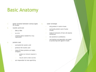
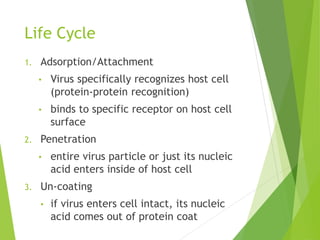
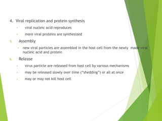
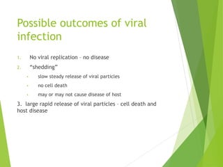
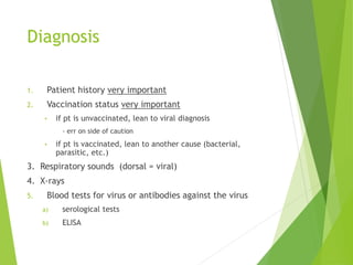
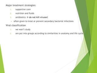
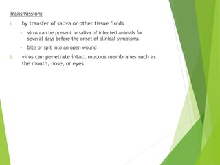
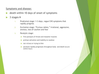
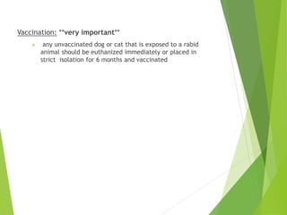
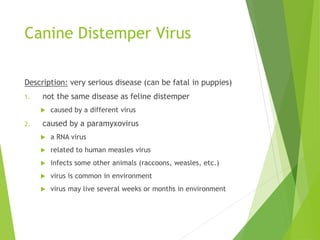
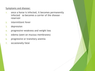
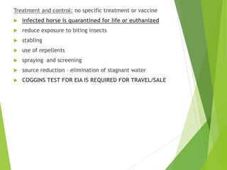
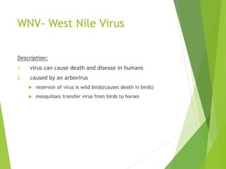
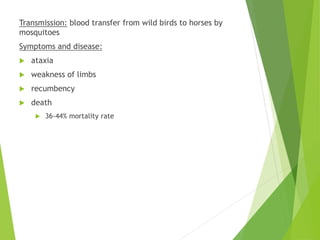
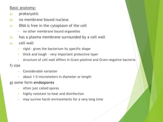
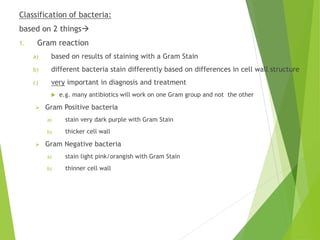
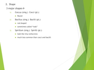
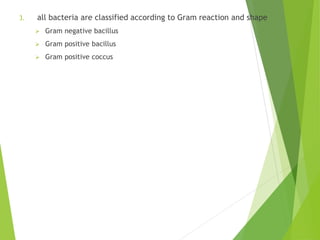
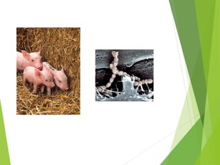
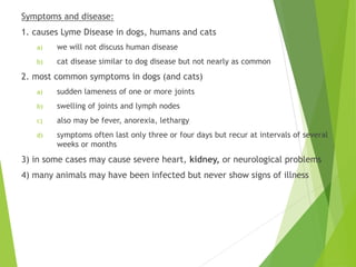
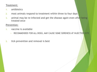
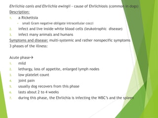
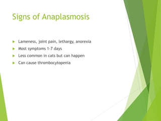
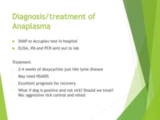
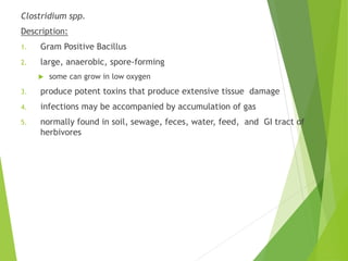
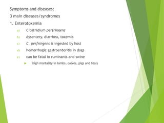
Microbiology:
Viruses
Description
1. non-cellular infectious particle
2. obligate intracellular parasites
• can only live and reproduce inside other cells
Basic Anatomy
1. great variation between various types
of viruses
2. nucleic acid core
• DNA or RNA
• ss or ds
• contains genes needed for virus
replication
3. protein coat
• surrounds the nucleic acid
• protects the nucleic acid
• many of these proteins are highly
antigenic
• produce an immune response in
host
• may be used to make vaccines
• are responsible for host specificity
4. outer envelope
• only present in some viruses
• surrounds protein coat of some
viruses
• made of remnants of host cell plasma
membrane
• not sensitive to antibiotics
• vaccinations are generally very good
at protecting host against viruses
Life Cycle
1. Adsorption/Attachment
• Virus specifically recognizes host cell
(protein-protein recognition)
• binds to specific receptor on host cell
surface
2. Penetration
• entire virus particle or just its nucleic
acid enters inside of host cell
3. Un-coating
• if virus enters cell intact, its nucleic
acid comes out of protein coat
4. Viral replication and protein synthesis
• viral nucleic acid reproduces
• more viral proteins are synthesized
5. Assembly
• new viral particles are assembled in the host cell from the newly made viral
nucleic acid and protein
6. Release
• virus particle are released from host cell by various mechanisms
• may be released slowly over time (“shedding”) or all at once
• may or may not kill host cell
Possible outcomes of viral
infection
1. No viral replication – no disease
2. “shedding”
• slow steady release of viral particles
• no cell death
• may or may not cause disease of host
3. large rapid release of viral particles – cell death and
host disease
Diagnosis
1. Patient history very important
2. Vaccination status very important
• if pt is unvaccinated, lean to viral diagnosis
- err on side of caution
• if pt is vaccinated, lean to another cause (bacterial,
parasitic, etc.)
3. Respiratory sounds (dorsal = viral)
4. X-rays
5. Blood tests for virus or antibodies against the virus
a) serological tests
b) ELISA
Major treatment strategies:
1. supportive care
2. nutrition and fluids
3. antibiotics  do not kill viruses!
 often given to treat or prevent secondary bacterial infections
Viral classification
1. we won’t study
2. are put into groups according to similarities in anatomy and life cycle
Common Veterinary Viral
Infections:
FeLV- Feline Leukemia Virus
Description:
• often fatal
• a retrovirus – small ss RNA virus
• virus is easily killed in the environment
• Prevalence had decreased significantly due to vaccine
Transmission:
1. direct contact through saliva and nasal secretions of
infected cats
2. Virus found mostly in saliva but can be in nasal secretions,
urine, feces and milk of infected cats
3. Cat to cat transfer from bite wounds, mutual grooming
and rarely with shared litter boxes and feeding dishes
4. Queen to kitten
- either before they are born or from nursing
- most kittens successfully eliminate the virus (so retest
young kittens)
Persistently infected cats may shed virus for many years in
body secretions
• shared litter boxes, toys, food dishes
• nose to nose contact, sneezing
Symptoms and disease:
1. It is the most common cause of cancer in cats
2. Can cause blood disorders and may lead to a stage of
immune deficiency that leads to more infections
3. Common bacteria, viruses, protozoa and fungi that
don’t affect healthy cats can cause severe illness in
infected cats
4. In early stages of infection the cat may show no signs
( rec test all cats)
5. Sick cats show, poor condition, enlarged ln, fever,
inflammation of gums, infections , persistent
diarrhea, eye issues, neurologic disorders and may
have abortion or repro issues
Diagnosis:
1. ELISA test: in house, detects free FeLV particles in blood
2. IFA test: at the lab, detects virus particles in white blood
cells and is indication of more advanced disease, cats
positive for this test usually remain infected for life
Treatment: supportive care, antibiotics for infections and may
require blood transfusion
Vaccination: effective, but not 100%
Prognosis : median survival time 2.5 years, careful monitoring
and treatment can prolong it
FIV: Feline Immunodeficiency
Virus
Description: feline form of HIV
 a retrovirus
 Attacks the immune system
 Median survival time 5 years
Transmission:
- bite wounds from infected cats so free roaming, aggressive
male cats are most frequently infected
- Housemates that don’t fight should not spread the disease,
casual, non aggressive contact does spread the virus
- kittens may be infected in the birth canal or from infected
milk
Symptoms and disease:
1. Early stage of the disease is often missed but shows fever
and enlarged LN
2. Signs of immunodeficiency and deterioration of cats
health
3. Persistent fever, loss of appetite, gingivitis, persistent
diarrhea, neurologic disorders
4. Cancer and blood disorders
Diagnosis: dependent of cat mounting an immune response to
virus, if no antibodies have been formed, the tests may be
negative
1. ELISA
2. Western blot
3. IFA assay
Positive for FIV
 Few if any cats eliminate the infection so presence of
antibodies indicates cat is infected with FIV
 False positives can occur so retesting is important
 Kittens born to infected mothers may show positive for
several months but few of these kittens will become
infected
 Kittens younger than 6 months that test positive for FIV
should be retested at 60 day intervals until 6 months of
age
 FIV vaccine causes cats to test positive so rec microchip
these cats
Negative FIV
 Cat has not produced antibodies to FIV
 In most cases suggests cat is not infected
 Usually takes 12 weeks for antibodies to appear
 RETEST every 60 days
 PCR testing may help
FIV: treatment, management,
prognosis
 No cure but many of these cats live normal lives if
managed properly
 Spayed or neutered, fed balanced diet, kept inside to
prevent transmission and exposure to other diseases or
bacteria
 Test all cats before introducing into the household
 Prevent CAT BITES
 Before introducing a new cat into a household that
previously held a FIV cat, clean or replace all cat toys,
bedding, food bowls etc
FPV: Feline Panleukopenia
Virus
Uncommon due to effective vaccines
Description:
 feline distemper
 a parvovirus (very small ssDNA virus) can use dog parvo
test to screen for it.
 virus can survive extreme temperatures and humidity
for years
 can also survive many common disinfectants
Transmission:
1. very contagious disease, seen in pet shops, shelters
and feral cat colonies
2. Virus shed in urine, stool, and nasal secretions
3. Infected cat usually shed for a short period, 1-2 days,
but virus can survive up to 1 year in environment
4. sharing food, water, bedding, litter boxes with other
cats
5. mostly by contact with feces of another cat
6. also by direct contact with infected cat
7. in utero from infected mother to offspring
 cerebellar hyperplasia in offspring, or abortion
Symptoms and disease:
1. very severe and often fatal – some cats die suddenly
2. Leukopenia is a common in bloodwork
3. depression, loss of appetite, fever, vomiting, diarrhea
4. pregnant cat may have stillborn kittens
5. Kittens are most severely affected, usually 3-5 months of
age
6. Virus infects and kills cells that are rapidly growing and
dividing such as in bone marrow, intestines and the
developing fetus
Prevention: vaccinations very effective disease rare
FP treatment/prevention
 Recovery of cats under 8 weeks of age is poor
 Supportive care since no antibiotics kill virus
 Correct dehydration, provide nutrition, use antibiotics
to prevent secondary infection
 Some recovered cats can shed virus for up to 6 weeks
 Recovered cats are immune
Prevention
- Vaccinate with the first vaccine between 6-8 weeks and
up to 16 weeks of age
FIP: Feline Infectious
Peritonitis
**very serious and often fatal**
Description:
 caused by feline coronavirus
 there are a variety of feline coronaviruses, most of which
do not cause severe disease
 a benign strain can mutate to the disease forming virus and
cause illness
 1-5% of cats with a feline coronavirus develop FIP
 therefore you have asymptomatic carriers of benign
Coranaviruses
 diagnostic tests do not distinguish between carriers of
harmless virus and those with FIP
Transmission:
1. Any cat carrying any coronavirus is at risk for developing
FIP
2. Weak immune systems like kittens, cats with FeLV and
geriatric cats are more likely to develop FIP, most cats
that develop it are under 2 years of age
3. FIP is not a highly contagious disease, by the time clinical
disease is present only a small amount of virus is being
shed
4. Coronavirus is found in large quantities in saliva and feces
of cats during acute infection so it can be spread cat to
cat and with exposure to feces
5. Most common transmission female cats pass the virus to
kittens between 5 and 8 weeks of age
 Much more prevalent in multi cat, shelters and catteries
FIP symptoms
 Initially mild URI symptoms or mild intestinal disease
and show diarrhea
 Loss of appetite, weight loss, depression, fever
 Two major forms
1. Wet
- Fluid in abdomen and less commonly in chest, become
pot-bellied , fever, weight loss, anorexia
1. Dry ( slower)
- Chronic weight loss, depression, anemia, persistent fever
not responsive to antibiotics
FIP testing
 Testing for FIP
- No simple diagnostic test
- ELISA, IFA detect coronavirus antibodies but can’t
differentiate the FIP causing coronavirus
- PCR test is being developed to help in diagnosis
FIP
 FIP Treatment : none
 FIP prevention:
1. Minimize exposure
2. Litter boxes clean and away from food and water,
regular cleaning and disinfection
3. Prevent overcrowding, adequate vaccinations, good
food
FCV: Feline Calicivirus
Description:
 a calicivirus (small naked ss RNA virus)
 part of the feline respiratory disease complex
 feline rhinotracheitis virus
 feline herpes virus also
 feline respiratory disease complex accounts for 85-90%
of upper respiratory infections in cats (URI’s)
 virus can survive outside the body for 8 to 10 days
 in dishes, litter boxes, clothing
Transmission:
1. contact with nasal, ocular, or oral secretions and feces
of infected cats
2. by direct contact or contact with a contaminated
object (feed bowl, toy, etc.)
Symptoms and disease: ( incubation 2-14 days)
1. mild flu-like, like a cold
• sneezing, runny nose, fever. Loss of appetite, sores around
mouth, nose, lips, upper respiratory disease, possibly
arthritis (lameness)
2. usually not serious in adults: recover but may have
gingivitis for life
3. may be fatal in kittens
Diagnosis/treatment/prevention
 Diagnose: swabs from eyes, nose or mouth sent out to
lab, interpret results carefully as healthy cats can test
positive and recent vaccination with a modified live
strain can test positive
 Treatment: nursing care to keep cat eating and prevent
secondary infection
 Prevention: no vaccine 100%, nasal form of vaccine
leads to more rapid protection, start vaccines at 6-8
weeks. Vaccine often in combination with distemper
Feline Viral Rhinotracheitis:
Herpevirus
 Caused by feline herpesvirus type – 1
 Major cause of upper respiratory disease and is the most
common cause of conjunctivitis
 Cat becomes infected by direct contact, spread in saliva
and in discharges from eyes and nose of infected cat
 Infection when cat comes into direct contact with an
infected cat or object with virus particles ( clothing,
food, water dishes, furniture)
 Infection usually lasts 10-20 days but shows disease in 2-
5 days ( incubation period) and can infect other cats
during this incubation period
 ALL CATS WILL BECOME CARRIERS AND MANY WILL BE
LATENT INFECTIONS THAT STRESS WILL CAUSE TO FLARE
UP
 VIRUS CAN SURVIVE IN ENVIRONMENT AS LONG AS IT
STAYS MOIST USUALLY A FEW HOURS
 ON SKIN ABOUT A HALF AN HOUR
 BLEACH SOLUTIONS CAN KILL THE VIRUS AND FURNITURE
SHAMPOOS WILL KILL VIRUS AS WELL
 Most severe in young
 Sneezing, nasal congestion, conjuctivits, discharge from
eyes, keratitis: inflammation/scarring/dry eye of
cornea
 Diagnose by collecting swabs and sending to lab
Treatment of FVR
 Treat symptomatically, manage chronic cases
 Topical eye meds
 Check for dry eye /ulcers and treat accordingly
 Steam bathroom to open up nasal or airway congestion
 Polyprenyl immunostimulant to boost cats immune
system
 Can infect other cats from incubation period and up to 3
weeks
 Prevent with vaccination: not 100% usually combined
with calici and distemper
Rabies Virus
Description:
1. virus can infect all mammalian species
2. virus selectively attacks the nervous system
3. viral incubation period from 2 weeks to several
months
Transmission:
1. by transfer of saliva or other tissue fluids
• virus can be present in saliva of infected animals for
several days before the onset of clinical symptoms
• bite or spit into an open wound
2. virus can penetrate intact mucous membranes such as
the mouth, nose, or eyes
Symptoms and disease:
 death within 10 days of onset of symptoms
 3 stages
1. Prodromal stage: 1-3 days, vague CNS symptoms that
rapidly progress
2. Excitation stage: “Furious rabies,” irrational, aggressive,
anxious, loss of caution and fear
3. Paralytic stage:
 first paralysis of throat and masseter muscles
 profuse salivation and inability to swallow
 not vicious or trying to bite
 paralysis rapidly progresses throughout body and death occurs
within hours
Vaccination: **very important**
 any unvaccinated dog or cat that is exposed to a rabid
animal should be euthanized immediately or placed in
strict isolation for 6 months and vaccinated
Canine Distemper Virus
Description: very serious disease (can be fatal in puppies)
1. not the same disease as feline distemper
 caused by a different virus
2. caused by a paramyxovirus
 a RNA virus
 related to human measles virus
 infects some other animals (raccoons, weasles, etc.)
 virus is common in environment
 virus may live several weeks or months in environment
Transmission:
1. highly contagious
2. spread by aerosol-droplet route upper respiratory
discharge is inhaled by another dog
3. direct contact
4. possibly by contact with contaminated objects
Symptoms and disease:
1. fever
2. coughing, labored breathing
3. conjunctivitis with ocular discharge
4. runny nose
5. vomiting, diarrhea
6. dogs may become anorectic
7. hyperkeratosis of foot pads and nose**
8. may be severe neurological symptoms esp. late in infection
9. dog will eventually die or have permanent neurological damage
Prevention:
1. vaccination is very effective and should be routine for every dog
2. routine disinfection effective
3. removal from population of dogs to prevent spread
Canine Parvovirus
Description: very serious disease high death rate
1. caused by parvovirus
2. ss DNA virus
3. very tough and resistant
4. survives for long time in environment
5. most common viral disease of dogs
6. mainly affects puppies up to 1 year of age
7. virus attacks and kills rapidly dividing cells
 intestines
 lymph nodes
 bone marrow
 heart muscle
Transmission:
1. Fecal oral
2. virus discharged in feces
3. dogs eat fecal contaminated material
Symptoms and disease:
1. diarrhea- often bloody
2. vomiting
3. leucopenia, neutropenia
4. fever, lethargy
5. anorexia
6. may cause “sudden” death in puppies due to infection of heart muscle
“Kennel Cough”
Description:
1. infectious tracheobronchitis
2. can be caused by about 13 different infectious agents
3. usually caused by one of two viruses
 CAV-2: Canine Adenovirus-2
 CPI: Canine Parainfluenza Virus
4. usually mild and self-limiting
5. can progress into fatal pneumonia
6. often leads to secondary infection with Bordetella
(bacterium)
 may cause most of the symptoms
Transmission:
1. highly contagious
2. often infects dogs in close confinement with other
dogs kennels, shelters, pet stores, hospitals
3. oronasal contact
Symptoms and disease:
1. cough
2. anorexia
3. sometimes nasal discharge
4. symptoms usually fade in 5-20 days
Treatment and prevention: vaccinations not totally effective
– many possible causes of kennel cough
EIA – Equine Infectious Anemia
“Swamp Fever”
Description:
1. persistent viral disease of horses-
• acute or chronic
2. caused by a virus related to human HIV virus
3. caused by an arbovirus
• arthropod borne virus
• carried from animal to animal by mosquitoes and other blood
sucking insects
Transmission: blood transfer from one animal to another
 usually by blood sucking insects
Symptoms and disease:
1. once a horse is infected, it becomes permanently
infected so becomes a carrier of the disease –
reservoir
2. intermittent fever
3. depression
4. progressive weakness and weight loss
5. edema (seen on mucous membranes)
6. progressive or transitory anemia
7. occasionally fatal
Treatment and control: no specific treatment or vaccine
 infected horse is quarantined for life or euthanized
 reduce exposure to biting insects
 stabling
 use of repellents
 spraying and screening
 source reduction – elimination of stagnant water
 COGGINS TEST FOR EIA IS REQUIRED FOR TRAVEL/SALE
WNV- West Nile Virus
Description:
1. virus can cause death and disease in humans
2. caused by an arbovirus
 reservoir of virus is wild birds(causes death in birds)
 mosquitoes transfer virus from birds to horses
Transmission: blood transfer from wild birds to horses by
mosquitoes
Symptoms and disease:
 ataxia
 weakness of limbs
 recumbency
 death
 36-44% mortality rate
Treatment and control:
 no vaccine
 prevent exposure to mosquitoes
 see EIA controls
Equine Viral Encephalitides“Sleeping
Sickness”
Description:
1. most common forms:
 EEE Eastern Equine Encephalitis
 WEE Western Equine Encephalitis
 VEE Venezuelan Equine Encephalitis
 WNV is also considered an equine viral encephalitis
2. arboviruses are the most common cause
a) Togaviruses most pathogenic
b) can be non-viral causes of equine encephalitis also
3. birds and rodents are most common reservoirs of virus
Transmission: from reservoir animal to horses by mosquito bites
Symptoms and disease:
1. CNS symptoms – see WNV
2. mortality rates are high
a) EEE – 75-100%
b) WEE – 30-50%
c) VEE – 40-80%
Treatment and control:
 vaccine available
 prevent exposure to mosquitoes
Bacteria
Description:
1. Kingdom Eubacteria
2. living cells
3. usually free-living but may be obligate intracellular
parasites (Rickettsia)
Basic anatomy:
a) prokaryotic
b) no membrane bound nucleus
c) DNA is free in the cytoplasm of the cell
• no other membrane bound organelles
d) has a plasma membrane surrounded by a cell wall
e) cell wall
• rigid – gives the bacterium its specific shape
• thick and tough - very important protective layer
• structure of cell wall differs in Gram positive and Gram negative bacteria
f) size
• Considerable variation
• about 1-5 micrometers in diameter or length
g) some form endospores
• often just called spores
• highly resistant to heat and disinfection
• may survive harsh environments for a very long time
Classification of bacteria:
based on 2 things
1. Gram reaction
a) based on results of staining with a Gram Stain
b) different bacteria stain differently based on differences in cell wall structure
c) very important in diagnosis and treatment
 e.g. many antibiotics will work on one Gram group and not the other
 Gram Positive bacteria
a) stain very dark purple with Gram Stain
b) thicker cell wall
 Gram Negative bacteria
a) stain light pink/orangish with Gram Stain
b) thinner cell wall
2. Shape
3 major shapes
1) Coccus (sing.) Cocci (pl.)
 Round
2) Bacillus (sing.) Bacilli (pl.)
 rod shaped
 sometimes called “rods”
3) Spirillum (sing.) Spirilli (pl.)
 look like tiny corkscrews
 much less common than cocci and bacilli
3. all bacteria are classified according to Gram reaction and shape
 Gram negative bacillus
 Gram positive bacillus
 Gram positive coccus
Life Cycle:
1. very simple
2. no immature or juvenile stages
3. divide by binary fission
 basically one adult divides into two new adults
Diagnosis:
1. Involves collection of specimen from patient
 must be done aseptically
2. May involve direct microscopic evaluation of live bacteria
3. Usually requires culture of the bacterium on culture medium
4. Usually requires Gram Staining and other lab tests to identify
5. May use antibody tests
6. Culture may be sent to another lab for definitive diagnosis
Major treatment strategies:
1. antibiotics – antimicrobial agents
2. each antibiotic is effective against specific groups of
microorganisms
 called the spectrum of the antibiotic
3. many bacteria have become resistant to specific
antibiotics
 should test cultures for antibiotic sensitivity
Some common veterinary bacterial infections:
1. Staphylococcus spp.
2. Description:
• Gram Positive Coccus
• occurs in clumps – looks like clumps of grapes
• extremely resistant to destruction in the environment
• ubiquitous – they are everywhere
• normally present on skin- can infect body through skin lesions or through
mucous membranes
• most common Staphylococcus aureus
Symptoms and diseases:
often isolated from pyogenic lesions
• infected wounds
• dermatitis
• otitis
• conjunctivitis
• cystitis
… many others
Mastitis especially in ruminants- causes decrease in milk
production and visible change in quality of milk
common cause of skin and subcutaneous infections
common cause of urinary infections
Gram stain of Staph
2. Streptococcus spp.
Description:
• Gram Positive Coccus
• occurs in chains or pairs – looks like beads on a string
• normally found on mucous membranes
 Tonsils
 Skin
 intestinal tract
 bovine udder
… others
• some are fecal
• all but the fecal strep do not survive long in environment
Transmission:
 through direct contact or aerosol
 often through milker’s hands and milking machines
Symptoms and diseases:
1. can cause pyogenic infections and mastitis in all mammals
2. Dairy Cows and Goats
a) one of the most common causes of mastitis
b) can be acute or chronic
c) can cause great economic losses in dairy industry
3. Horses
a) causes “Strangles”
b) also called “Equine Distemper”
c) Streptococcus equi
d) causes pus-like discharge from nose and swollen throat
and lower jaw
• hard to swallow – “strangles”
• lymph nodes in neck fill with pus and often burst
• fever, depression, etc.
e) diagnosis – culture S. equi from pus from nostrils
f) usually not fatal – horse usually recovers
g) very contagious within a herd
• passed easily by food, water, tack
• direct or indirect contact
h) vaccine available
• nasal aerosol vaccine is very good
i) not zoonotic
Swine
a) Streptococcus suis
b) can cause meningitis or septicemia
c) meningitis – infects CNS
• lameness, convulsions, paralysis, death
• death may be sudden
d) septicemia – blood infection
• “fading piglet syndrome”
• mostly in weanling pigs
• pneumonia often leading to sudden death
e) zoonotic
• passed to humans through wounds on skin or on mucus membranes
• can cause bacterial meningitis or TSS (Toxic Shock Syndrome) in humans.
*Caution* – may have asymptomatic carrier pigs
3. Borrelia burgdorferi – Lyme Disease
Description:
• a Gram Positive Spirullium
• slow-growing bacterium
• does not grow well in most environments
 except grows very well in some animal hosts
 reservoir host – voles and mice
 transport hosts -“deer ticks” or “black-legged ticks”
a) in Northeast mostly Ixodes scapularis
b) pick up organisms from mice and voles and transport them to humans,
cats, dogs, other animals
c)mostly found in grasslands and woodlands and like to feed on deer,
sheep, horses and rodents
Symptoms and disease:
1. causes Lyme Disease in dogs, humans and cats
a) we will not discuss human disease
b) cat disease similar to dog disease but not nearly as common
2. most common symptoms in dogs (and cats)
a) sudden lameness of one or more joints
b) swelling of joints and lymph nodes
c) also may be fever, anorexia, lethargy
d) symptoms often last only three or four days but recur at intervals of several
weeks or months
3) in some cases may cause severe heart, kidney, or neurological problems
4) many animals may have been infected but never show signs of illness
Treatment:
1. antibiotics
2. most animals respond to treatment within three to four days
3. animal may be re-infected and get the disease again even after being
treated once
Prevention:
1. vaccine is available
• RECOMMENDED FOR ALL DOGS, MAY CAUSE SOME SORENESS AT INJECTION SITE
2. tick prevention and removal is best
Ehrlichia canis and Ehrlichia ewingii - cause of Ehrlichiosis (common in dogs)
Description:
1. a Rickettsia
• small Gram negative obligate intracellular cocci
2. infect and live inside white blood cells (leukotrophic disease)
3. infect many animals and humans
Symptoms and disease: multi-systemic and rather nonspecific symptoms
3 phases of the illness:
Acute phase
1. mild
2. lethargy, loss of appetite, enlarged lymph nodes
3. low platelet count
4. joint pain
5. usually dog recovers from this phase
6. lasts about 2 to 4 weeks
7. during this phase, the Ehrlichia is infecting the WBC’s and the spleen
Subclinical phase
1. dogs appear to be normal
2. this stage can last up to several years
3. organism is hiding in the spleen
Chronic phase
1. dog gets sick again
2. may have abnormal bleeding (low platelet count)
3. kidney damage, loss of blood cells
4. may think the dog has leukemia at this Stage
5. may be neurological symptoms and arthritis
Transmission: ticks - mainly the brown dog tick (Rhipicephalus spp.) and other
ticks as well
Diagnosis and treatment:
1. fever, enlarged lymph nodes, bleeding
2. low platelet counts
3. blood test
4. treatment with tetracycline type antibiotics
Prevention: tick control
Anaplasmosis in Dogs
 Tick born disease caused by Anaplasma phagocytophilum
 Transmitted by deer tick and western black legged tick
 Lesser form Anaplasma platys transmitted by brown dog
tick
Signs of Anaplasmosis
 Lameness, joint pain, lethargy, anorexia
 Most symptoms 1-7 days
 Less common in cats but can happen
 Can cause thrombocytopenia
Diagnosis/treatment of
Anaplasma
 SNAP or Accuplex test in hospital
 ELISA, IFA and PCR sent out to lab
Treatment
- 2-4 weeks of doxycycline just like lyme disease
- May need NSAIDS
- Excellent prognosis for recovery
- What if dog is positive and not sick? Should we treat?
Rec aggressive tick control and retest
Clostridium spp.
Description:
1. Gram Positive Bacillus
2. large, anaerobic, spore-forming
 some can grow in low oxygen
3. produce potent toxins that produce extensive tissue damage
4. infections may be accompanied by accumulation of gas
5. normally found in soil, sewage, feces, water, feed, and GI tract of
herbivores
Symptoms and diseases:
3 main diseases/syndromes
1. Enterotoxemia
a) Clostridium perfringens
b) dysentery. diarrhea, toxemia
c) C. perfringens is ingested by host
d) hemorrhagic gastroenteritis in dogs
e) can be fatal in ruminants and swine
 high mortality in lambs, calves, pigs and foals
2. Tetanus
a) Clostridium tetani
b) all species of domestic animals (esp. in horses)
c) bacterium usually enters body through puncture wound
d) muscular spasms
 esp. in head and neck
 lockjaw (masseter muscle)
e) death usually by dehydration
3. Botulism
a) Clostridium botulinum
b) C. botulinum produces a very powerful neurotoxin
c) can be ingested or enter host through a wound
d) ataxia, recumbency, paralysis of hind quarters
Escherichia coli
Description:
1. small Gram Negative Rods (or coccobacilli)
2. normally part of intestinal flora but can cause disease under the right
circumstances
3. fecal-oral route is most common means of infection
Symptoms and disease:
1. cause of diarrhea, vomiting, gastroenteritis in many types of animals
2. dehydration and loss of electrolytes may cause death
3. especially dangerous to suckling and weanling pigs and neonatal calves
4. may also cause urinary tract infections or respiratory infections
5. also may cause mastitis in dairy cows
Salmonella, Klebsiella, and Proteus
 similar to E.coli
 all these are called enteric organisms
Pasteurella multocida
Description:
1. small Gram Negative Bacilli (or coccobacilli)
2. causes a wide variety of diseases in many different animals
Symptoms and disease:
1. serious infection in animals and humans
2. wound infection in dogs and cats
• normally found in mouth of dogs and cats
• opportunistic pathogen
3. respiratory tract infections in most animals
4. snuffles in rabbits
• common and VERY IMPORTANT
• pneumonia and upper respiratory infection
• sneezing, coughing, runny eyes and nose
5. cause of swine plague
• pneumonia-like symptoms
• high fever, cough, anorexia
• almost always a secondary infection
Treatment: Penicillin and other antibiotics
Zoonotic:
1. through bites and scratches from animal
2. pneumonia in humans
• usually not fatal in humans
Bordetella
Description:
1. small Gram Negative Bacilli (and coccobacilli)
2. normally found in upper respiratory tract of dogs and swine
3. often causes secondary infection in dogs with distemper
4. spread by droplet inhalation
Symptoms and disease:
1. a cause of kennel cough or bronchopneumonia in dogs
2. often associated with distemper
3. honking, unproductive cough
Leptospirosis in Dogs
 Caused by Leptospira
 Causes serious damage to kidney and liver and may be
fatal in severe cases
 ZOONOTIC
 Passed in urine and dogs are exposed in rivers, streams,
puddles that have become infected with urine or
rodents or other dogs
 Become infected and develop lepto if mm or any wound
come in contact with infected urine, urine
contaminated soil, water, food or bedding or bite from
infected animal or eating infected tissues , can also be
passed through placenta
Signs of leptospirosis
 No signs, mild illness, severe illness and death
 Fever of unknown origin, increased thirst, increased
urine, loss of appetite, lethargy, jaundice,
 Can cause kidney failure with or without liver failure
 Can cause bleeding disorders, blood tinged vomit, urine
or stool, nosebleeds, swollen legs or excess fluid in
abdomen
Treatment and prevention of
leptospirosis
 Antibiotics and supportive care
 Early and aggressive treatment usually works
 Avoid contact with urine
 VACCINATE
Fungi
Description:
1. study of fungi called mycology
• myco means fungi
2. belong to Kingdom Fungi
3. includes molds and yeasts
4. yeasts – unicellular fungi
5. molds – multicellular fungi
• form tubular filaments called hyphae
• tangled mass of hyphae is called a mycelium
• produce fuzzy looking colonies of growth
• strictly aerobic
• all are Gram Positive
6. reproduce by:
1. fragmentation of cells
2. budding of cells (yeast)
3. producing spores
 mold spores are highly resistant
 not as highly resistant as bacterial spores
 round or oval microscopic structures
7. essentially all are zoonotic
Types of diseases
1. fungi are saprophytes so their pathogenicity is
accidental
2. cause disease in three ways
a) systemic or subcutaneous infection
1) these are called deep mycoses
2) not that common
3) fungus directly invades healthy tissue
4) are usually normally present in animal and causes disease when host is very weak
b) Mycotoxicoses
1) fungi produce toxic substances that cause disease when
ingested
2) mainly caused by molds growing on feed stocks (stored grains)
3) usually causes subclinical symptoms of poor growth,
depression, salivation, kidney liver problems, etc.
c) cutaneous infections
1) called superficial mycoses
2) fungi causing these are called dermatophytes
3) fungi are living on dead tissues
 hair, nails, outer layer of skin
4) may not evoke immune response from host
5) may cause irritation
 itching, etc
Some common fungal infections:
1. Yeast infections- these are deep mycoses
Cryptococcus:
a) disease is called cryptococcosis
b) subacute or chronic disease of dogs, cats, farm animals and people
c) respiratory disease in dogs and cats
d) not sensitive to antibiotics
Dermatophytes:
a) do not penetrate the skin – live on superficial dead skin
b) often called “ringworm” - lesions are typically circular
c) diagnosis by:
 skin scrapings followed by microscopic exam and/or culture
 Wood’s lamp – 40% of ringworms are fluorescent under black light
d) treatment: topical or internal antimycotics
Microsporum canis
1. main cause of ringworm in dogs and cats
2. lesions can be all over body, but are mostly on head, ears, and eyes
3. lesions are circular areas of hair loss with scaling and crusting
2020 microbiology viruses final

2020 microbiology viruses final

  • 1.
  • 2.
    Description 1. non-cellular infectiousparticle 2. obligate intracellular parasites • can only live and reproduce inside other cells
  • 3.
    Basic Anatomy 1. greatvariation between various types of viruses 2. nucleic acid core • DNA or RNA • ss or ds • contains genes needed for virus replication 3. protein coat • surrounds the nucleic acid • protects the nucleic acid • many of these proteins are highly antigenic • produce an immune response in host • may be used to make vaccines • are responsible for host specificity 4. outer envelope • only present in some viruses • surrounds protein coat of some viruses • made of remnants of host cell plasma membrane • not sensitive to antibiotics • vaccinations are generally very good at protecting host against viruses
  • 4.
    Life Cycle 1. Adsorption/Attachment •Virus specifically recognizes host cell (protein-protein recognition) • binds to specific receptor on host cell surface 2. Penetration • entire virus particle or just its nucleic acid enters inside of host cell 3. Un-coating • if virus enters cell intact, its nucleic acid comes out of protein coat
  • 5.
    4. Viral replicationand protein synthesis • viral nucleic acid reproduces • more viral proteins are synthesized 5. Assembly • new viral particles are assembled in the host cell from the newly made viral nucleic acid and protein 6. Release • virus particle are released from host cell by various mechanisms • may be released slowly over time (“shedding”) or all at once • may or may not kill host cell
  • 6.
    Possible outcomes ofviral infection 1. No viral replication – no disease 2. “shedding” • slow steady release of viral particles • no cell death • may or may not cause disease of host 3. large rapid release of viral particles – cell death and host disease
  • 7.
    Diagnosis 1. Patient historyvery important 2. Vaccination status very important • if pt is unvaccinated, lean to viral diagnosis - err on side of caution • if pt is vaccinated, lean to another cause (bacterial, parasitic, etc.) 3. Respiratory sounds (dorsal = viral) 4. X-rays 5. Blood tests for virus or antibodies against the virus a) serological tests b) ELISA
  • 8.
    Major treatment strategies: 1.supportive care 2. nutrition and fluids 3. antibiotics  do not kill viruses!  often given to treat or prevent secondary bacterial infections Viral classification 1. we won’t study 2. are put into groups according to similarities in anatomy and life cycle
  • 9.
    Common Veterinary Viral Infections: FeLV-Feline Leukemia Virus Description: • often fatal • a retrovirus – small ss RNA virus • virus is easily killed in the environment • Prevalence had decreased significantly due to vaccine
  • 10.
    Transmission: 1. direct contactthrough saliva and nasal secretions of infected cats 2. Virus found mostly in saliva but can be in nasal secretions, urine, feces and milk of infected cats 3. Cat to cat transfer from bite wounds, mutual grooming and rarely with shared litter boxes and feeding dishes 4. Queen to kitten - either before they are born or from nursing - most kittens successfully eliminate the virus (so retest young kittens) Persistently infected cats may shed virus for many years in body secretions • shared litter boxes, toys, food dishes • nose to nose contact, sneezing
  • 11.
    Symptoms and disease: 1.It is the most common cause of cancer in cats 2. Can cause blood disorders and may lead to a stage of immune deficiency that leads to more infections 3. Common bacteria, viruses, protozoa and fungi that don’t affect healthy cats can cause severe illness in infected cats 4. In early stages of infection the cat may show no signs ( rec test all cats) 5. Sick cats show, poor condition, enlarged ln, fever, inflammation of gums, infections , persistent diarrhea, eye issues, neurologic disorders and may have abortion or repro issues
  • 12.
    Diagnosis: 1. ELISA test:in house, detects free FeLV particles in blood 2. IFA test: at the lab, detects virus particles in white blood cells and is indication of more advanced disease, cats positive for this test usually remain infected for life Treatment: supportive care, antibiotics for infections and may require blood transfusion Vaccination: effective, but not 100% Prognosis : median survival time 2.5 years, careful monitoring and treatment can prolong it
  • 13.
    FIV: Feline Immunodeficiency Virus Description:feline form of HIV  a retrovirus  Attacks the immune system  Median survival time 5 years Transmission: - bite wounds from infected cats so free roaming, aggressive male cats are most frequently infected - Housemates that don’t fight should not spread the disease, casual, non aggressive contact does spread the virus - kittens may be infected in the birth canal or from infected milk
  • 14.
    Symptoms and disease: 1.Early stage of the disease is often missed but shows fever and enlarged LN 2. Signs of immunodeficiency and deterioration of cats health 3. Persistent fever, loss of appetite, gingivitis, persistent diarrhea, neurologic disorders 4. Cancer and blood disorders Diagnosis: dependent of cat mounting an immune response to virus, if no antibodies have been formed, the tests may be negative 1. ELISA 2. Western blot 3. IFA assay
  • 15.
    Positive for FIV Few if any cats eliminate the infection so presence of antibodies indicates cat is infected with FIV  False positives can occur so retesting is important  Kittens born to infected mothers may show positive for several months but few of these kittens will become infected  Kittens younger than 6 months that test positive for FIV should be retested at 60 day intervals until 6 months of age  FIV vaccine causes cats to test positive so rec microchip these cats
  • 16.
    Negative FIV  Cathas not produced antibodies to FIV  In most cases suggests cat is not infected  Usually takes 12 weeks for antibodies to appear  RETEST every 60 days  PCR testing may help
  • 17.
    FIV: treatment, management, prognosis No cure but many of these cats live normal lives if managed properly  Spayed or neutered, fed balanced diet, kept inside to prevent transmission and exposure to other diseases or bacteria  Test all cats before introducing into the household  Prevent CAT BITES  Before introducing a new cat into a household that previously held a FIV cat, clean or replace all cat toys, bedding, food bowls etc
  • 18.
    FPV: Feline Panleukopenia Virus Uncommondue to effective vaccines Description:  feline distemper  a parvovirus (very small ssDNA virus) can use dog parvo test to screen for it.  virus can survive extreme temperatures and humidity for years  can also survive many common disinfectants
  • 19.
    Transmission: 1. very contagiousdisease, seen in pet shops, shelters and feral cat colonies 2. Virus shed in urine, stool, and nasal secretions 3. Infected cat usually shed for a short period, 1-2 days, but virus can survive up to 1 year in environment 4. sharing food, water, bedding, litter boxes with other cats 5. mostly by contact with feces of another cat 6. also by direct contact with infected cat 7. in utero from infected mother to offspring  cerebellar hyperplasia in offspring, or abortion
  • 20.
    Symptoms and disease: 1.very severe and often fatal – some cats die suddenly 2. Leukopenia is a common in bloodwork 3. depression, loss of appetite, fever, vomiting, diarrhea 4. pregnant cat may have stillborn kittens 5. Kittens are most severely affected, usually 3-5 months of age 6. Virus infects and kills cells that are rapidly growing and dividing such as in bone marrow, intestines and the developing fetus Prevention: vaccinations very effective disease rare
  • 21.
    FP treatment/prevention  Recoveryof cats under 8 weeks of age is poor  Supportive care since no antibiotics kill virus  Correct dehydration, provide nutrition, use antibiotics to prevent secondary infection  Some recovered cats can shed virus for up to 6 weeks  Recovered cats are immune Prevention - Vaccinate with the first vaccine between 6-8 weeks and up to 16 weeks of age
  • 22.
    FIP: Feline Infectious Peritonitis **veryserious and often fatal** Description:  caused by feline coronavirus  there are a variety of feline coronaviruses, most of which do not cause severe disease  a benign strain can mutate to the disease forming virus and cause illness  1-5% of cats with a feline coronavirus develop FIP  therefore you have asymptomatic carriers of benign Coranaviruses  diagnostic tests do not distinguish between carriers of harmless virus and those with FIP
  • 23.
    Transmission: 1. Any catcarrying any coronavirus is at risk for developing FIP 2. Weak immune systems like kittens, cats with FeLV and geriatric cats are more likely to develop FIP, most cats that develop it are under 2 years of age 3. FIP is not a highly contagious disease, by the time clinical disease is present only a small amount of virus is being shed 4. Coronavirus is found in large quantities in saliva and feces of cats during acute infection so it can be spread cat to cat and with exposure to feces 5. Most common transmission female cats pass the virus to kittens between 5 and 8 weeks of age  Much more prevalent in multi cat, shelters and catteries
  • 24.
    FIP symptoms  Initiallymild URI symptoms or mild intestinal disease and show diarrhea  Loss of appetite, weight loss, depression, fever  Two major forms 1. Wet - Fluid in abdomen and less commonly in chest, become pot-bellied , fever, weight loss, anorexia 1. Dry ( slower) - Chronic weight loss, depression, anemia, persistent fever not responsive to antibiotics
  • 25.
    FIP testing  Testingfor FIP - No simple diagnostic test - ELISA, IFA detect coronavirus antibodies but can’t differentiate the FIP causing coronavirus - PCR test is being developed to help in diagnosis
  • 26.
    FIP  FIP Treatment: none  FIP prevention: 1. Minimize exposure 2. Litter boxes clean and away from food and water, regular cleaning and disinfection 3. Prevent overcrowding, adequate vaccinations, good food
  • 27.
    FCV: Feline Calicivirus Description: a calicivirus (small naked ss RNA virus)  part of the feline respiratory disease complex  feline rhinotracheitis virus  feline herpes virus also  feline respiratory disease complex accounts for 85-90% of upper respiratory infections in cats (URI’s)  virus can survive outside the body for 8 to 10 days  in dishes, litter boxes, clothing
  • 28.
    Transmission: 1. contact withnasal, ocular, or oral secretions and feces of infected cats 2. by direct contact or contact with a contaminated object (feed bowl, toy, etc.) Symptoms and disease: ( incubation 2-14 days) 1. mild flu-like, like a cold • sneezing, runny nose, fever. Loss of appetite, sores around mouth, nose, lips, upper respiratory disease, possibly arthritis (lameness) 2. usually not serious in adults: recover but may have gingivitis for life 3. may be fatal in kittens
  • 29.
    Diagnosis/treatment/prevention  Diagnose: swabsfrom eyes, nose or mouth sent out to lab, interpret results carefully as healthy cats can test positive and recent vaccination with a modified live strain can test positive  Treatment: nursing care to keep cat eating and prevent secondary infection  Prevention: no vaccine 100%, nasal form of vaccine leads to more rapid protection, start vaccines at 6-8 weeks. Vaccine often in combination with distemper
  • 30.
    Feline Viral Rhinotracheitis: Herpevirus Caused by feline herpesvirus type – 1  Major cause of upper respiratory disease and is the most common cause of conjunctivitis  Cat becomes infected by direct contact, spread in saliva and in discharges from eyes and nose of infected cat  Infection when cat comes into direct contact with an infected cat or object with virus particles ( clothing, food, water dishes, furniture)  Infection usually lasts 10-20 days but shows disease in 2- 5 days ( incubation period) and can infect other cats during this incubation period  ALL CATS WILL BECOME CARRIERS AND MANY WILL BE LATENT INFECTIONS THAT STRESS WILL CAUSE TO FLARE UP
  • 31.
     VIRUS CANSURVIVE IN ENVIRONMENT AS LONG AS IT STAYS MOIST USUALLY A FEW HOURS  ON SKIN ABOUT A HALF AN HOUR  BLEACH SOLUTIONS CAN KILL THE VIRUS AND FURNITURE SHAMPOOS WILL KILL VIRUS AS WELL  Most severe in young  Sneezing, nasal congestion, conjuctivits, discharge from eyes, keratitis: inflammation/scarring/dry eye of cornea  Diagnose by collecting swabs and sending to lab
  • 32.
    Treatment of FVR Treat symptomatically, manage chronic cases  Topical eye meds  Check for dry eye /ulcers and treat accordingly  Steam bathroom to open up nasal or airway congestion  Polyprenyl immunostimulant to boost cats immune system  Can infect other cats from incubation period and up to 3 weeks  Prevent with vaccination: not 100% usually combined with calici and distemper
  • 33.
    Rabies Virus Description: 1. viruscan infect all mammalian species 2. virus selectively attacks the nervous system 3. viral incubation period from 2 weeks to several months
  • 34.
    Transmission: 1. by transferof saliva or other tissue fluids • virus can be present in saliva of infected animals for several days before the onset of clinical symptoms • bite or spit into an open wound 2. virus can penetrate intact mucous membranes such as the mouth, nose, or eyes
  • 35.
    Symptoms and disease: death within 10 days of onset of symptoms  3 stages 1. Prodromal stage: 1-3 days, vague CNS symptoms that rapidly progress 2. Excitation stage: “Furious rabies,” irrational, aggressive, anxious, loss of caution and fear 3. Paralytic stage:  first paralysis of throat and masseter muscles  profuse salivation and inability to swallow  not vicious or trying to bite  paralysis rapidly progresses throughout body and death occurs within hours
  • 36.
    Vaccination: **very important** any unvaccinated dog or cat that is exposed to a rabid animal should be euthanized immediately or placed in strict isolation for 6 months and vaccinated
  • 37.
    Canine Distemper Virus Description:very serious disease (can be fatal in puppies) 1. not the same disease as feline distemper  caused by a different virus 2. caused by a paramyxovirus  a RNA virus  related to human measles virus  infects some other animals (raccoons, weasles, etc.)  virus is common in environment  virus may live several weeks or months in environment
  • 38.
    Transmission: 1. highly contagious 2.spread by aerosol-droplet route upper respiratory discharge is inhaled by another dog 3. direct contact 4. possibly by contact with contaminated objects
  • 39.
    Symptoms and disease: 1.fever 2. coughing, labored breathing 3. conjunctivitis with ocular discharge 4. runny nose 5. vomiting, diarrhea 6. dogs may become anorectic 7. hyperkeratosis of foot pads and nose** 8. may be severe neurological symptoms esp. late in infection 9. dog will eventually die or have permanent neurological damage
  • 40.
    Prevention: 1. vaccination isvery effective and should be routine for every dog 2. routine disinfection effective 3. removal from population of dogs to prevent spread
  • 41.
    Canine Parvovirus Description: veryserious disease high death rate 1. caused by parvovirus 2. ss DNA virus 3. very tough and resistant 4. survives for long time in environment 5. most common viral disease of dogs 6. mainly affects puppies up to 1 year of age 7. virus attacks and kills rapidly dividing cells  intestines  lymph nodes  bone marrow  heart muscle
  • 42.
    Transmission: 1. Fecal oral 2.virus discharged in feces 3. dogs eat fecal contaminated material Symptoms and disease: 1. diarrhea- often bloody 2. vomiting 3. leucopenia, neutropenia 4. fever, lethargy 5. anorexia 6. may cause “sudden” death in puppies due to infection of heart muscle
  • 43.
    “Kennel Cough” Description: 1. infectioustracheobronchitis 2. can be caused by about 13 different infectious agents 3. usually caused by one of two viruses  CAV-2: Canine Adenovirus-2  CPI: Canine Parainfluenza Virus 4. usually mild and self-limiting 5. can progress into fatal pneumonia 6. often leads to secondary infection with Bordetella (bacterium)  may cause most of the symptoms
  • 44.
    Transmission: 1. highly contagious 2.often infects dogs in close confinement with other dogs kennels, shelters, pet stores, hospitals 3. oronasal contact Symptoms and disease: 1. cough 2. anorexia 3. sometimes nasal discharge 4. symptoms usually fade in 5-20 days Treatment and prevention: vaccinations not totally effective – many possible causes of kennel cough
  • 45.
    EIA – EquineInfectious Anemia “Swamp Fever” Description: 1. persistent viral disease of horses- • acute or chronic 2. caused by a virus related to human HIV virus 3. caused by an arbovirus • arthropod borne virus • carried from animal to animal by mosquitoes and other blood sucking insects Transmission: blood transfer from one animal to another  usually by blood sucking insects
  • 46.
    Symptoms and disease: 1.once a horse is infected, it becomes permanently infected so becomes a carrier of the disease – reservoir 2. intermittent fever 3. depression 4. progressive weakness and weight loss 5. edema (seen on mucous membranes) 6. progressive or transitory anemia 7. occasionally fatal
  • 47.
    Treatment and control:no specific treatment or vaccine  infected horse is quarantined for life or euthanized  reduce exposure to biting insects  stabling  use of repellents  spraying and screening  source reduction – elimination of stagnant water  COGGINS TEST FOR EIA IS REQUIRED FOR TRAVEL/SALE
  • 48.
    WNV- West NileVirus Description: 1. virus can cause death and disease in humans 2. caused by an arbovirus  reservoir of virus is wild birds(causes death in birds)  mosquitoes transfer virus from birds to horses
  • 49.
    Transmission: blood transferfrom wild birds to horses by mosquitoes Symptoms and disease:  ataxia  weakness of limbs  recumbency  death  36-44% mortality rate
  • 50.
    Treatment and control: no vaccine  prevent exposure to mosquitoes  see EIA controls
  • 51.
    Equine Viral Encephalitides“Sleeping Sickness” Description: 1.most common forms:  EEE Eastern Equine Encephalitis  WEE Western Equine Encephalitis  VEE Venezuelan Equine Encephalitis  WNV is also considered an equine viral encephalitis 2. arboviruses are the most common cause a) Togaviruses most pathogenic b) can be non-viral causes of equine encephalitis also 3. birds and rodents are most common reservoirs of virus
  • 52.
    Transmission: from reservoiranimal to horses by mosquito bites Symptoms and disease: 1. CNS symptoms – see WNV 2. mortality rates are high a) EEE – 75-100% b) WEE – 30-50% c) VEE – 40-80% Treatment and control:  vaccine available  prevent exposure to mosquitoes
  • 53.
    Bacteria Description: 1. Kingdom Eubacteria 2.living cells 3. usually free-living but may be obligate intracellular parasites (Rickettsia)
  • 54.
    Basic anatomy: a) prokaryotic b)no membrane bound nucleus c) DNA is free in the cytoplasm of the cell • no other membrane bound organelles d) has a plasma membrane surrounded by a cell wall e) cell wall • rigid – gives the bacterium its specific shape • thick and tough - very important protective layer • structure of cell wall differs in Gram positive and Gram negative bacteria f) size • Considerable variation • about 1-5 micrometers in diameter or length g) some form endospores • often just called spores • highly resistant to heat and disinfection • may survive harsh environments for a very long time
  • 55.
    Classification of bacteria: basedon 2 things 1. Gram reaction a) based on results of staining with a Gram Stain b) different bacteria stain differently based on differences in cell wall structure c) very important in diagnosis and treatment  e.g. many antibiotics will work on one Gram group and not the other  Gram Positive bacteria a) stain very dark purple with Gram Stain b) thicker cell wall  Gram Negative bacteria a) stain light pink/orangish with Gram Stain b) thinner cell wall
  • 56.
    2. Shape 3 majorshapes 1) Coccus (sing.) Cocci (pl.)  Round 2) Bacillus (sing.) Bacilli (pl.)  rod shaped  sometimes called “rods” 3) Spirillum (sing.) Spirilli (pl.)  look like tiny corkscrews  much less common than cocci and bacilli
  • 57.
    3. all bacteriaare classified according to Gram reaction and shape  Gram negative bacillus  Gram positive bacillus  Gram positive coccus
  • 58.
    Life Cycle: 1. verysimple 2. no immature or juvenile stages 3. divide by binary fission  basically one adult divides into two new adults Diagnosis: 1. Involves collection of specimen from patient  must be done aseptically 2. May involve direct microscopic evaluation of live bacteria 3. Usually requires culture of the bacterium on culture medium 4. Usually requires Gram Staining and other lab tests to identify 5. May use antibody tests 6. Culture may be sent to another lab for definitive diagnosis
  • 59.
    Major treatment strategies: 1.antibiotics – antimicrobial agents 2. each antibiotic is effective against specific groups of microorganisms  called the spectrum of the antibiotic 3. many bacteria have become resistant to specific antibiotics  should test cultures for antibiotic sensitivity
  • 60.
    Some common veterinarybacterial infections: 1. Staphylococcus spp. 2. Description: • Gram Positive Coccus • occurs in clumps – looks like clumps of grapes • extremely resistant to destruction in the environment • ubiquitous – they are everywhere • normally present on skin- can infect body through skin lesions or through mucous membranes • most common Staphylococcus aureus
  • 61.
    Symptoms and diseases: oftenisolated from pyogenic lesions • infected wounds • dermatitis • otitis • conjunctivitis • cystitis … many others Mastitis especially in ruminants- causes decrease in milk production and visible change in quality of milk common cause of skin and subcutaneous infections common cause of urinary infections
  • 62.
  • 63.
    2. Streptococcus spp. Description: •Gram Positive Coccus • occurs in chains or pairs – looks like beads on a string • normally found on mucous membranes  Tonsils  Skin  intestinal tract  bovine udder … others • some are fecal • all but the fecal strep do not survive long in environment Transmission:  through direct contact or aerosol  often through milker’s hands and milking machines
  • 64.
    Symptoms and diseases: 1.can cause pyogenic infections and mastitis in all mammals 2. Dairy Cows and Goats a) one of the most common causes of mastitis b) can be acute or chronic c) can cause great economic losses in dairy industry 3. Horses a) causes “Strangles” b) also called “Equine Distemper” c) Streptococcus equi
  • 66.
    d) causes pus-likedischarge from nose and swollen throat and lower jaw • hard to swallow – “strangles” • lymph nodes in neck fill with pus and often burst • fever, depression, etc. e) diagnosis – culture S. equi from pus from nostrils f) usually not fatal – horse usually recovers g) very contagious within a herd • passed easily by food, water, tack • direct or indirect contact h) vaccine available • nasal aerosol vaccine is very good i) not zoonotic
  • 67.
    Swine a) Streptococcus suis b)can cause meningitis or septicemia c) meningitis – infects CNS • lameness, convulsions, paralysis, death • death may be sudden d) septicemia – blood infection • “fading piglet syndrome” • mostly in weanling pigs • pneumonia often leading to sudden death e) zoonotic • passed to humans through wounds on skin or on mucus membranes • can cause bacterial meningitis or TSS (Toxic Shock Syndrome) in humans. *Caution* – may have asymptomatic carrier pigs
  • 69.
    3. Borrelia burgdorferi– Lyme Disease Description: • a Gram Positive Spirullium • slow-growing bacterium • does not grow well in most environments  except grows very well in some animal hosts  reservoir host – voles and mice  transport hosts -“deer ticks” or “black-legged ticks” a) in Northeast mostly Ixodes scapularis b) pick up organisms from mice and voles and transport them to humans, cats, dogs, other animals c)mostly found in grasslands and woodlands and like to feed on deer, sheep, horses and rodents
  • 70.
    Symptoms and disease: 1.causes Lyme Disease in dogs, humans and cats a) we will not discuss human disease b) cat disease similar to dog disease but not nearly as common 2. most common symptoms in dogs (and cats) a) sudden lameness of one or more joints b) swelling of joints and lymph nodes c) also may be fever, anorexia, lethargy d) symptoms often last only three or four days but recur at intervals of several weeks or months 3) in some cases may cause severe heart, kidney, or neurological problems 4) many animals may have been infected but never show signs of illness
  • 71.
    Treatment: 1. antibiotics 2. mostanimals respond to treatment within three to four days 3. animal may be re-infected and get the disease again even after being treated once Prevention: 1. vaccine is available • RECOMMENDED FOR ALL DOGS, MAY CAUSE SOME SORENESS AT INJECTION SITE 2. tick prevention and removal is best
  • 73.
    Ehrlichia canis andEhrlichia ewingii - cause of Ehrlichiosis (common in dogs) Description: 1. a Rickettsia • small Gram negative obligate intracellular cocci 2. infect and live inside white blood cells (leukotrophic disease) 3. infect many animals and humans Symptoms and disease: multi-systemic and rather nonspecific symptoms 3 phases of the illness: Acute phase 1. mild 2. lethargy, loss of appetite, enlarged lymph nodes 3. low platelet count 4. joint pain 5. usually dog recovers from this phase 6. lasts about 2 to 4 weeks 7. during this phase, the Ehrlichia is infecting the WBC’s and the spleen
  • 74.
    Subclinical phase 1. dogsappear to be normal 2. this stage can last up to several years 3. organism is hiding in the spleen Chronic phase 1. dog gets sick again 2. may have abnormal bleeding (low platelet count) 3. kidney damage, loss of blood cells 4. may think the dog has leukemia at this Stage 5. may be neurological symptoms and arthritis
  • 75.
    Transmission: ticks -mainly the brown dog tick (Rhipicephalus spp.) and other ticks as well Diagnosis and treatment: 1. fever, enlarged lymph nodes, bleeding 2. low platelet counts 3. blood test 4. treatment with tetracycline type antibiotics Prevention: tick control
  • 77.
    Anaplasmosis in Dogs Tick born disease caused by Anaplasma phagocytophilum  Transmitted by deer tick and western black legged tick  Lesser form Anaplasma platys transmitted by brown dog tick
  • 78.
    Signs of Anaplasmosis Lameness, joint pain, lethargy, anorexia  Most symptoms 1-7 days  Less common in cats but can happen  Can cause thrombocytopenia
  • 79.
    Diagnosis/treatment of Anaplasma  SNAPor Accuplex test in hospital  ELISA, IFA and PCR sent out to lab Treatment - 2-4 weeks of doxycycline just like lyme disease - May need NSAIDS - Excellent prognosis for recovery - What if dog is positive and not sick? Should we treat? Rec aggressive tick control and retest
  • 80.
    Clostridium spp. Description: 1. GramPositive Bacillus 2. large, anaerobic, spore-forming  some can grow in low oxygen 3. produce potent toxins that produce extensive tissue damage 4. infections may be accompanied by accumulation of gas 5. normally found in soil, sewage, feces, water, feed, and GI tract of herbivores
  • 81.
    Symptoms and diseases: 3main diseases/syndromes 1. Enterotoxemia a) Clostridium perfringens b) dysentery. diarrhea, toxemia c) C. perfringens is ingested by host d) hemorrhagic gastroenteritis in dogs e) can be fatal in ruminants and swine  high mortality in lambs, calves, pigs and foals
  • 82.
    2. Tetanus a) Clostridiumtetani b) all species of domestic animals (esp. in horses) c) bacterium usually enters body through puncture wound d) muscular spasms  esp. in head and neck  lockjaw (masseter muscle) e) death usually by dehydration
  • 83.
    3. Botulism a) Clostridiumbotulinum b) C. botulinum produces a very powerful neurotoxin c) can be ingested or enter host through a wound d) ataxia, recumbency, paralysis of hind quarters
  • 84.
    Escherichia coli Description: 1. smallGram Negative Rods (or coccobacilli) 2. normally part of intestinal flora but can cause disease under the right circumstances 3. fecal-oral route is most common means of infection Symptoms and disease: 1. cause of diarrhea, vomiting, gastroenteritis in many types of animals 2. dehydration and loss of electrolytes may cause death 3. especially dangerous to suckling and weanling pigs and neonatal calves 4. may also cause urinary tract infections or respiratory infections 5. also may cause mastitis in dairy cows
  • 85.
    Salmonella, Klebsiella, andProteus  similar to E.coli  all these are called enteric organisms
  • 86.
    Pasteurella multocida Description: 1. smallGram Negative Bacilli (or coccobacilli) 2. causes a wide variety of diseases in many different animals Symptoms and disease: 1. serious infection in animals and humans 2. wound infection in dogs and cats • normally found in mouth of dogs and cats • opportunistic pathogen 3. respiratory tract infections in most animals 4. snuffles in rabbits • common and VERY IMPORTANT • pneumonia and upper respiratory infection • sneezing, coughing, runny eyes and nose 5. cause of swine plague • pneumonia-like symptoms • high fever, cough, anorexia • almost always a secondary infection
  • 88.
    Treatment: Penicillin andother antibiotics Zoonotic: 1. through bites and scratches from animal 2. pneumonia in humans • usually not fatal in humans
  • 89.
    Bordetella Description: 1. small GramNegative Bacilli (and coccobacilli) 2. normally found in upper respiratory tract of dogs and swine 3. often causes secondary infection in dogs with distemper 4. spread by droplet inhalation Symptoms and disease: 1. a cause of kennel cough or bronchopneumonia in dogs 2. often associated with distemper 3. honking, unproductive cough
  • 91.
    Leptospirosis in Dogs Caused by Leptospira  Causes serious damage to kidney and liver and may be fatal in severe cases  ZOONOTIC  Passed in urine and dogs are exposed in rivers, streams, puddles that have become infected with urine or rodents or other dogs  Become infected and develop lepto if mm or any wound come in contact with infected urine, urine contaminated soil, water, food or bedding or bite from infected animal or eating infected tissues , can also be passed through placenta
  • 92.
    Signs of leptospirosis No signs, mild illness, severe illness and death  Fever of unknown origin, increased thirst, increased urine, loss of appetite, lethargy, jaundice,  Can cause kidney failure with or without liver failure  Can cause bleeding disorders, blood tinged vomit, urine or stool, nosebleeds, swollen legs or excess fluid in abdomen
  • 93.
    Treatment and preventionof leptospirosis  Antibiotics and supportive care  Early and aggressive treatment usually works  Avoid contact with urine  VACCINATE
  • 94.
    Fungi Description: 1. study offungi called mycology • myco means fungi 2. belong to Kingdom Fungi 3. includes molds and yeasts 4. yeasts – unicellular fungi 5. molds – multicellular fungi • form tubular filaments called hyphae • tangled mass of hyphae is called a mycelium • produce fuzzy looking colonies of growth • strictly aerobic • all are Gram Positive
  • 95.
    6. reproduce by: 1.fragmentation of cells 2. budding of cells (yeast) 3. producing spores  mold spores are highly resistant  not as highly resistant as bacterial spores  round or oval microscopic structures 7. essentially all are zoonotic
  • 96.
    Types of diseases 1.fungi are saprophytes so their pathogenicity is accidental 2. cause disease in three ways a) systemic or subcutaneous infection 1) these are called deep mycoses 2) not that common 3) fungus directly invades healthy tissue 4) are usually normally present in animal and causes disease when host is very weak
  • 97.
    b) Mycotoxicoses 1) fungiproduce toxic substances that cause disease when ingested 2) mainly caused by molds growing on feed stocks (stored grains) 3) usually causes subclinical symptoms of poor growth, depression, salivation, kidney liver problems, etc. c) cutaneous infections 1) called superficial mycoses 2) fungi causing these are called dermatophytes 3) fungi are living on dead tissues  hair, nails, outer layer of skin 4) may not evoke immune response from host 5) may cause irritation  itching, etc
  • 98.
    Some common fungalinfections: 1. Yeast infections- these are deep mycoses Cryptococcus: a) disease is called cryptococcosis b) subacute or chronic disease of dogs, cats, farm animals and people c) respiratory disease in dogs and cats d) not sensitive to antibiotics
  • 99.
    Dermatophytes: a) do notpenetrate the skin – live on superficial dead skin b) often called “ringworm” - lesions are typically circular c) diagnosis by:  skin scrapings followed by microscopic exam and/or culture  Wood’s lamp – 40% of ringworms are fluorescent under black light d) treatment: topical or internal antimycotics Microsporum canis 1. main cause of ringworm in dogs and cats 2. lesions can be all over body, but are mostly on head, ears, and eyes 3. lesions are circular areas of hair loss with scaling and crusting