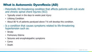
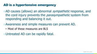
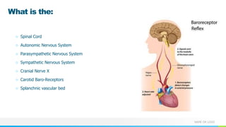
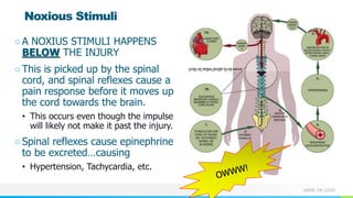
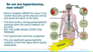
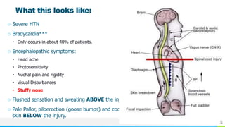
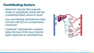
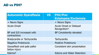
2. NAME OR LOGO
â Define autonomic dysreflexia
(AD)
â Understand pathophysiology
of AD
â Identify the characteristics of
AD
â Understand challenges in the
identification, prevention and
management of AD
â Discuss prehospital
considerations in the acute
management of AD.
2
Objectives
3. NAME OR LOGO
What is Autonomic Dysreflexia (AD)
â Potentially life threatening condition that affects patients with sub acute
and chronic spinal chord injuries (SCI)
⢠Typically onset in the days to weeks post injury
⢠Lifelong Condition
⢠About 80 % of patients paralyzed above T-6 will develop this condition.
â Is a condition that causes symptoms related to life-threatening
hypertension such as:
⢠Stroke
⢠Pulmonary Edema
⢠Seizures and encephalopathic symptoms
⢠Coma
⢠Death
4. NAME OR LOGO
Brief History
â Documented on ancient papyrus 5000 years ago as an âAilment not to be treatedâ
â Described again in 1890
â Head and Riddoch first described autonomic dysreflexia in 1917
â Significance and dangers described by:
⢠Whitteridge and Guttman 1946-1947
⢠Thompson and Whitham 1948
⢠Pollock et al. 1951
⢠Bors and French 1952
⢠Schreibert 1955
⢠Arieff et al. 1962
â Most current guidelines build on the 2001 Paralyzed Veterans Association recommendations âAcute
Management of Autonomic Dysreflexiaâ
5. NAME OR LOGO
Interesting side note
âAutonomic Dysreflexia is a
prominent condition suffered
by the fictional forensic
expert, Lincoln âLinkâ Rhyme
in the 1999 movie, The Bone
Collector.
â(They didnât do a very good
job of portraying it)
6. NAME OR LOGO
Who can get AD? (Epidemiology)
â Spinal Cord Injury (SCI) patients
â Roughly estimated 1,462,220 SCI (all types) in US
alone*
â Tetraplegics and high level parapalegics:
⢠300-500K above T-6
⢠12,500 â 20,000 new cases each year.
⢠Majority are above T6, very rarely between T10 and
T6.
⢠91% with complete injury vs 27% with incomplete
injury 27%
⢠Occurs in first few days to weeks.
â More males than females (4:1)
⢠Because of lifestyle instead of physiological
differences.
â * source: The Reeves Foundation
7. NAME OR LOGO
AD is a hypertensive emergency
âAD causes (allows) an abnormal sympathetic response, and
the cord injury prevents the parasympathetic system from
responding and balancing it out.
âAwareness and simple measures can prevent AD.
⢠Most of these measures are BLS
âUntreated AD can be rapidly fatal.
9. NAME OR LOGO
What is the:
â Spinal Cord
â Autonomic Nervous System
â Parasympathetic Nervous System
â Sympathetic Nervous System
â Cranial Nerve X
â Carotid Baro-Receptors
â Splanchnic vascular bed
10. NAME OR LOGO
A noxious stimuli causes a
sympathetic nervous response.
Parasympathetic NS typically
act in opposition to the
sympathetic NS.
Normally a complementary and
synergistic response, causing
a balance of sympathetic and
parasympathetic responses
In AD, this does not happen.
11. NAME OR LOGO
⢠This parasympathetic
response is primarily
mediated through
cranial nerve X (the
vagus nerve ), and the
S2, S3, and S4 spinal
nerves.
⢠This is important when
you consider that in AD,
there is usually an injury
between these two
parasympathetic
centers at T6 (or
above).
12. NAME OR LOGO
Noxious Stimuli
âA NOXIUS STIMULI HAPPENS
BELOW THE INJURY
âThis is picked up by the spinal
cord, and spinal reflexes cause a
pain response before it moves up
the cord towards the brain.
⢠This occurs even though the impulse
will likely not make it past the injury.
âSpinal reflexes cause epinephrine
to be excretedâŚcausing
⢠Hypertension, Tachycardia, etc.
13. NAME OR LOGO
So we are hypertensive,
now what?
â Barrow receptors ABOVE the injury in the
carotid sinus pick up the pressure increase
and sound the alarm to the brain.
â The brain sends a strong parasympathetic
response down the cord to balance out
the HTN.
BUT THE CORD INJURY STOPS THIS
MESSAGE.
â The hypertension continues unopposed
â The only significant parasympathetic
response is from the Vagus Nerve causing
bradycardia.
14. NAME OR LOGO
What this looks like:
â Severe HTN
â Bradycardia***
⢠Only occurs in about 40% of patients.
â Encephalopathic symptoms:
⢠Head ache
⢠Photosensitivity
⢠Nuchal pain and rigidity
⢠Visual Disturbances
⢠Stuffy nose
â Flushed sensation and sweating ABOVE the injury
â Pale Pallor, piloerection (goose bumps) and cool
skin BELOW the injury.
15. NAME OR LOGO
Contributing factors
â Splanchnic Vascular Bed responds
readily to sympathetic stimuli and has
a substantial blood volume to shunt.
â Low Level Resting catecholamine state
common with SCI as a compensatory
mechanism
â âorphanedâ catecholamine receptors
below the level of the injury become
hyper-responsive to catecholamines.
16. NAME OR LOGO
SymptomsâŚ
â Rise in SBP 20-30 mm Hg or more
above baseline.
⢠Remember that a SCI baseline is low
(approx. 80-100 mm Hg).
⢠Blood Pressures can rise to over 200
mm Hg systolic.
â Head ache
â Photosensitivity
â Nuchal pain and rigidity
â Visual Disturbances
â Stuffy nose
17. NAME OR LOGO
Adverse Sequala
âCNS complications (Most
frequent)
⢠Stroke, SZ,
⢠Retinal Detachment
âCardiovascular
⢠MI
⢠CHF
âPulmonary
⢠Pulmonary Edema
20. NAME OR LOGO
GU Causes
âBladder Distension
⢠Kinked Catheter
⢠Blocked Catheter
⢠Overfull Catheter
⢠Needs to be cathed
âUTI
⢠Very common with Caths
âKidney Stones
21. NAME OR LOGO
GI Problems
â âNeurogenic Bowelâ
⢠AKAâ âReflex Bowelâ
â Constipation
â Hemorrhoids
â Missing or overly rough bowell care
⢠Digital impaction or stimulation.
22. NAME OR LOGO
Skin Causes
â Pressure Sores
â Wrinkles in Sheets
â Position
â Tight or bunched up clothing
â Ingrown or infected toenail
â Blisters
â Burns
â Other injury or infection
23. NAME OR LOGO
Sexual Activity
â âPressureâ of sexual activity can cause AD
â KEY POINT: Many SCI patients require the use for
Viagra (or similar Rx) to engage with sex.
â This is a contraindication for NTG and NTG
paste.
24. NAME OR LOGO
AD, Pregnancy and Child Birth
â Can be very difficult to differentiate PIH/Pre-
eclampsia from AD
â Considerations:
⢠The risk of urinary tract infection is twice as high for a
pregnant woman as for a non-pregnant woman.
⢠Increased incidence of constipation
https://sciparenting.com/2014/04/29/pregnancy/
26. NAME OR LOGO
AD vs PIH?
Autonomic Dysreflexia VS PIH/Pre-
Eclampsia/Exclamsia
+ Neuro Signs + Neuro Signs
Acute Onset Acute Onset or Delayed
Recognition?
BP and S/S increased with
contractions
BP Consistently elevated
Bradycardia or Tachycardia Tachycardia
Negative Proteinuria Positive Proteinuria
Gooseflesh and pale pallor
below injury
Consistent skin presentation
Edema? Edema and Water Retention
28. NAME OR LOGO
In a nutshell, an ounce of prevention is worth a pound
of cureâŚ
âBowell and Bladder care
âFoley management
âPrevention of pressure sores and compression
âPrevention of constriction of clothing
âProper positioning
âProper analgesia
âFrequent BP Monitoring
29. NAME OR LOGO
Baclofen
â Baclofen is an anti-spasmodic
â Umps into the CSF in the sub arachnoid space of
the spinal chord
Withdrawal can mimic or precipitate AD
37. NAME OR LOGO
Treatment
â Sit the patient up if possible
â Relieve the noxious cause firstâŚ.
⢠CHECK THE CATHETER
⢠CHECK THE POSITION
⢠CHECK THE BELTS AND SHEETS
â Then treat the remaining hypertension (Typically if symptomatic AND above 150 mm Hg)
⢠Short acting anti-hypertensives
⢠NTG
⢠Calcium Channel Blockers - Procardia
38. NAME OR LOGO
Nitroglycerine
â Nitropaste
⢠0.5-1.5 inches TD
â Nitroglycerine Sub Lingual
⢠1-2 sprays Q 5 minutes
â *** Note that men with spinal cord injury often
use cGMP-specific phosphodiesterase type 5
(PDE5) inhibitors (eg, sildenafil, vardenafil,
tadalafil.) for sexual dysfunction. Use of nitrates
is contraindicated in these patients.
â Especially effective in Bladder mediated AD
39. NAME OR LOGO
Nifedipine (Procardia)
â 10 mg SubLingal
â Calcium Channel Blocker
â Short acting
â Sublingual + Bite and chew
40. NAME OR LOGO
Captopril
â ACE inhibitor
â May be absorbed slowly sublingually
â About 15 min to onset
â Dose: 25 mg SL
â Not as commonly used in the US
41. NAME OR LOGO
Beta blockers
â Off Label and exploratory use
â Metoprolol has been used in prevention for AD
refractory to other conditions.
â Strong concern for unopposed Alpha Adrenergic
vasoconstriction
â Moral: Prehospital use of Betablockers for AD is a
âlast resortâ on medical control only.
â Some limited evidence of combining Alpha and
Beta Adrenergic blockade for chronic (non-
emergent) management/prevention of AD.
42. NAME OR LOGO
Analgesia?
â For refractory hypertension, consider analgesia.
â Opioids provide MAY some central nervous system
analgesia
â Effectiveness is dubious, but acceptable in
refractory and severe AD.
Autonomic dysreflexia (AD) is a potentially life-threatening medical emergency that affects people with spinal cord injuries at the T6 level or higher. Although rare, some people with T7 and T8 injuries can develop AD. For most people, AD can be easily treated as well as prevented. The key is knowing your baseline blood pressure, triggers, and symptoms.
When triggered, AD requires quick and correct action or there may be serious consequence such as a stroke. Because many health professionals are not familiar with this condition, it is important for people who are at risk for AD, including the people close to them, to recognize the symptoms and know how to act.
Spinal Cord Injury itself was first discussed in a surgical papyrus dated over 5000 years ago. It described two cases of high cord injury (AKA: Quadriplegia or Tetraplegia) as an âailment not to be treatedâ. Until the advent of modern medicine, however, these patients seldom survived more than a few weeks. This has changed, with a typical patient surviving his injury for many, many years. With this ever-increasing life span, more and more conditions and complications of SCI are being discovered and managed. AD is one of these conditions.
Consortium for Spinal Cord Medicine, "Acute Management of Autonomic Dysreflexia: Adults with Spinal Cord Injury Presenting to Health-Care Facilities." Clinical Practice Guidelines 2nd editionJul 2001 29 MAR 2007 <http://www.pva.org/site/DocServer/AD2.pdf?docID=565>.AD was first reported in modern medical literature in 1890, and has presumably been present for as long as there have been a spinal cord injured patient, although prior to the advent of modern medicine the lifespan of these patients was probably very short. Fortunately, prevention, patient education, and home health care greatly reduce the field presentation of this syndrome. In fact many patients encountered may be more familiar with this condition than most health care providers.
Campagnolo, Denise I. "Autonomic Dysreflexia in Spinal Cord Injury." www.emedicine.com. 05 OCT 2006. 29 Mar 2007 <http://www.emedicine.com/pmr/topic217.htm>.
The occurrence of autonomic dysreflexia increases as the patient evolves out of spinal shock. With the return of sacral reflexes, the possibility of autonomic dysreflexia increases.Â
Spinal Cord
Autonomic Nervous System
Parasympathetic Nervous System
Sympathetic Nervous System
Cranial Nerve X
Carotid Baro Receptors
splanchnic vascular bedProbably the term most will be the least familiar with is the splanchnic vascular bed.The splanchnic circulation is a complex system. A number of important functions depend on its normal operation, including digestion and absorption within the gut, maintenance of the mucosal barrier, and successful healing of surgical anastomoses. Blood flow is approximately 30% of total cardiac output.
A noxious stimuli causes a sympathetic nervous response.
Parasympathetic NS typically act in opposition to the sympathetic NS.
Normally a complementary and synergistic response, causing a balance of sympathetic and parasympathetic responses
This parasympathetic response is primarily mediated through cranial nerve X ( the vagus nerve ), and the S2, S3, and S4 spinal nerves
In individuals with intact central and peripheral nervous systems, a noxious stimulus results initially in a sympathetic response, leading to elevation in heart rate and blood pressure primarily through spinal reflexes.Â
This response is modulated by the central nervous system and peripheral baroreceptors through the parasympathetic nervous system; this results in heart rate and blood pressure control both through direct responses by the vagus nerve and through inhibitory spinal cord signals. An appropriate balance of sympathetic and parasympathetic outflow is attained and modulated by both the central and peripheral nervous systems.
In individuals with intact central and peripheral nervous systems, a noxious stimulus results initially in a sympathetic response, leading to elevation in heart rate and blood pressure primarily through spinal reflexes. This response is modulated by the central nervous system and peripheral baroreceptors through the parasympathetic nervous system; this results in heart rate and blood pressure control both through direct responses by the vagus nerve and through inhibitory spinal cord signals. An appropriate balance of sympathetic and parasympathetic outflow is attained and modulated by both the central and peripheral nervous systems.
In those with a spinal cord injury at the level of T6 and above, a noxious (or otherwise strong) stimulus below the level of injury results in an unbalanced physiologic response. The strong stimulus causes a peripheral sympathetic response through spinal reflexes, resulting in vasoconstriction below the level of injury. This reflex response ascends and descends the spinal cord and paraspinal sympathetic ganglia, causing both direct vasoconstriction through activation of perivascular receptors and systemic/indirect vasoconstriction through stimulation of the adrenal medulla, resulting in epinephrine and norepinephrine release into the systemic circulation. This therefore results in hypertension, primarily through splanchnic and peripheral vasoconstriction.
The baroreceptors in the carotid sinus and aortic arch convey appropriate responses to hypertension through the petrosal ganglion to the nucleus ambiguous and result in strong vagal (CN X) outflow, bradycardia, and vasodilatation above the level of injury. The central nervous system cannot directly detect the strong or noxious signal below the level of injury (owing to the lack continuity of the ascending sensory fibers from the underlying spinal cord injury), and, therefore, responds to hypertension by sending a strong inhibitory response through the spinal cord aimed at reducing the sympathetic response. However, because of the lack of spinal cord continuity, the descending inhibitory response only travels as far as the level of neurologic injury and does not cause the desired response in the sympathetic fibers below the injury; therefore, the hypertension remains uncontrolled.
As a result, there is flushing and sweating only above the level of injury, bradycardia, pupillary constriction, and nasal congestion (unopposed parasympathetic responses); and below the level of injury, there is pale, cool skin and piloerection due to sympathetic tone and lack of the descending inhibitory parasympathetic modulation. [3]  (However, a study by Solinsky et al of 78 male patients with SCI who had incidents of autonomic dysfunction found that out of 445 episodes, relative tachycardia occurred in 68.0%, far more frequently than relative bradycardia [0.3%]. [28] )
T6 is of particular importance in the pathogenesis of autonomic dysreflexia. The splanchnic vascular bed is one of the bodyâs largest reserves of circulatory volume and is controlled primarily by the greater splanchnic nerve. This important nerve derives its innervation from T5-T9. Lesions to the spinal cord at or above T6 allow the strong and uninhibited sympathetic tone to constrict the splanchnic vascular bed, causing systemic hypertension. Lesions below T6 generally allow enough descending inhibitory parasympathetic control to modulate the splanchnic tone and prevent hyperteA patient with autonomic dysreflexia may have one or more of the following findings on physical examination:
Significant rise in systolic and diastolic blood pressure greater than 20 mm Hg systolic or 10 mm Hg
diastolic above baseline (The sudden rise in blood pressure in autonomic dysreflexia is usually associated
with bradycardia. Normal systolic blood pressure for an individual with spinal cord injury above T6 is 90-110
mm Hg; blood pressure of 20-40 mm Hg above the reference range for such patients may be a sign of
autonomic dysreflexia. However, patients with autonomic dysreflexia may display no symptoms, despite
elevated blood pressure.)
Profuse sweating above the level of lesion - Especially in the face, neck, and shoulders; rarely occurs
below the level of the lesion because of sympathetic activity
Goose bumps below the level of the lesion
Flushing of the skin above the level of the lesion - Especially in the face, neck, and shoulders; this is a
frequent symptom
Blurred vision
Nasal congestion â A common symptomnsion.
T6 is of particular importance in the pathogenesis of autonomic dysreflexia. The splanchnic vascular bed is one of the bodyâs largest reserves of circulatory volume and is controlled primarily by the greater splanchnic nerve. This important nerve derives its innervation from T5-T9. Lesions to the spinal cord at or above T6 allow the strong and uninhibited sympathetic tone to constrict the splanchnic vascular bed, causing systemic hypertension. Lesions below T6 generally allow enough descending inhibitory parasympathetic control to modulate the splanchnic tone and prevent hypertension.
The underlying pathophysiological changes that occur in the spinal cord and in the periphery that cause autonomic dysreflexia have not been fully elucidated in a human model. It has been postulated that peripheral alpha-adrenergic receptors associated with blood vessels become hyperresponsive below the level of the spinal cord lesion. This hyperresponsiveness is secondary to a low resting catecholamine state associated with spinal cord injury. The orphaned receptors have a decreased threshold to react to adrenergic stimuli and react with an increased responsiveness.The splanchnic vascular bed demonstrates an autoregulatory capacity similar to that seen in other vascular beds such as the renal and cerebral circulations. All of the splanchnic vasculature with the exclusion of the capillaries receive sympathetic innervation.
Warning signs vary from person to person: Some people have all of the signs, some only one or two; signs may be obvious in some and more subtle in others. So it's important for each person who is at risk of dysreflexia to get to know their own bodies and know how dysreflexia affects them.
The most important sign is a sudden, major increase in blood pressure of about 20 to 40 mm Hg (millimeters of mercury) higher than your normal blood pressure.
Know your normal blood pressure! Since people with SCI usually have a low resting blood pressure (80 or 90 systolic for a cervical injury), a rise to 130 or 140 systolicânormal in someone without SCIâcould be dangerously high for a person above T6.
Pounding headache.
Sweating above the level of the SCI.
Flushed or red skin (especially in face and neck).
Goose bumps.
Tightness in the chest, blurry vision, anxiety or jittersâoften reported as feeling like a panic attack.
Stuffy nose.
While all these signs and symptoms are uncomfortable and can be very annoying or upsetting, the increased blood pressure is what makes this a medical emergency.
After spinal cord injury, the bowel will no longer work like before the injury. If the injury is located at T12 or higher, the bowel will empty by a reflex. It will be called a "reflex bowel." This means that when the rectal vault is full of stool, it will increase the pressure and then stool will be pushed out. The key to continence is to empty the rectal vault before it becomes too full and pushes stool out at an unacceptable time.If the injury is at L1 or below it will not cause a reflex to happen. This type of bowel is called the "non-reflex bowel." There are differences between reflex and non reflex bowels and the type of program that works best.The bowel program for a "reflex bowel" is a dil (digital stimulation) and/or a suppository depending on a person's level of feeling near his/her anus/rectum.
After a SCI, men may get erections in one of three ways.
Psychogenic: For many men after SCI, having an erection by just thinking about something sexy may not work so well. This is because the area of the spinal cord responsible for erections is located between T11 and L2. Therefore, if your level of SCI is above this level, the message (sexy thoughts) from your brain cannot get through the damaged part of your spinal cord.
Reflexogenic: Some men with SCI may still get an erection when the catheter is being changed or when their penis is being cleaned or wiped or even by just pulling clothing or blankets over the body. This stimulation to the penis may actually produce an erection even if you really didnât want that to happen. Itâs important for you to pay close attention to what causes an erection as that may be useful for future occasions.
Spontaneous: Some men after SCI may still get an erection when their bladder is full. Depending on how long the erection lasts will help you determine its usefulness for sexual activity.
The ability to get a reflexogenic erection or spontaneous erection is controlled by nerves found in the lowest part of the spinal cord segments (S2-3-4).
There are no obstacles that prevent a woman with spinal cord injury from becoming pregnant. Studies show that a woman with spinal cord injury has the same prospects to give birth to a completely healthy child as a woman without spinal cord injury.  However, certain complications may be more common, such as an increased risk of blood clot, urinary tract infection, autonomic dysreflexia and pressure ulcers.The general recommendation is that SCI patients who desire to or become pregnant should be followed by a high risk center/specialist. Childbirth is a major triggering factor, but various conditions associated with pregnancy increase the risk, such as urinary tract infections, constipation, uterine pressure and Braxton Hicks contractions.Also, siode note: Women with complete injuries above T10 usually do not experience the ânormalâ labor pain associated with contractions. However, the body often displays other signs that something is going on, such as vague stomach discomfort, increased spasticity, nasal congestion, slight rise in blood pressure or even autonomic dysreflexia. Labor commonly begins with a change in vaginal discharge that is often blood-tinged. However, in some cases labor may begin without the woman realizing it. It is therefore important to be extra alert for body signals that something is happening and contact labor and delivery at the hospital, or the pregnancy center if you suspect that labor is about to begin. It is important for the pregnant woman to feel secure in the situation.
Preeclampsia and autonomic dysreflexia cause similar symptoms. It is therefore important to make the right diagnosis to determine which condition is present. It may be worth noting that blood pressure tends to normalize between contractions in an individual suffering from autonomic dysreflexia during childbirth. In contrast, high blood pressure is more persistent in individuals with preeclampsia. Both conditions can be life-threatening if left untreated.
Bladder
Intermittent catheterization should be regular and timely; only clean catheters should be used. Indwelling catheters
should be changed routinely and regularly checked for blockage or kinking.
Bowels
A regular bowel program is essential for the prevention of constipation, impaction, and ileus. Prior to a bowel
procedure, an anal block helps prevent autonomic dysreflexia.[21] Topical lidocaine may be of help.
Labor and delivery
Spinal anesthesia can help to prevent autonomic dysreflexia.
Pressure ulcers
Routine weight shifts and skin checks are necessary to prevent ulceration. Any skin breakdown should be
addressed early by a knowledgeable wound care team or physician.
A retrospective study by Del Fabro et al indicated that an intrathecal baclofen (ITB) pump can reduce the
incidence of autonomic dysreflexia in patients with SCI. The investigators found that prior to ITB pump placement,
25 out of the 34 patients (73.5%) in the study experienced autonomic dysreflexia, compared with two patients
(5.9%) following pump placement.[22]
Pathophysiology of Baclofen (Goldfrankâs 2015)
Inhibitory neurotransmitter that acts as a GABABÂ receptor agonist
Has both presynaptic and postsynaptic inhibitory properties
Presynaptic: prevents Ca2+Â influx
Postsynaptic: increases K+Â efflux
Inhibition results in decreased muscle tone and muscle spasms
Withdrawal likely results from loss of chronic inhibitory effect on postsynaptic receptors
Withdrawal typically occurs 24-48 hours after discontinuation of the drug or a reduction in dose
Intrathecal Baclofen Pumps
Catheter is placed into the cerebrospinal fluid (CSF) in the subarachnoid space
Catheter is connected to a pump which is typically inserted into the lower abdominal wall. This pump has a reservoir that can be filled with baclofen
Pump delivers baclofen in small amounts into the CSF causing inhibition at the spinal level
Causes of Baclofen Withdrawal (Stetkarova 2010)
Baclofen dose reduction or cessation
Intrathecal Pump Issues
Catheter dislodgement, kinking or migration
Reservoir underfilling: can occur if reservoir âmissedâ during medication refill leading to placement of medication in âpocketâ around pump
Pump device malfunction
Presentation
Baclofen withdrawal mimics symptoms seen with other CNS depressant withdrawal syndromes
History
The patient may report a reduction in baclofen dose or cessation of the drug
Increased spasticity
Fever
Myalgias
Neuropsychiatric Symptoms: confusion, altered mental status, visual hallucinations
Physical Examination
Vital Sign Alterations
Hypertension
Tachycardia
Hyperthermia
Muscle rigidity
Seizures
Note:Â Consider baclofen withdrawal in any patient who presents with vital sign abnormalities and has an intrathecal pump.Baclofen
Currently, baclofen is the pharmacologic agent of choice for the treatment of SCI-induced spasticity. Baclofen is a derivative of the inhibitory neurotransmitter gamma-aminobutyric acid (GABA), which binds to GABAB receptors of the lamina I-IV of the spinal cord where primary sensory fibers terminate [12]. Binding of baclofen to the presynaptic terminal of the gabaergic interneuron leads to membrane hyperpolarization, which in turn leads to a restriction of the influx of calcium into the presynaptic terminal and endogenous transmitter release is reduced [13, 14]. Baclofen is rapidly absorbed after oral administration and has a therapeutic half-life of about 3 to 4 hours. However, the use of this pharmacological agent can be limited by its adverse effects, which include sedation, fatigue, drowsiness, ataxia, and mental confusion [15â19], which can actually reduce the effectiveness of rehabilitation programs [11].
Intrathecal baclofen
In addition to the oral route of administration, baclofen has been shown to effectively manage spasticity when administered intrathecally [20â23]. Intrathecal administration is a long-term treatment with continuous, intra-spinal administration via a catheter connected to an implantable pump. The rationale for this modality of baclofen administration has been to deliver the drug directly into the spinal fluid to allow higher concentrations in the spinal cord using lower dosages than the oral route, thereby optimizing the beneficial effects of baclofen while minimizing the well-established side effects. There is currently one registered clinical trial that is examining intrathecal baclofen for the management of spasticity (Table 1). However, this clinical trial is not recruiting subjects yet. Predominate indications for intrathecal baclofen are intractable and generalized spasticity of the legs, which may also involve the trunk musculature [24]. Importantly, although continuous intrathecal infusion of baclofen can be effective in the treatment of severe spasticity, there is concern for baclofen tolerance in many individuals that remains a critical issue [25]. Another report indicates that abrupt withdrawal from intrathecal baclofen can lead to the onset of a potentially life-threatening condition, referred to as intrathecal baclofen withdrawal syndrome. This syndrome can manifest as a form of autonomic dysreflexia or malignant hyperthermia [26].
Bladder related AD episodes treated with nitroglycerin ointment had a faster onset of action (10.8 minutes vs. 15.9 minutes), faster time to reach a safe target blood pressure (16.5 minutes vs 20.9 minutes), and greater decrease in SBP (84.3mmHg vs. 68.6mmHg) than non-bladder related episodes (P=0.19, 0.23, and 0.02 respectively) than opiods