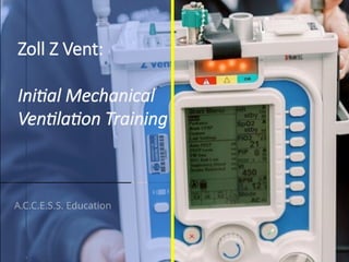
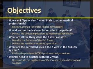
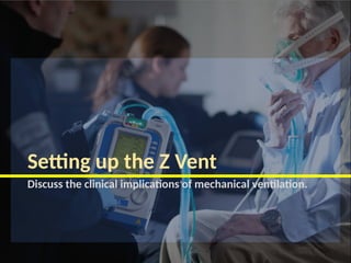
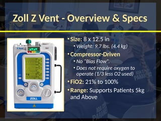
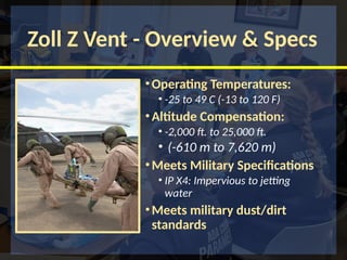
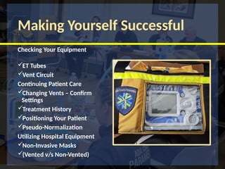
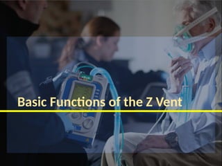
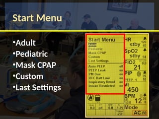
ZOLL Medical
Zoll Z Vent:
Initial Mechanical
Ventilation Training
A.C.C.E.S.S. Education
Objectives
• How can I “Speak Vent” when I talk to other medical
professionals?
• Review Common Ventilator related terminology
• How does mechanical ventilation affect the patient?
• Discuss the clinical implications of mechanical ventilation.
• What are all the things that the Z Vent can do?
• Describe the features of the Zoll Z Vent.
• Discuss the ventilator modes of operation
• What are the permitted uses if the Z Vent in the ACCESS
system?
• Review the pertinent ACCESS protocols and procedures.
• I think I need to practice with the Z Vent.
• Demonstrate the application of the Z vent in a simulated patient
scenario.
2
Ventilator Terminology
Review Common Ventilator related terminology
Tidal Volume (Vt) and Minute
Volume (Mv)
•Tidal Volume (Vt): The volume (measured in cc) of air
delivered with each breath.
•The appropriate tidal volume depends on numerous factors,
most notably the patient’s ideal body weight (IBW).
Vt is based on IBW
•Minute Volume (Mv): The volume (measured in liters)
of air delivered over a minute.
Vt x BPM = Mv
Use Height (I/PBW) for Tidal Volume
Adult Male Adult Female Pediatric
Brief Exercise (3 minutes)
•MV for 10 Breaths/minute and Vt 500 cc
• 5 liters / minute
•MV for 20 Breaths/minute and Vt 500 cc
• 10 liters a minute
•MV for 30 Breaths/minute and Vt 500 cc
• 15 liters a minute
•“I now appreciate that 15 Lpm via a non-rebreather
mask may not meet the minute ventilation of patients
in extremis; this explains how a non-rebreather can
collapse with inspiration and why many patients feel
suffocated with a mask over their face.” (Levitan, 2015)
Fraction of Inspired Oxygen (FIO2)
Fraction of Delivered Oxygen (FDO2)
•FIO2
• The lowest possible fraction of inspired oxygen (FIO2) necessary to
meet oxygenation goals should be used.
• This will decrease the likelihood that adverse consequences of
supplemental oxygen will develop, such as absorption atelectasis,
accentuation of hypercapnia, airway injury, and parenchymal injury
 Start at 100% FIO2 and titrate to main SpO2 at (94% - 99%)
•FDO2
• fractional delivered oxygen, and it describes the amount of oxygen
that reaches the body's capillary beds and perfuses its cells. FdO2 is a
factor that determines oxygenation,
Dead Space
Volume of a breath or ventilation that does not participate
in gas exchange.
Total Dead Space has 3 components
• Anatomic / Physiologic - (approximately 2ml/kg)
• Mechanical - (equipment dependent)
Dead Space
Dead Space
Dead Space increases the required Tidal
Volume
Dead Space increases retained CO2
Setting up the Z Vent
Discuss the clinical implications of mechanical ventilation.
Zoll Z Vent - Overview & Specs
•Size: 8 x 12.5 in
• Weight: 9.7 lbs. (4.4 kg)
•Compressor-Driven
• No “Bias Flow”.
• Does not require oxygen to
operate (1/3 less O2 used)
•FiO2: 21% to 100%
•Range: Supports Patients 5kg
and Above
Zoll Z Vent - Overview & Specs
•Operating Temperatures:
• -25 to 49 C (-13 to 120 F)
•Altitude Compensation:
• -2,000 ft. to 25,000 ft.
• (-610 m to 7,620 m)
•Meets Military Specifications
• IP X4: Impervious to jetting
water
•Meets military dust/dirt
standards
Making Yourself Successful
Checking Your Equipment
ET Tubes
Vent Circuit
Continuing Patient Care
Changing Vents – Confirm
Settings
Treatment History
Positioning Your Patient
Pseudo-Normalization
Utilizing Hospital Equipment
Non-Invasive Masks
(Vented v/s Non-Vented)
SpO2 and Charging
Charging & Caution
•Battery: 10 Hour battery
run time
•ACP will charge at 50%
•Fully charged in 2 hours
Important Point
The power cable connection
is one of the most fragile
parts of the Zoll Z Vent® .
Please treat with care.
Adult vs Pedi Circuits
Top view
Ventilator Setup
Vt: 200ml to
2000ml
(20kg and >)
Vt: 50ml to
300ml
(5kg to 30kg)
Oxygen Input (High/Low)
Airway Pressure
Transducer
Exhalation Valve
Gas Output
Where to put the HEPA Filter?
Fresh Gas / Emergency Air
Intake
Parameter and MENU Buttons
NO!!!
Yes!!!
Parameter and MENU Buttons
• Primary Parameters
•Single Press
• Secondary Parameters
•Multiple Presses
• Context Menus
•Press and Hold
Ventilator Start Up Sequence
Vent Book Start-Up Process Start Menu
Ventilator Start Up Sequence
Touch - Turn - Confirm
•Touch – The
Parameter
Buttons
•Turn - The Dial to
Adjust the Value
Confirm - To
Make the Change
Alarms
Red: High Priority
Yellow: Low Priority
Green: No Alarm / Normal
Operations
• No Audio Alarm for first 2 min
• Cancel Button will silence
alarms (30s)
• Bells signify alarm parameters
• High and Low values can be
changed.
Start Menu
•Adult
•Pediatric
•Mask CPAP
•Custom
•Last Settings
Smart Help
•SmartHelpTM
• On-screen prompts guide users
through alarm resolution
•Critical Controls on One
Screen
• No need to toggle through
multiple screens to change
primary parameters or monitor
patient status
• Digital LCD, rather than analog
controls, for precision and ease of
use
Pop-Up Messages
Remember
•1. First conduct the "Ventilator
Operational Test"
•2. THEN Set patient-specific settings
•3. Visually confirm proper
functioning by attaching to a test
lung prior to attaching to the patient.
When in doubt….
Intermission
Basic Functions of the Z Vent
Start Menu
•Adult
•Pediatric
•Mask CPAP
•Custom
•Last Settings
HR and SPO2
•Not used in the
ACCESS system
•Use the Monitor
instead
FIO2: Oxygen
Concentration
•“Fraction of inspired
oxygen”
•Zoll Z Vent: measured
as a %
•ACCESS Protocols
start at 100% and
titrated to 50%
•Exception: Matching
pre-existing settings
PIP: Peak Inspiratory
Pressure
•The highest level of
pressure applied to the
lungs during inhalation.
•PIP is the combination
of the PEEP and
pressure above PEEP
•PIP is set in pressure
modes
•PIP is measured in
volume modes
PIP Alarms
Standard alarm
settings should be:
•High pressure
alarm: 10 cmH2O
above peak airway
pressure.
•Low pressure
alarm: 5 cmH2O
below peak airway
pressure.
PEEP: Peak
Inspiratory Pressure
•PIP is the combination
of the PEEP and
pressure above PEEP
•PIP is set in pressure
modes
•PIP is measured in
volume modes
•Defaults to 5 cmH2O
https://www.youtube.com/watch?v=CDEYWok94uQ&t=2s
Vt: Tidal Volume
•The amount of air delivered
with each breath.
•Vt is set in volume modes
•Based on predicted ideal
body weight (IBW).
•Usual Vt: 6-8 ml/Kg
depending on disease state
and protocols
•Vt is measured in pressure
modes.
BPM: Breaths
per minute
•For most adult
patients, a respiratory
rate between 10 and
16 breaths per minute
is a reasonable
starting point
•Standard Rages are
noted in the vent
book
BPM Alarms
In invasive ventilation (A/C V
and SIMV), a high respiratory
rate alarm may indicate poor
sedation or a too sensitive
trigger.
In non-invasive ventilation
(cpap and bipap) you may have
to adjust upper alarm limits
due to pt’s resp. distress.
Inspiratory Time (Ti or I-Time)
• Inspiratory Time (Ti or I-Time)
• The time of the breath where the patient is inhaling or the
time it takes to deliver the set Tidal Volume (VT).
• Increasing the inspiratory time can give the Patient a
longer time to get air and O2 in and improve oxygenation
& ventilation
• Too long of an inspiratory time may inhibit exhalation and
cause the CO2 to rise (monitor EtCO2)
Affected by
resp rate
Inspiratory and Expiratory Time
(I:E Ratio)
• During spontaneous breathing, the normal I:E ratio is 1:2.5
indicating that for normal patients the exhalation time is
about twice as long as inhalation time.
• If exhalation time is too short “breath stacking” occurs
resulting in an increase in end-expiratory pressure also
called auto-PEEP.
Ti: 1.25
Te: 3.75
Ti + Te = 5 sec
ZOLL Medical
I: E Ratio
Inspiration
1 Seconds
Exhalation
2 Seconds
I:E Ratio of 1:2
Oxygenation and Ventilation
Need to improve Oxygenation? PEEP + FIO2
Need to improve Ventilation?  Vt + BPM
Trapping Air?  Increase Exhalation (I:E Ratio)
Rise Time (RT )
• The speed at which inspiratory pressure increases, to reach
the set target pressure (PIP).
• Adjustments in rise time can improve patient
comfort/tolerability with Non-Invasive modes.
• Rise times generally go from 100ms to 600ms, with
settings of 1 through 10
•2.0 is default
Trigger Level
•The amount of negative pressure a patient must
generate to get a breath delivered.
•Triggering, can be either time or patient cycling.
•Can be set from -0.5 cmH2O to -6.0 cmH2O
•-0.5 is the easiest for the patient to initiate and -
6.0 is the hardest.
•-2.0 is default
Cycle Percent
•The event that ends the inspiratory time, and
exhalation begins.
•Like triggering there can be either time or patient
driven.
•25% is default
Plateau
Pressure
•The plateau pressure is
measured at end-
inspiration (no flow) by
pressing P-PLAT 0.5 to
1 second.
• Should be <30 cm H2O
•Should stay consistent
ZOLL Medical
Plateau Pressure
 Plateau Pressure = End of Flow/Compliance
 Peak Pressure = Airflow/Airways
PEEP of 5 cm H2O
PIP vs. PEEP vs. Pplat
PEEP of 5 cm H2O
Name 1 of the 3 Hidden functions
and where can they be Found?
• FIO2 O2 Reservoir
• PIP Trigger
• BPM Control Parameter
Cycle Off %
Spont. Ti Limit
Intermission
Modes of Operation
Modes of Operation
Modes: Volume v/s Pressure
Pressure Targeted
Modes
• Pressure Set
• Volume Variable
Volume Targeted
Modes
• Volume set
• Pressure Variable
“Pressure targeted" breathing modes
are NOT approved functions for
ACCESS providers. These modes
require an RT (or similar) to attend.
Modes: CMV
•Controlled Mandatory Ventilation
•
Modes: CMV (V)
Even though a breath trigger is
attempted, there is no “assist”
from the vent.
Modes: A/C (V)
ALL breaths are the same set
volume
Modes: SIMV (V)
trigger
Full Volume
“Synchronized”
Breath
CPAP and Bi-Level Modes
CPAP
2-5 cmH2O,
Titrated to 10 cmH2O
Bi-Level
ePAP: 3-10 cm H2O
iPAP: 9-20 cm H2O
iPAP is always 5-10 cm H2O >
ePAP
Z-Vent provides :
• Leak compensation during
noninvasive ventilation
• Tubing compliance
compensation
CPAP and Bi-Level Modes
CPAP
Constant Positive
Pressure
Bi-Level
Inhalation and
Exhalation
Pressures
CPAP and BL are intended for
ventilatory support, NOT
ventilation.
In-Line Nebulized Treatment
80 cc
Cardiac Arrest – Custom Setting
D O P E R S
Documentation
Questions?
ZOLL Medical
Scenario Review
Scenario #1
•Disp. to prison for Low SPO2,
arrived to find pt. in cardiac arrest.
•Recent Hx of Pneumonia
•No other Hx
•Pt Defib x1, CPR, ETT, In line suction
and neb.
•POST-ROSC you decide to place the
patient on the vent.
76
Scenario #1
• Pt is estimated at 5’10, 200 lbs
•What is your starting:
•FIO2
•PEEP
•Vt
•BPM
•I:E
•Preferred Mode
Scenario #1
• Pt is estimated at 5’10, 200 lbs
•B/P 100/40
•HR 128
•Resp 4 spont. + Ventilation
•What is your :
•POST ETT/ROSC Sedation?
•Other Concerns?
Scenario #1
• During transport:
•SPO2 72% and dropping.
•HR is 52/min and dropping.
•ETCO2 is 24mmHg and dropping.
•What is your first steps?
Scenario #1
• During transport:
•Pt Rearrests
•What is your next steps?
Now to
Skills…

ACCESS - CE- 2024 08 Z-Vent presentation.pptx

  • 1.
    ZOLL Medical Zoll ZVent: Initial Mechanical Ventilation Training A.C.C.E.S.S. Education
  • 2.
    Objectives • How canI “Speak Vent” when I talk to other medical professionals? • Review Common Ventilator related terminology • How does mechanical ventilation affect the patient? • Discuss the clinical implications of mechanical ventilation. • What are all the things that the Z Vent can do? • Describe the features of the Zoll Z Vent. • Discuss the ventilator modes of operation • What are the permitted uses if the Z Vent in the ACCESS system? • Review the pertinent ACCESS protocols and procedures. • I think I need to practice with the Z Vent. • Demonstrate the application of the Z vent in a simulated patient scenario. 2
  • 3.
    Ventilator Terminology Review CommonVentilator related terminology
  • 4.
    Tidal Volume (Vt)and Minute Volume (Mv) •Tidal Volume (Vt): The volume (measured in cc) of air delivered with each breath. •The appropriate tidal volume depends on numerous factors, most notably the patient’s ideal body weight (IBW). Vt is based on IBW •Minute Volume (Mv): The volume (measured in liters) of air delivered over a minute. Vt x BPM = Mv
  • 5.
    Use Height (I/PBW)for Tidal Volume Adult Male Adult Female Pediatric
  • 7.
    Brief Exercise (3minutes) •MV for 10 Breaths/minute and Vt 500 cc • 5 liters / minute •MV for 20 Breaths/minute and Vt 500 cc • 10 liters a minute •MV for 30 Breaths/minute and Vt 500 cc • 15 liters a minute •“I now appreciate that 15 Lpm via a non-rebreather mask may not meet the minute ventilation of patients in extremis; this explains how a non-rebreather can collapse with inspiration and why many patients feel suffocated with a mask over their face.” (Levitan, 2015)
  • 8.
    Fraction of InspiredOxygen (FIO2) Fraction of Delivered Oxygen (FDO2) •FIO2 • The lowest possible fraction of inspired oxygen (FIO2) necessary to meet oxygenation goals should be used. • This will decrease the likelihood that adverse consequences of supplemental oxygen will develop, such as absorption atelectasis, accentuation of hypercapnia, airway injury, and parenchymal injury  Start at 100% FIO2 and titrate to main SpO2 at (94% - 99%) •FDO2 • fractional delivered oxygen, and it describes the amount of oxygen that reaches the body's capillary beds and perfuses its cells. FdO2 is a factor that determines oxygenation,
  • 9.
    Dead Space Volume ofa breath or ventilation that does not participate in gas exchange. Total Dead Space has 3 components • Anatomic / Physiologic - (approximately 2ml/kg) • Mechanical - (equipment dependent)
  • 10.
  • 11.
    Dead Space Dead Spaceincreases the required Tidal Volume Dead Space increases retained CO2
  • 12.
    Setting up theZ Vent Discuss the clinical implications of mechanical ventilation.
  • 13.
    Zoll Z Vent- Overview & Specs •Size: 8 x 12.5 in • Weight: 9.7 lbs. (4.4 kg) •Compressor-Driven • No “Bias Flow”. • Does not require oxygen to operate (1/3 less O2 used) •FiO2: 21% to 100% •Range: Supports Patients 5kg and Above
  • 14.
    Zoll Z Vent- Overview & Specs •Operating Temperatures: • -25 to 49 C (-13 to 120 F) •Altitude Compensation: • -2,000 ft. to 25,000 ft. • (-610 m to 7,620 m) •Meets Military Specifications • IP X4: Impervious to jetting water •Meets military dust/dirt standards
  • 15.
    Making Yourself Successful CheckingYour Equipment ET Tubes Vent Circuit Continuing Patient Care Changing Vents – Confirm Settings Treatment History Positioning Your Patient Pseudo-Normalization Utilizing Hospital Equipment Non-Invasive Masks (Vented v/s Non-Vented)
  • 16.
  • 17.
    Charging & Caution •Battery:10 Hour battery run time •ACP will charge at 50% •Fully charged in 2 hours
  • 18.
    Important Point The powercable connection is one of the most fragile parts of the Zoll Z Vent® . Please treat with care.
  • 19.
    Adult vs PediCircuits Top view Ventilator Setup Vt: 200ml to 2000ml (20kg and >) Vt: 50ml to 300ml (5kg to 30kg) Oxygen Input (High/Low) Airway Pressure Transducer Exhalation Valve Gas Output
  • 21.
    Where to putthe HEPA Filter?
  • 22.
    Where to putthe HEPA Filter?
  • 23.
    Fresh Gas /Emergency Air Intake
  • 25.
    Parameter and MENUButtons NO!!! Yes!!!
  • 26.
    Parameter and MENUButtons • Primary Parameters •Single Press • Secondary Parameters •Multiple Presses • Context Menus •Press and Hold
  • 27.
    Ventilator Start UpSequence Vent Book Start-Up Process Start Menu
  • 28.
  • 29.
    Touch - Turn- Confirm •Touch – The Parameter Buttons •Turn - The Dial to Adjust the Value Confirm - To Make the Change
  • 30.
    Alarms Red: High Priority Yellow:Low Priority Green: No Alarm / Normal Operations • No Audio Alarm for first 2 min • Cancel Button will silence alarms (30s) • Bells signify alarm parameters • High and Low values can be changed.
  • 31.
  • 32.
    Smart Help •SmartHelpTM • On-screenprompts guide users through alarm resolution •Critical Controls on One Screen • No need to toggle through multiple screens to change primary parameters or monitor patient status • Digital LCD, rather than analog controls, for precision and ease of use
  • 33.
  • 34.
    Remember •1. First conductthe "Ventilator Operational Test" •2. THEN Set patient-specific settings •3. Visually confirm proper functioning by attaching to a test lung prior to attaching to the patient.
  • 35.
  • 36.
  • 37.
  • 38.
  • 39.
    HR and SPO2 •Notused in the ACCESS system •Use the Monitor instead
  • 40.
    FIO2: Oxygen Concentration •“Fraction ofinspired oxygen” •Zoll Z Vent: measured as a % •ACCESS Protocols start at 100% and titrated to 50% •Exception: Matching pre-existing settings
  • 41.
    PIP: Peak Inspiratory Pressure •Thehighest level of pressure applied to the lungs during inhalation. •PIP is the combination of the PEEP and pressure above PEEP •PIP is set in pressure modes •PIP is measured in volume modes
  • 42.
    PIP Alarms Standard alarm settingsshould be: •High pressure alarm: 10 cmH2O above peak airway pressure. •Low pressure alarm: 5 cmH2O below peak airway pressure.
  • 43.
    PEEP: Peak Inspiratory Pressure •PIPis the combination of the PEEP and pressure above PEEP •PIP is set in pressure modes •PIP is measured in volume modes •Defaults to 5 cmH2O
  • 44.
  • 45.
    Vt: Tidal Volume •Theamount of air delivered with each breath. •Vt is set in volume modes •Based on predicted ideal body weight (IBW). •Usual Vt: 6-8 ml/Kg depending on disease state and protocols •Vt is measured in pressure modes.
  • 46.
    BPM: Breaths per minute •Formost adult patients, a respiratory rate between 10 and 16 breaths per minute is a reasonable starting point •Standard Rages are noted in the vent book
  • 47.
    BPM Alarms In invasiveventilation (A/C V and SIMV), a high respiratory rate alarm may indicate poor sedation or a too sensitive trigger. In non-invasive ventilation (cpap and bipap) you may have to adjust upper alarm limits due to pt’s resp. distress.
  • 48.
    Inspiratory Time (Tior I-Time) • Inspiratory Time (Ti or I-Time) • The time of the breath where the patient is inhaling or the time it takes to deliver the set Tidal Volume (VT). • Increasing the inspiratory time can give the Patient a longer time to get air and O2 in and improve oxygenation & ventilation • Too long of an inspiratory time may inhibit exhalation and cause the CO2 to rise (monitor EtCO2) Affected by resp rate
  • 49.
    Inspiratory and ExpiratoryTime (I:E Ratio) • During spontaneous breathing, the normal I:E ratio is 1:2.5 indicating that for normal patients the exhalation time is about twice as long as inhalation time. • If exhalation time is too short “breath stacking” occurs resulting in an increase in end-expiratory pressure also called auto-PEEP. Ti: 1.25 Te: 3.75 Ti + Te = 5 sec
  • 50.
    ZOLL Medical I: ERatio Inspiration 1 Seconds Exhalation 2 Seconds I:E Ratio of 1:2
  • 51.
    Oxygenation and Ventilation Needto improve Oxygenation? PEEP + FIO2 Need to improve Ventilation?  Vt + BPM Trapping Air?  Increase Exhalation (I:E Ratio)
  • 52.
    Rise Time (RT) • The speed at which inspiratory pressure increases, to reach the set target pressure (PIP). • Adjustments in rise time can improve patient comfort/tolerability with Non-Invasive modes. • Rise times generally go from 100ms to 600ms, with settings of 1 through 10 •2.0 is default
  • 53.
    Trigger Level •The amountof negative pressure a patient must generate to get a breath delivered. •Triggering, can be either time or patient cycling. •Can be set from -0.5 cmH2O to -6.0 cmH2O •-0.5 is the easiest for the patient to initiate and - 6.0 is the hardest. •-2.0 is default
  • 54.
    Cycle Percent •The eventthat ends the inspiratory time, and exhalation begins. •Like triggering there can be either time or patient driven. •25% is default
  • 55.
    Plateau Pressure •The plateau pressureis measured at end- inspiration (no flow) by pressing P-PLAT 0.5 to 1 second. • Should be <30 cm H2O •Should stay consistent
  • 56.
    ZOLL Medical Plateau Pressure Plateau Pressure = End of Flow/Compliance  Peak Pressure = Airflow/Airways PEEP of 5 cm H2O
  • 57.
    PIP vs. PEEPvs. Pplat PEEP of 5 cm H2O
  • 58.
    Name 1 ofthe 3 Hidden functions and where can they be Found? • FIO2 O2 Reservoir • PIP Trigger • BPM Control Parameter Cycle Off % Spont. Ti Limit
  • 59.
  • 60.
  • 61.
  • 62.
    Modes: Volume v/sPressure Pressure Targeted Modes • Pressure Set • Volume Variable Volume Targeted Modes • Volume set • Pressure Variable
  • 63.
    “Pressure targeted" breathingmodes are NOT approved functions for ACCESS providers. These modes require an RT (or similar) to attend.
  • 64.
  • 65.
    Modes: CMV (V) Eventhough a breath trigger is attempted, there is no “assist” from the vent.
  • 66.
    Modes: A/C (V) ALLbreaths are the same set volume
  • 67.
    Modes: SIMV (V) trigger FullVolume “Synchronized” Breath
  • 68.
    CPAP and Bi-LevelModes CPAP 2-5 cmH2O, Titrated to 10 cmH2O Bi-Level ePAP: 3-10 cm H2O iPAP: 9-20 cm H2O iPAP is always 5-10 cm H2O > ePAP Z-Vent provides : • Leak compensation during noninvasive ventilation • Tubing compliance compensation
  • 69.
    CPAP and Bi-LevelModes CPAP Constant Positive Pressure Bi-Level Inhalation and Exhalation Pressures CPAP and BL are intended for ventilatory support, NOT ventilation.
  • 70.
  • 71.
    Cardiac Arrest –Custom Setting
  • 72.
    D O PE R S
  • 73.
  • 74.
  • 75.
  • 76.
    Scenario #1 •Disp. toprison for Low SPO2, arrived to find pt. in cardiac arrest. •Recent Hx of Pneumonia •No other Hx •Pt Defib x1, CPR, ETT, In line suction and neb. •POST-ROSC you decide to place the patient on the vent. 76
  • 77.
    Scenario #1 • Ptis estimated at 5’10, 200 lbs •What is your starting: •FIO2 •PEEP •Vt •BPM •I:E •Preferred Mode
  • 78.
    Scenario #1 • Ptis estimated at 5’10, 200 lbs •B/P 100/40 •HR 128 •Resp 4 spont. + Ventilation •What is your : •POST ETT/ROSC Sedation? •Other Concerns?
  • 79.
    Scenario #1 • Duringtransport: •SPO2 72% and dropping. •HR is 52/min and dropping. •ETCO2 is 24mmHg and dropping. •What is your first steps?
  • 80.
    Scenario #1 • Duringtransport: •Pt Rearrests •What is your next steps?
  • 81.

Editor's Notes

  • #4 Tidal Volume (Vt) Is the volume of air that is exchanged in one breath.  Decreases in tidal volume can result from external pressure (i.e. Pneumothorax, hemothorax, tension pneumothorax) by effectively reducing lung volume. Dynamic hyperinflation also known as “breath stacking” is caused by the inability to completely exhale and can lead to “auto-positive end expiratory pressure (auto-PEEP).”  This may be due to inadequate exhalation time, airflow obstruction, or both. This condition leads to decreasing tidal volumes and can cause hemodynamic compromise.  Minute ventilation (Mv or VE): Tidal volume multiplied by the respiratory rate (normal is 60cc/kg/min), usually expressed in liters.  The body regulates carbon dioxide through changes in minute ventilation.  Increases in carbon dioxide leads to increased respiratory rate and/or tidal volume and increased minute ventilation (amount of air exchanged during one minute of ventilation).  Invasive mechanical ventilation is typically applied using a ventilator and either endotracheal intubation or tracheostomy. Potential harms include ventilator-associated pneumonia, tracheal stenosis, barotrauma, and need for sedation and paralysis leading to prolonged ICU stays, deconditioning, and delirium. All Vt is based on Ideal Body Weight
  • #5 In 2000, the landmark ARDSNet trial was published. This looked at patients with Acute Lung Injury(ALI) and Acute Respiratory Distress Syndrome (ARDS), and compared traditional tidal volumes versus lower tidal volumes. Simply put, ALI/ARDS are lung conditions characterized by difficulties in oxygenation and ventilation due to widespread pulmonary inflammation. The ARDSNet trial found that these patients had much better outcomes if their tidal volumes were restricted to 6 ml/kg (Predicted Body Weight), rather than the more traditional 12 ml/kg. This new lower tidal volume goal has generally become the standard of care for ventilated patients. Using tidal volumes of 6-8 ml/kg of ideal body weight directly improved morbidity and mortality. This discovery revolutionized tidal volume strategies and has become the gold standard of adult tidal volume ranges to decrease the risk of VILI from volutrauma/barotrauma. To do this we use the NIH estimate for predicted body weight (PBW) by gender. Where do these numbers come from? Something commonly called “ARDSNet” or the NHLBI ARDS Network The Acute Respiratory Distress Syndrome Network (ARDSNet) is a consortium of clinical centers and a coordinating center that designs and tests novel therapies for the treatment of acute lung injury (ALI) and acute respiratory distress syndrome (ARDS). Even though the ARDSNET program was replaced by PETALS, the guidelines for Vt in patients to prevent lung injury (including ARDS) remain the gold standard today. The ARDS Network was established as a contract program in 1994 and renewed in 2005 following two national competitions. The goal of the Network was to efficiently test promising agents, devices, or management strategies to improve the care of patients with ARDS. During its 20 years of service, 5,527 patients were enrolled in 10 randomized controlled trials and one observational study. In some of the most highly cited articles in critical care, network investigators reported improved survival with lung protective ventilation and shortened duration of mechanical ventilation with conservative fluid management. Additional trials informed best practices by suggesting no role for routine use of corticosteroids, beta agonists, pulmonary artery catheterization, or early full calorie enteral nutrition. The ARDS Network also developed new outcome measures (ventilator free days) and promoted innovative and efficient techniques (factorial designs and coenrollment) to speed the discovery of new treatment approaches for patients with ARDS.  On June 30th, 2014, the Network contract came to a close. It is replaced by PETAL, the NHLBI-funded Prevention and Treatment of Acute Lung Injury (PETAL). This Network will begin work on prevention and early treatment of ARDS. For more information, see http://petalnet.org. PETAL continued through the pandemic and finished in 2023. Several hospitals will continue researching therapies for ARDS through participation in the National Institute of Allergy and Infectious Diseases (NIAID)-funded STRIVE Network, NHLBI-funded ARDS, Pneumonia, Sepsis Network (APS Consortium), and BARDA-funded ARDS clinical trials network.
  • #6 The morbidly obese do not have substantially larger lungs compared to patients of more normal mass. Use the charts!
  • #8 FIO2 The lowest possible fraction of inspired oxygen (FIO2) necessary to meet oxygenation goals should be used. This will decrease the likelihood that adverse consequences of supplemental oxygen will develop, such as absorption atelectasis, accentuation of hypercapnia, airway injury, and parenchymal injury Start at 100% FIO2 and titrate to main SpO2 at (94% - 99%) FDO2 fractional delivered oxygen, and it describes the amount of oxygen that reaches the body's capillary beds and perfuses its cells. FdO2 is a factor that determines oxygenation,
  • #10 VENT (W/ HEPA)  NEB  Corr. Tubing  Elbow Conn.  ETCO2  ETT. Estimated Dead Space 90 ml
  • #11 VENT (W/ HEPA)  NEB  Corr. Tubing  Elbow Conn.  ETCO2  ETT. Estimated Dead Space 90 ml
  • #13 Size: 8 x 12.5 in Weight: 9.7 lbs. (4.4 kg) Compressor-Driven No “Bias Flow”. Does not require oxygen to operate (1/3 less O2 used) FiO2: 21% to 100% Range: Supports Patients 5kg and Above
  • #14 Operating Temperatures: -25 to 49 C (-13 to 120 F) Altitude Compensation: -2,000 ft. to 25,000 ft. (-610 m to 7,620 m) Meets Military Specifications IP X4: Impervious to jetting water Meets military dust/dirt standards
  • #17 Even though the Zoll Z Vent® has an impressive 10 hours of use on battery power, it should be kept well-charged and in a state of readiness. Below are some important icons to help you understand the status of the Zoll Z Vent® .
  • #19 Two patient circuits are currently available for the Zoll Z Vent®, one for adults/pediatrics and one for pediatrics/infants.  The adult/pediatric patient circuits are intended for use when delivering tidal volume from 200 ml to Adult.  The infant/pediatric patient circuit is designed for use when delivering tidal volume from 50 ml to 300 ml.  There is no neonatal circuit currently as the Zoll Z Vent® is only approved for infants above 5 kg (11 pounds). Infant / Pediatric – 50ml to 300ml Tidal Volume ( 5kg to 30kg ) Pediatric / Adult – 200ml to 2000ml Tidal Volume ( 20kg and > ) While the Zoll Z Vent® does have an option for reusable patient circuits, the ACCESS system will only use disposable circuits for patient care.   The patient circuit should be open to room air Select a patient default Check for patient disconnect alarm Check manual breath Check High Airway Pressure alarm (3 breaths)
  • #23 Fresh Gas/Emergency Air Intake The Fresh Gas/Emergency Air Intake allows ambient air into the device’s internal compressor. The intake also acts as an anti-asphyxia valve that enables the patient to breathe ambient air should the ventilator fail. The Fresh Gas/Emergency Air Intake will accept a standard threaded NATO CBRN filter connection. The Fresh Gas/Emergency Air Intake  will take a standard 22mm connection common to HEPA filters or BVMs.  Carry Handle The carry handle is for...well...carrying the vent. Like on a picnic. Or to the Store. Or on a walk on the beach.
  • #24  The intake also acts as an anti-asphyxia valve that enables the patient to breathe ambient air should the ventilator fail. The Fresh Gas/Emergency Air Intake will accept a standard threaded NATO CBRN filter connection. The Fresh Gas/Emergency Air Intake  will take a standard 22mm connection common to HEPA filters or BVMs.  ZOLL also offers an Oxygen Reservoir Bag Assembly Kit to allow for low-flow oxygen use with the ventilator to provide supplemental oxygen to patients. Low-flow oxygen sources can be from a flow meter or an oxygen concentrator. Oxygen is delivered through the Fresh Gas/Emergency Air Intake when the device's internal compressor cycles to deliver a breath.
  • #25 HR Low and High Alarm SpO2 Low and High Alarm FIO2 O2 Used, FiO2 % O2 Reservoir PIP Low and High Alarm PEEP and Trigger Setting Vt Low and High Alarm BPM Low and High Alarm, I:E Ratio and Ti Mode Modes of Operation Menu: Alarm Configuration Power-Up Settings Pulse Oximeter Trigger Level O2 Reservoir Unit Information Contrast
  • #26 Primary Parameters : The main patient care settings are called "primary Parameters" on the ventilator. As the primary provider, this is where most of your adjustments will occur.  Secondary Parameters: In addition to the "primary" parameters, "secondary" parameters are additional settings or alarm thresholds associated with each primary parameter.  Context Menus: "Context Menus" are additional settings that further adjust the performance of the ventilator. They are usually not visible on the main screen and are accessed through a sub-menu related to the primary/secondary parameters. Some providers call these “hidden Menus”
  • #27 The "Ventilator Operational Test" confirms the ventilator's basic functions and the absence of any critical conditions. It does not confirm proper ventilator settings for your patient. You will: 1. First conduct the "Ventilator Operational Test" 2. Set patient-specific settings  3. Visually confirm proper functioning by attaching to a test lung prior to attaching to the patient. 
  • #30 Red: High Priority Yellow: Low Priority Green: No Alarm / Normal Operations Cancel Button will silence alarms (30s) Bells signify alarm parameters High and Low values can be changed.
  • #32 KEY POINT: The Alarm Messaging Center (AMC) prioritizes alarms based on the risk to the patient and always presents the alarm with the greatest risk to the patient first. There may be multiple alarms or issues. It will show you the alarms in order of priority.
  • #40 FIO2 Fraction of inspired oxygen (FiO2) is the percentage of oxygen in a gas mixture, or the concentration of oxygen that a person inhales. At room temperature, the FiO2 of air is 21%, and this percentage remains the same at all altitudes.  The lowest possible fraction of inspired oxygen (FIO2) necessary to meet oxygenation goals should be used. This will decrease the likelihood that adverse consequences of supplemental oxygen will develop, such as absorption atelectasis, accentuation of hypercapnia, airway injury, and parenchymal injury Start at 100% FIO2 and titrate to main SpO2 at (94% - 99%) FiO2: Fraction of Inspired Oxygen A fraction instead of a percentage 80% = 08 FiO2 21% = 0.21 FiO2 Typically < .6 if possible, as close to .21 as the patient will tolerate
  • #41 Peak Inspiratory Pressure (PIP): The greatest pressure within the lungs during inspiration.  Pressures above 35mmHg have been shown to cause pressure-related lung injury (barotrauma).  Ideally, pressures should remain at 30 mmHg and below.  Increased peak pressures are usually due to increases in resistance within the respiratory system (e.g., tension pneumothorax, inability for adequate exhalation, edema). 
  • #42 PIP Pressure alarms: Pressure alarms ensure that providers are alerted to pressures that fall outside of appropriate ranges and have potential to harm the patient via barotrauma (over-pressure) or under-ventilation (circuit disconnect or under-pressure).  Pressures will be determined by placing the patient on the vent for ~1-2 minutes and determining intrinsic peak inspiratory pressure (PIP) . Standard alarm settings should be: High pressure alarm: 10 cmH2O above peak airway pressure. Low pressure alarm: 5 cmH2O below peak airway pressure.
  • #43 Positive End Expiratory Pressure (PEEP):  Is the amount of positive pressure that is maintained at end-expiration.  It is expressed in centimeters of water (cmH2O).  The purpose of PEEP is to increase end-expiratory lung volume and reduce air-space closure at end-expiration.  Increases oxygenation and allows removal of CO2 due to more alveoli open (more possibility for gas exchange) ACCESS Range: 2 – 10 cmH2O Pressure Support (PS): Often confused with Pressure Control mode of ventilation. Delivers flow at a set pressure, generally to overcome resistance of the airway and ventilator circuit.  PS can also be used to support a spontaneously breathing patient, such as with Bi-PAP. This is NOT a specific mode, but is rather an adjunct to any of the vent modes. The application of positive pressure to the airways during expiration may keep alveoli open and prevent closure. Most patients are set on 5 of peep as a standard.
  • #45 “Low” Tidal Volume Range Most common ranges used Based on ARDSNET study to reduce the incidence of ARDS, VALI/VILI 4-6 ml/kg “Intermediate” Tidal Volume Range 7-10 ml/kg “High” Tidal Volume Range Rarely used 10-12 ml/kg
  • #46 Respiratory Rate, Breaths per Minute (f, RR or BPM) An optimal method for setting the respiratory rate has not been established. For most adult patients, a respiratory rate between 10 and 16 breaths per minute is a reasonable starting point Always assess the patient’s CO2 and adjust rate accordingly
  • #47 "I used CPAP/BiLEvel" and the darn Vent kept alarming because the patient was breathing too fast" The vent has user-adjustable alarm settings. In the BPM menu there is a small alarm icon with a high and low setting. You can adjust the alarm parameters as needed. See the graphic below.  (no we cannot make permanent adjustments to this setting)
  • #48 Flow: Is the velocity at which gas is delivered to the patient, expressed in liters per minute.  When the flow rate is set higher, the speed of gas delivery is faster and inspiratory time is shorter.
  • #49 Inspiratory (I) and Expiratory (E) time and I:E ratio: Is the time period over which the VT is delivered.  Setting a shorter inspiratory time (I) results in a faster inspiratory flow rate in volume cycled ventilation.  Average adult inspiratory time is 0.7 to 1 second. During spontaneous breathing, the normal I:E ratio is 1:2  1:3 (depending on text) indicating that for normal patients the exhalation time is about twice to three times as long as inhalation time. If exhalation time is too short “breath stacking” occurs resulting in an increase in end-expiratory pressure also called auto-PEEP.
  • #51 The key difference between oxygenation and ventilation is, oxygenation is an artificial process of providing oxygen when a patient is in a hypoxic state, while ventilation refers to the process of flowing air into and out of the lungs.
  • #52 Rise Time The speed at which inspiratory pressure increases, to reach the set target pressure (PIP). Adjustments in rise time can improve patient comfort/tolerability with Non-Invasive modes. Rise times generally go from 100ms to 600ms, with settings of 1 through 10
  • #53 Trigger Level: (AKA Sensitivity or trigger sensitivity): Effort, or negative pressure, required by the patient to trigger a machine breath, commonly set so that minimal effort (-1 to -2 cmH2O) is required to trigger a breath. Triggering, can be either time or patient cycling. Zoll: Can be set from -0.5 cmH2O to -6.0 cmH2O (-2.0)* -0.5 is the easiest for patient to initiate and -6.0 being the hardest.
  • #54 Cycle Percent (Cycle %) The event that ends the inspiratory time, and exhalation begins. Like triggering there can be either time or patient cycling. Patient cycling is a function of inspiratory flow and is very similar to how we actually breath, at the beginning of the breath flow is high (Peak Flow) and then tapers down/decelerates as the lungs fill. As the lung fills and flow decelerates from the peak flow, to a level that is some percentage of peak flow, this value is the Cycle % - So, when the flow decelerates to this level, flow stops and the exhalation valve opens. * (25%)
  • #55 Plateau Pressure: (PPLAT): The pressure applied to small airways and alveoli during positive-pressure mechanical ventilation.  It is measured during an inspiratory pause on the mechanical ventilator.  A mechanically ventilated patient with a plateau pressure greater than 30-35cm is at an increased risk for barotrauma. The plateau pressure is measured at end-inspiration (no flow) with an “inspiratory hold maneuver” (pressing and holding the manual breath) on the mechanical ventilator that is 0.5 to 1 second
  • #57 Peak inspiratory pressure (PIP) and positive end-expiratory pressure (PEEP) are both respiratory terms that relate to pressure levels in the lungs: Pplat should usually be kept at less than 28 cm H2O provided that pleural pressure/PEEP is not increased, and always should be kept as low as possible.
  • #61  Multiple modes of operation treat a wide range of patients Assist Control SIMV CPAP BiLevel (BL) Mode
  • #62 Pressure Targeted Modes:  Pressure set. Volume variable, terminates when airflow falls below threshold level.  Peak airway pressure is fixed, determined by set pressure level.  This is generally represented by a decelerating flow waveform. Increased Mean Airway Pressure Increased Period of Alveolar Recruitment May Protect Against Barotrauma Variable Tidal Volumes Uncontrolled Minute Volume High Initial Flow Volume Targeted Modes: Volume constant, Pressure Variable inspiration terminates when preset VT delivered.  Peak airway pressure is variable and increases as needed to deliver prescribed VT.  This is generally represented by a constant flow waveform. Guaranteed Tidal Volume Stable Minute Volume Low Initial Flow Lower Mean Airway Pressure Slower Recruitment, in Lungs with Poor Compliance
  • #63 KEY POINT Inverse I:E ratios and "pressure targeted" breathing modes are NOT approved functions for ACCESS providers.  These modes require an RT (or similar ) to assist and/or a critical care transport team. ACCESS Providers will be using "volume targeted mode"  Call your chain of command for further assistance if this becomes an issue.
  • #64 Continuous mandatory ventilation (CMV): Every breath is mandatory (i.e., inspiration is patient or machine triggered, but machine cycled). It is primary ventilator mode in patients who are apnic. •Doesn't permit normal spontaneous breathing, and can cause respiratory muscles to atrophy. •Used in a patient unable to initiate a breath •Spontaneously breathing patient must be sedated and/or paralyzed. •Frequent ABGs monitoring . AC/V: Although the work of breathing is not eliminated, this mode gives the respiratory muscles the greatest amount of rest because the patient needs only to create enough negative pressure to trigger the machine. An added advantage is that the patient can achieve the required minute ventilation by triggering additional breaths above the set back-up rate.
  • #65 Original mode of ventilation. Basically a mechanical BVM.
  • #66 AC/V: Delivers a preset number of mandatory breaths per minute.  The patient can take their own breaths in addition to mandatory breaths, with each spontaneous breath receiving the full preset tidal volume.  Airway pressures can vary during delivery.  Although the work of breathing is not eliminated, this mode gives the respiratory muscles the greatest amount of rest because the patient needs only to create enough negative pressure to trigger the machine. An added advantage is that the patient can achieve the required minute ventilation by triggering additional breaths above the set back-up rate.
  • #67 Synchronized Intermittent Mandatory Ventilation – Volume Controlled (SIMV) combines mandatory breaths and supported breaths. The ventilator delivers a preset number of mandatory breaths per minute.  Any breaths over the set rate will be supported with a fixed amount of pressure support.
  • #68 CPAP Pressure: Begin at 2-5 cmH2O, Titrated to 10 cmH2O MAX CPAP PRESSURE: 10 cmH2O (May call Medical Control to exceed) BiLevel Pressure (ALS only) ePAP 3-10 cm H2O iPAP – 9-20 cm H2O iPAP is always 5-10 cm H2O greater than ePAP (May call Medical Control to exceed)
  • #70 VENT (w/ HEPA)  NEB  Barrel Conn.  ETCO2  In line suction  ETT Estimated Dead Space 110 ml, (190 ml WITH HEPA FILTER)
  • #71 Weiss et al. had showed that healthcare professionals were able to accomplish more tasks, document more completely, and provide better patient care with the use of the MV in patients undergoing CPR with an advanced airway compared to that of BVM Rate and volume control is essential during cardiac arrest Frees up resources
  • #72 DOPE became DOPES became DOPERS Displacement: Verify that ETT is in place, patient not extubated/tube did not move during transfer. If the ETT has advanced – pull back to original length and attempt to bag; if tube has pulled farther out of trachea, DO NOT ATTEMPT TO ADVANCE the ETT without laryngoscopy or placement of bougie to verify tracheal placement. When advancing the bougie, feel for tracheal rings or stop/ resistance at the carina. If in doubt, remove the endotracheal tube and attempt BVM. If air movement is adequate, continue to bag ventilate the patient. Upon stabilization, consider alternative advanced airways (supraglottic airway or cricothyroidotomy). **If ETT moves freely, access for ETT bulb rupture via cuff manometer.9 Obstructions: Assess for secretions in ETT. Suction if indicated Assess for kinking in ETT . Pressure: Ensure that a tension pneumothorax / hemothorax has not developed (if the chest tube is in place, ensure it is properly suctioning, not kinked or clamped). If tension pneumo/hemothorax is suspected, perform immediate needle thoracentesis. Assess the need for escharotomy if circumferentially burned. Consider additional paralysis and sedation if patient does not tolerate ventilation. Equipment: Ensure that ventilator did not fail; O2 tank not empty. If ventilator is operational, trace all tubes to the patient connection (airway tube, transducer line, exhalation line) ensuring patency and connections. Stacked Breaths: Breath stacking, also known as air trapping or auto-positive end-expiratory pressure (auto-PEEP), is a common phenomenon in mechanically ventilated patients that occurs when a patient is unable to fully exhale before the next breath. This can happen when a patient is tachypneic or when the ventilator doesn't allow enough time for exhalation. As a result, more air builds up in the patient's lungs, which increases airway pressures and can lead to complications Most commonly noted with escalating PIP alarms or Pplat. Precurser to pneumo Disconnect pt while trouble shootong for a brif time to allow passive exhalation or in some cases active exhalation. Resistance to ventilation A sign of poor ventilation
  • #77 Refer to POST ROSC chart. Pt is estimated at 5’10, 200 lbs What is your starting: FIO2 100% PEEP 5 cm H20. Vt (438 ml  440 ml Vt) BPM 12 I:E 1:3.0 although with Hx of pneumona rasing this would be wise. Preferred Mode: SIMV (v) or A/C (v) post ROSC Refer to Vt chart (438 ml  440 ml Vt)
  • #78 Pt is estimated at 5’10, 200 lbs B/P 100/40 HR 128 Resp 4 spont. + Ventilation What is your : POST ETT/ROSC Sedation? 90 mcg Fent. And 5 mg Versed Other Concerns? Hypotension from PPV or drugs or simply sepsis. Consider Nor Epi and PDE at ready Re Arrest Obstruction of ETT with lung butter.  Pre-emptive suction.
  • #79 DOPERS Displacement: Verify that ETT is in place, patient not extubated/tube did not move during transfer. If the ETT has advanced – pull back to original length and attempt to bag; if tube has pulled farther out of trachea, DO NOT ATTEMPT TO ADVANCE the ETT without laryngoscopy or placement of bougie to verify tracheal placement. When advancing the bougie, feel for tracheal rings or stop/ resistance at the carina. If in doubt, remove the endotracheal tube and attempt BVM. If air movement is adequate, continue to bag ventilate the patient. Upon stabilization, consider alternative advanced airways (supraglottic airway or cricothyroidotomy). **If ETT moves freely, access for ETT bulb rupture via cuff manometer.9 Obstructions: Assess for secretions in ETT. Suction if indicated Assess for kinking in ETT . Pressure: Ensure that a tension pneumothorax / hemothorax has not developed (if the chest tube is in place, ensure it is properly suctioning, not kinked or clamped). If tension pneumo/hemothorax is suspected, perform immediate needle thoracentesis. Assess the need for escharotomy if circumferentially burned. Consider additional paralysis and sedation if patient does not tolerate ventilation. Equipment: Ensure that ventilator did not fail; O2 tank not empty. If ventilator is operational, trace all tubes to the patient connection (airway tube, transducer line, exhalation line) ensuring patency and connections. Stacked Breaths: Breath stacking, also known as air trapping or auto-positive end-expiratory pressure (auto-PEEP), is a common phenomenon in mechanically ventilated patients that occurs when a patient is unable to fully exhale before the next breath. This can happen when a patient is tachypneic or when the ventilator doesn't allow enough time for exhalation. As a result, more air builds up in the patient's lungs, which increases airway pressures and can lead to complications Most commonly noted with escalating PIP alarms or Pplat. Precurser to pneumo Disconnect pt while trouble shootong for a brif time to allow passive exhalation or in some cases active exhalation. Resistance to ventilation A sign of poor ventilation
  • #80 Initiate CPR Place vent into CPR mode