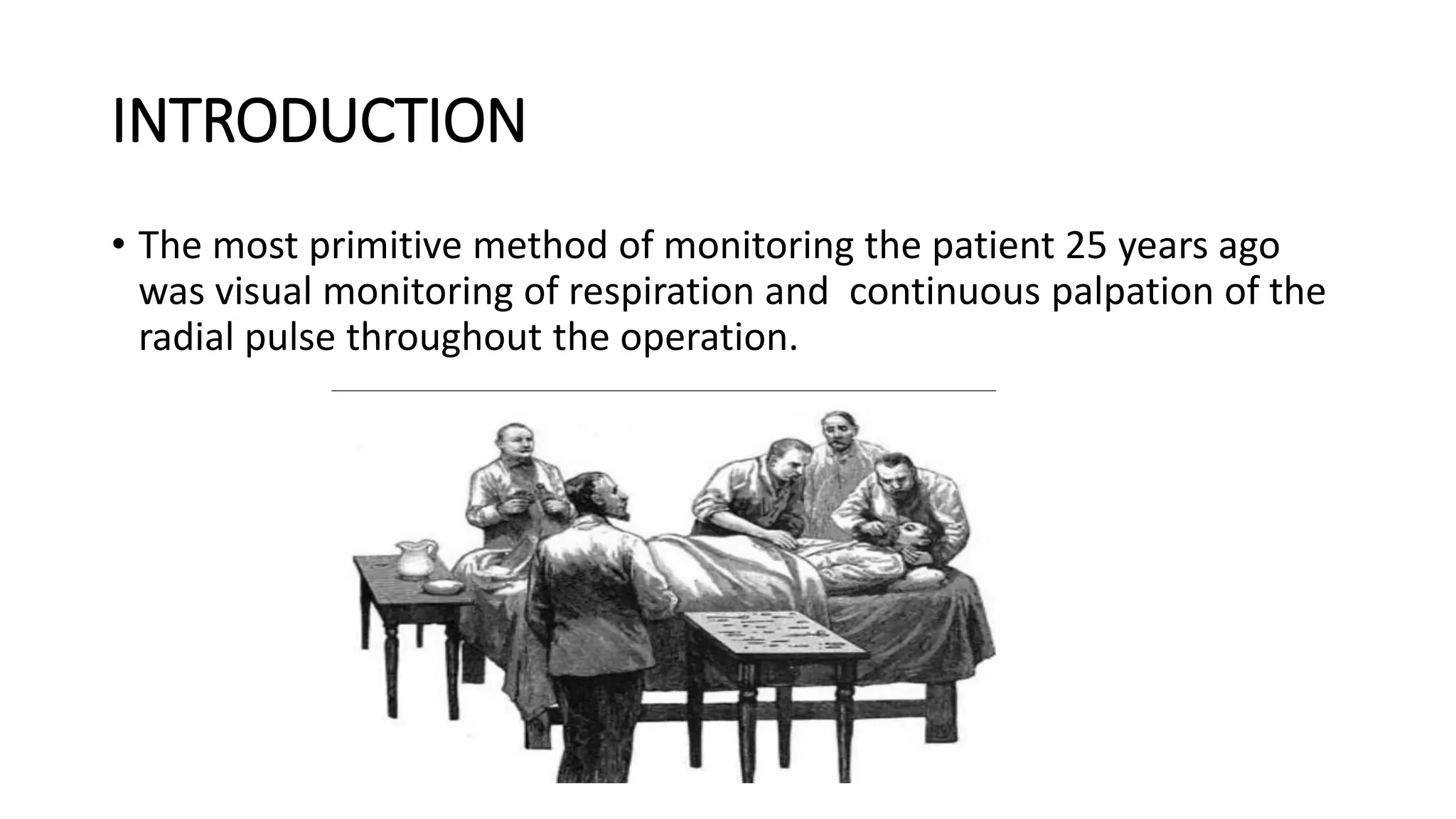
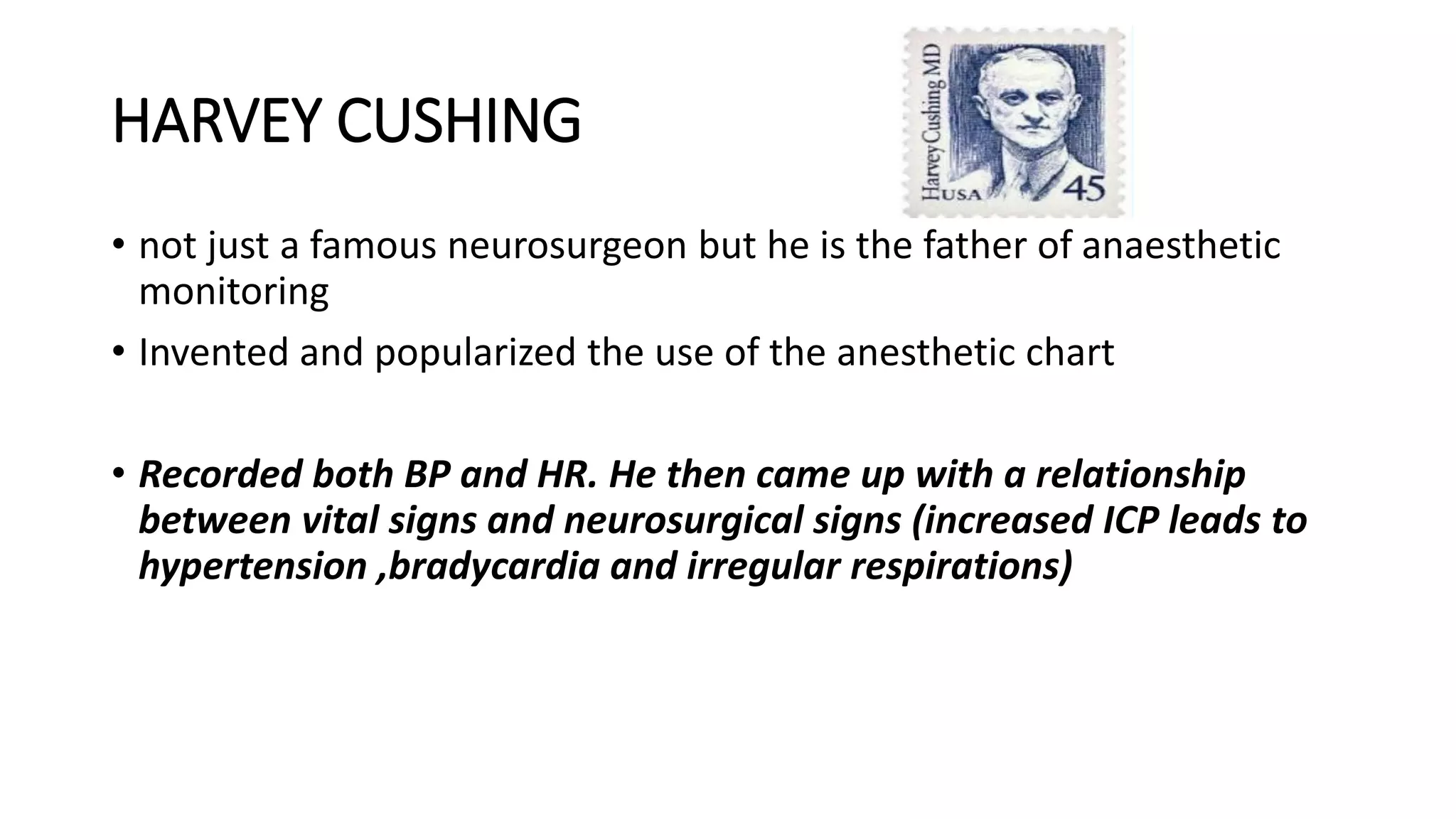
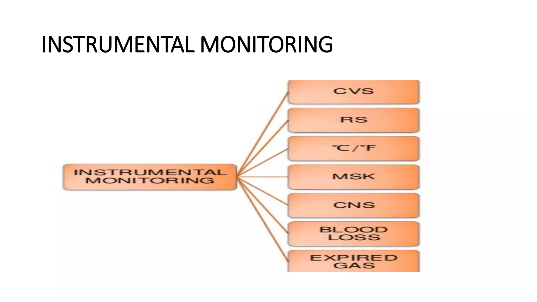
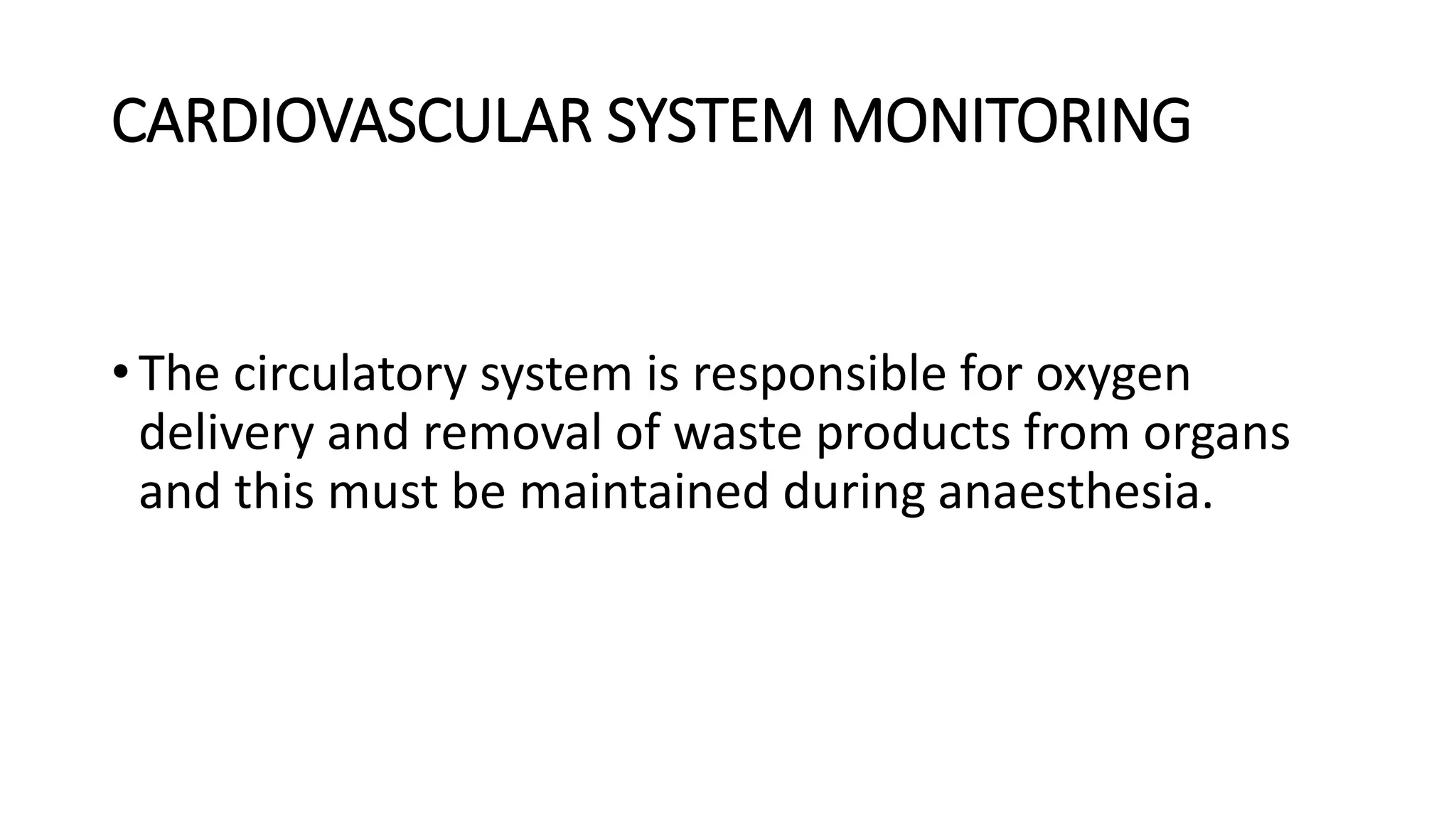
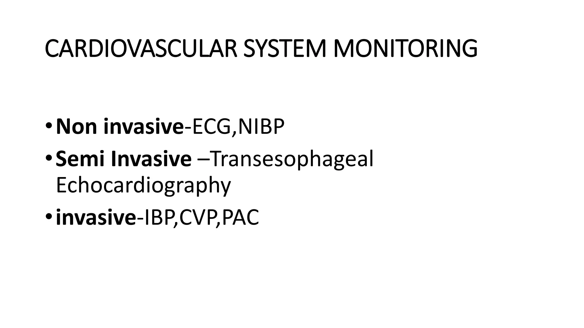
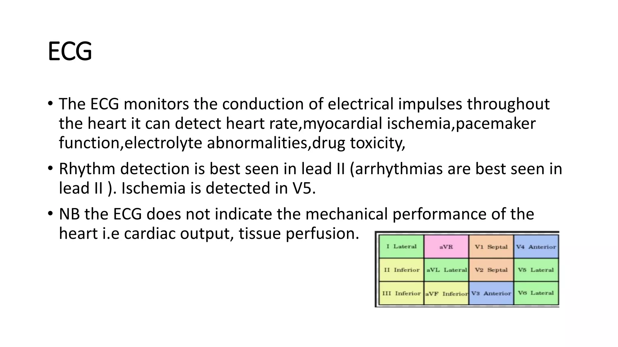
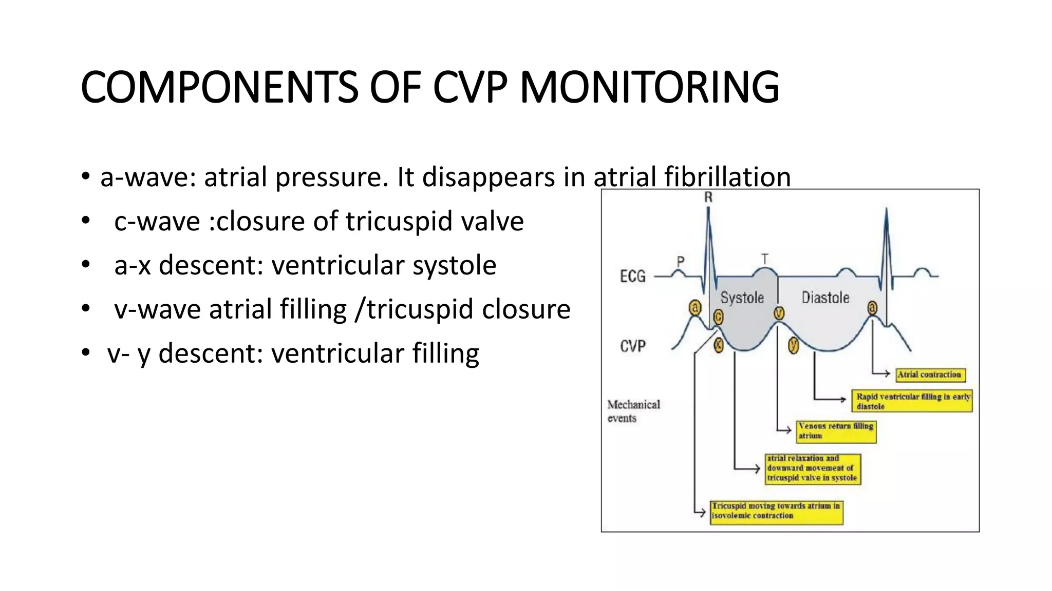
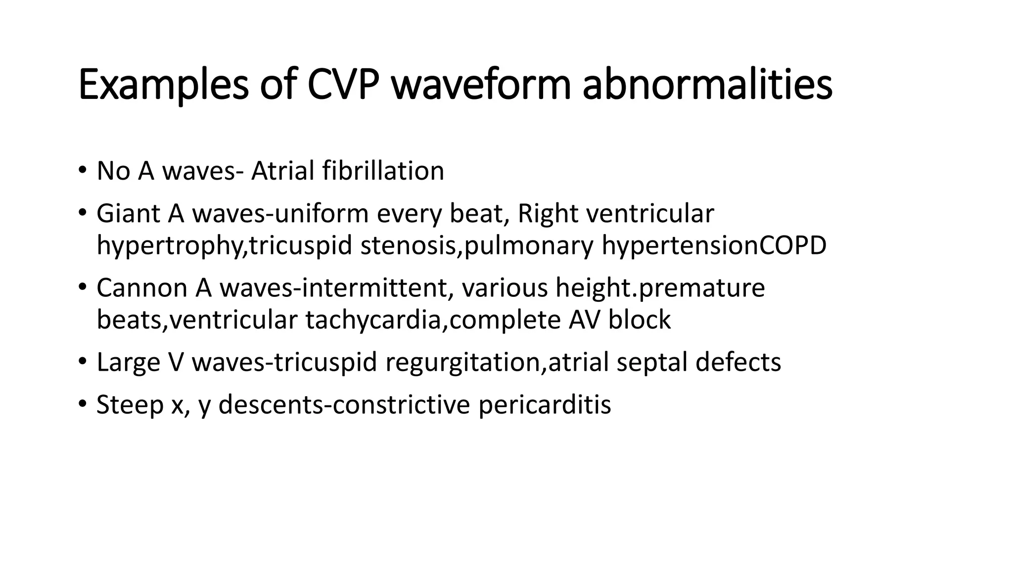
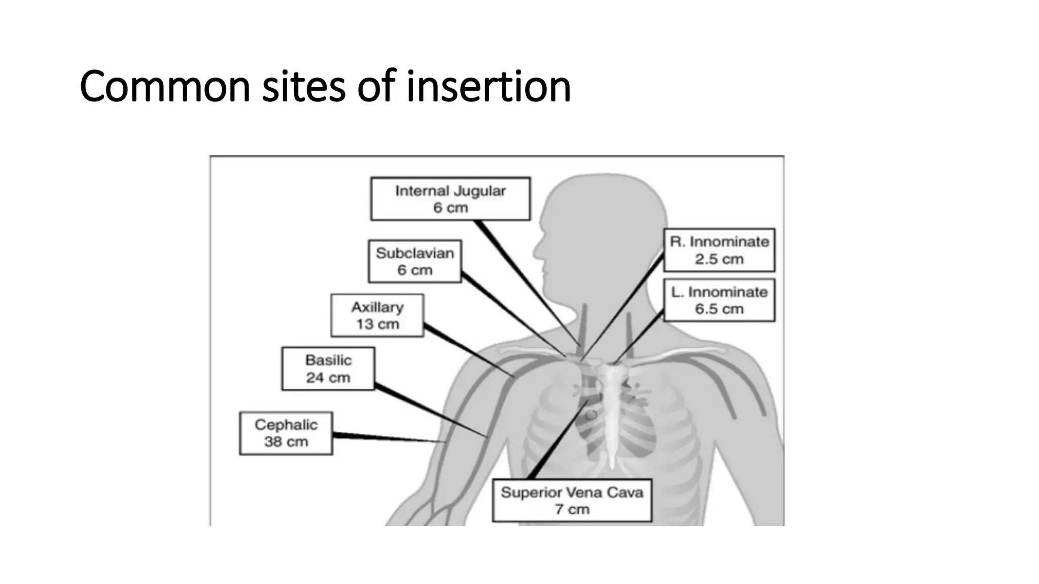
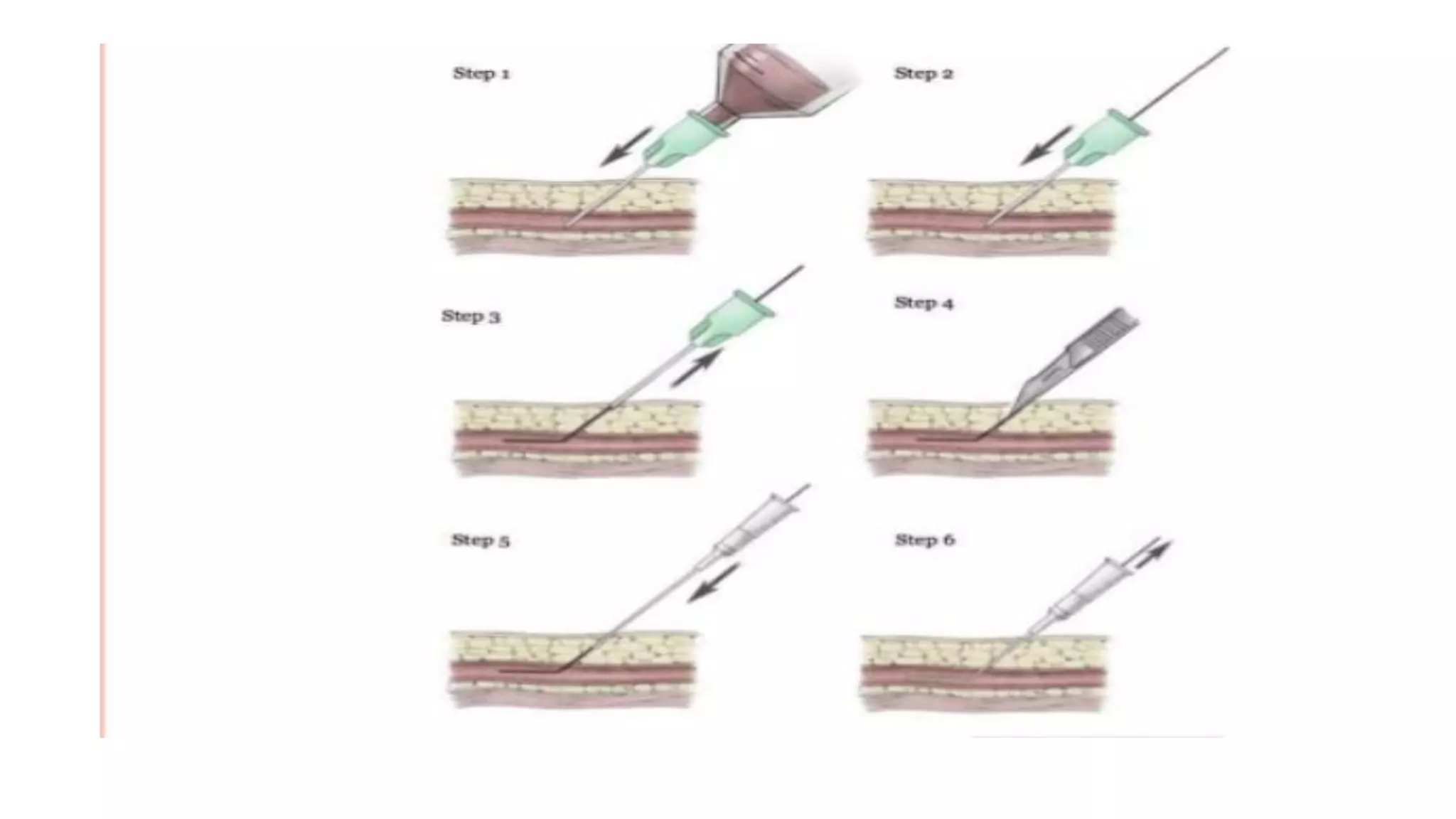
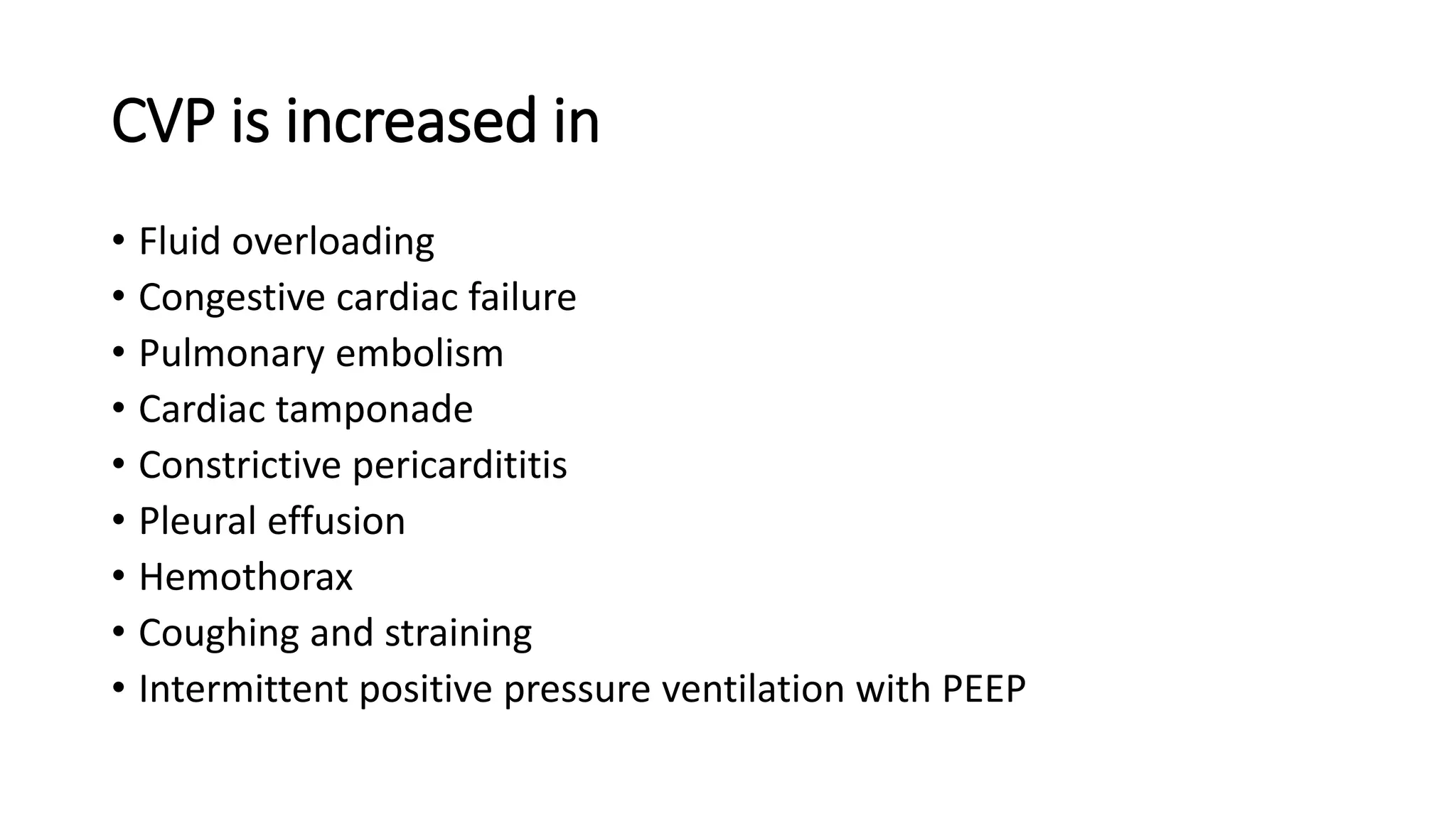
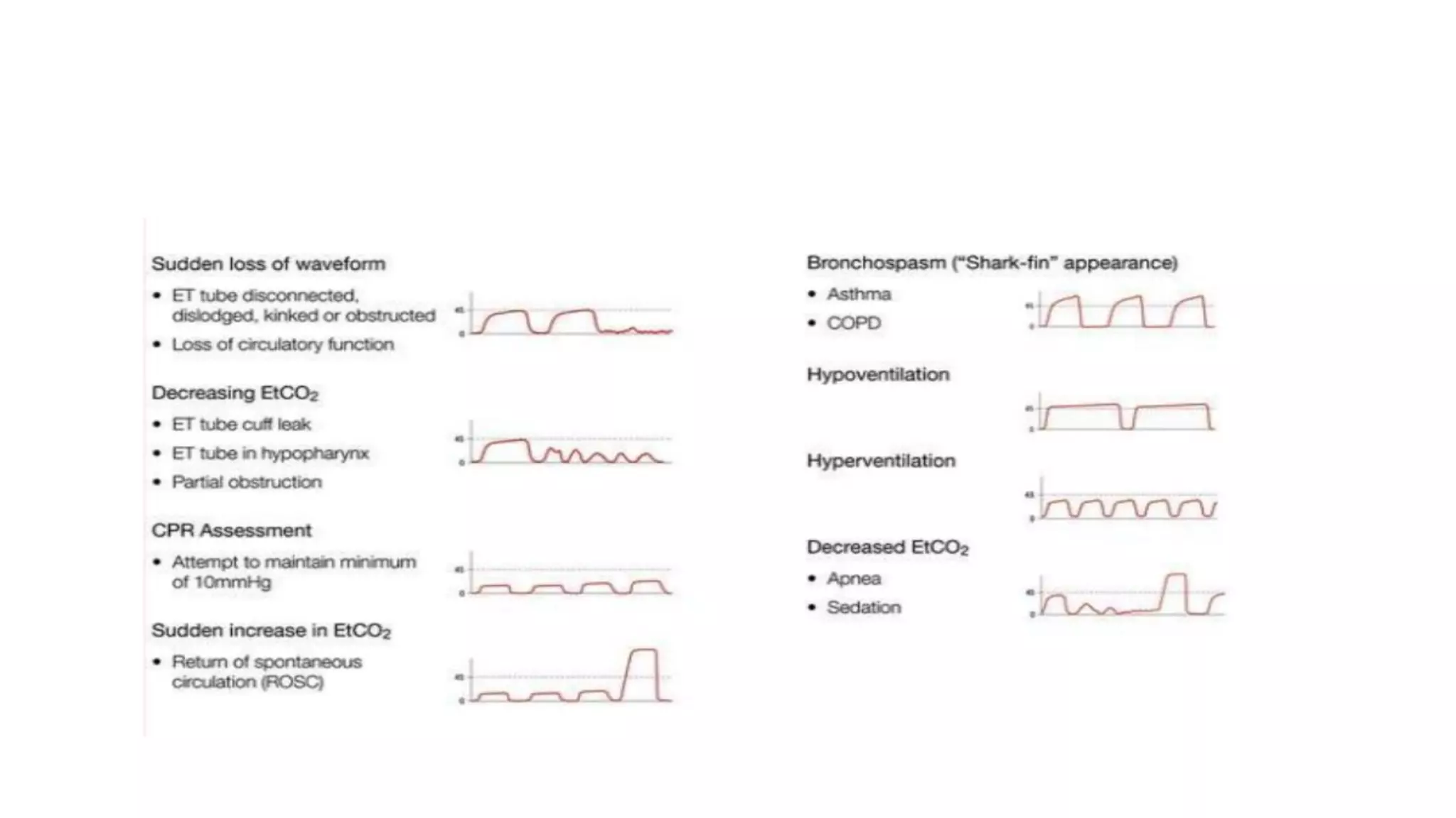
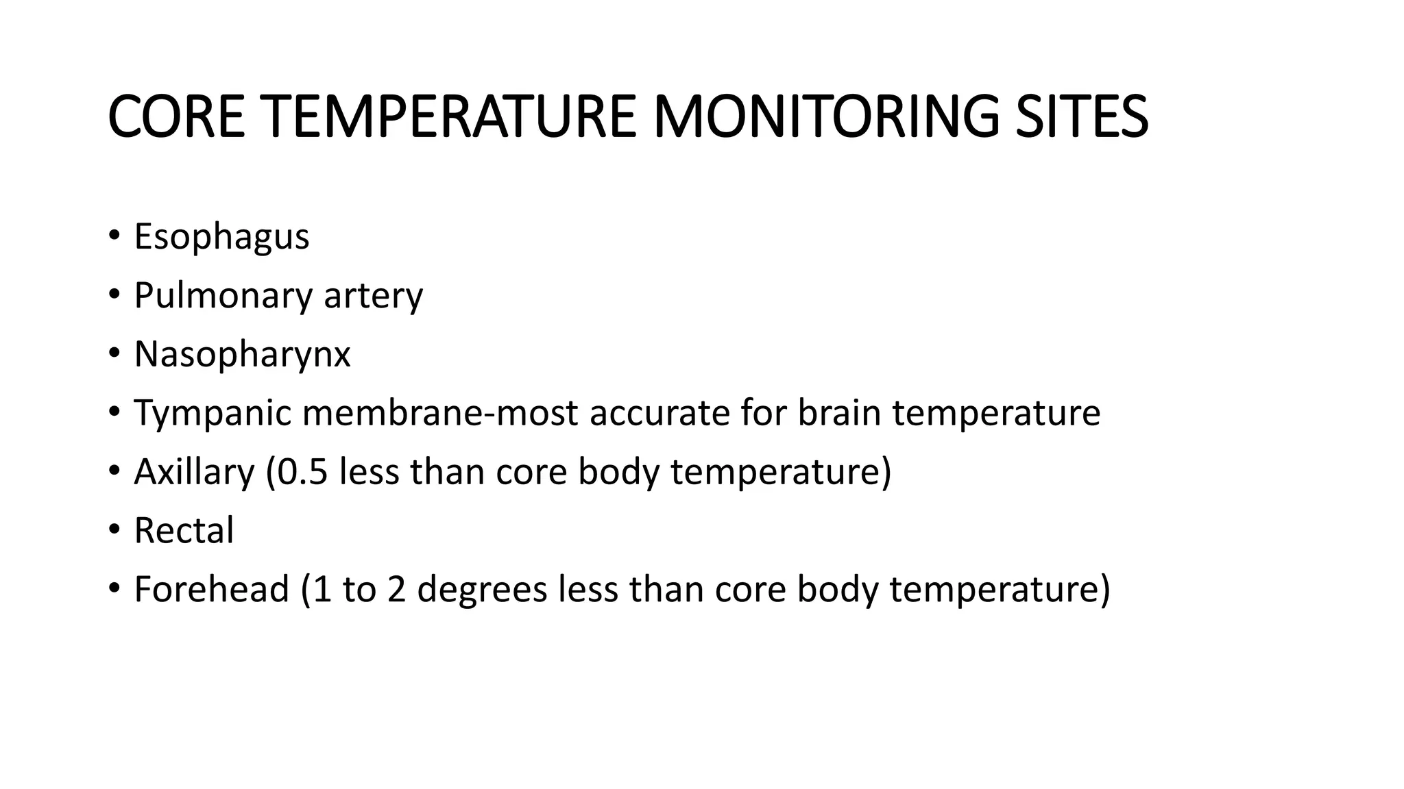
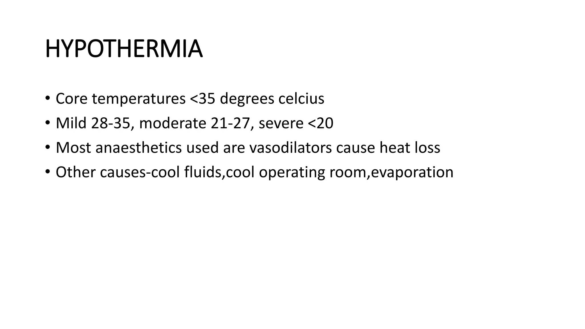
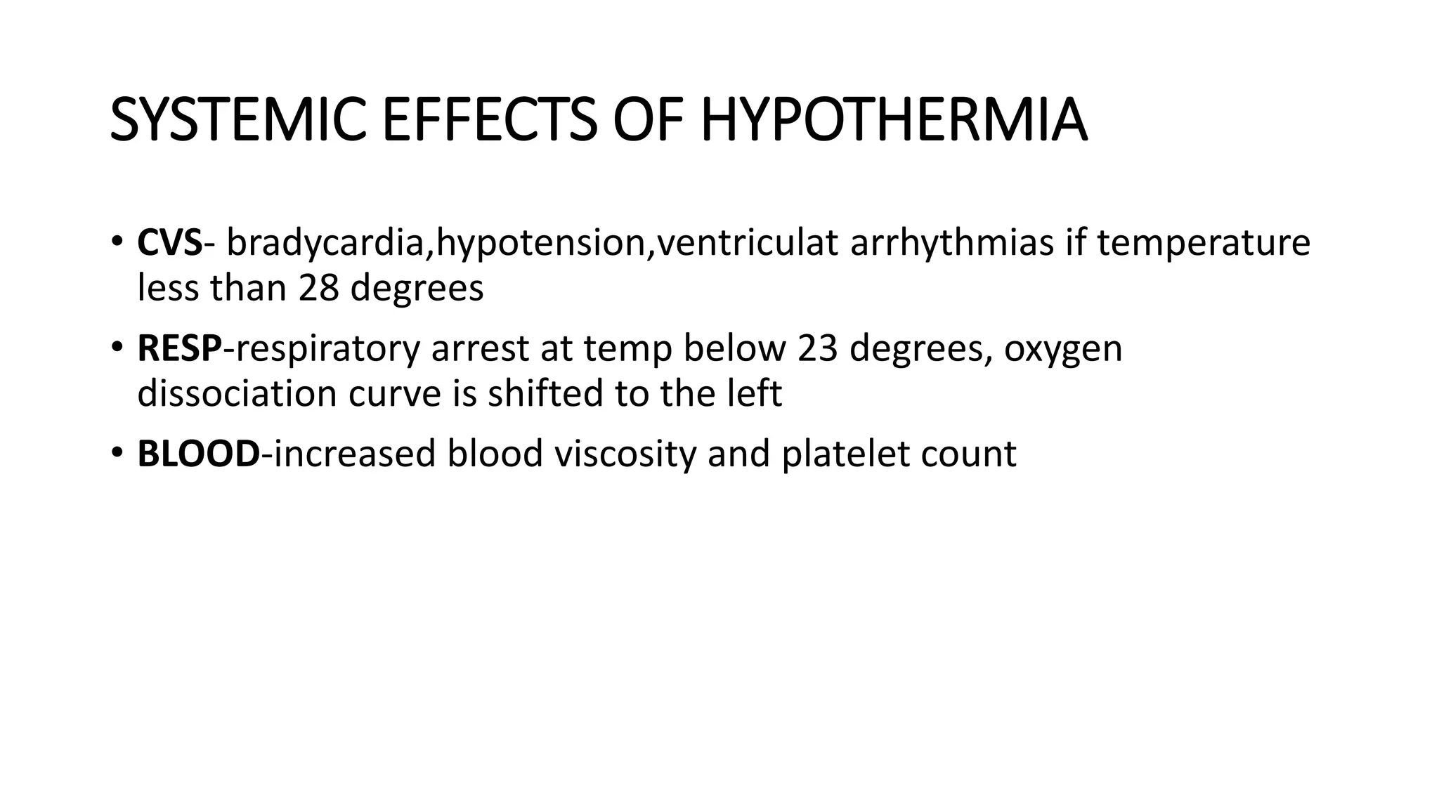
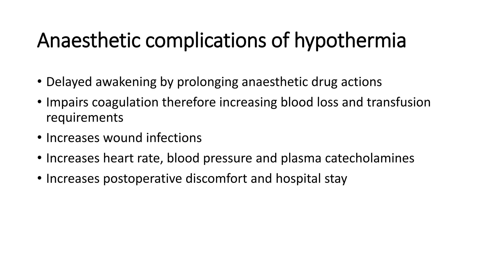
SlideShare a Scribd company logo
INTRAOPERATIVE
MONITORING
DR T. MASIYIWA
DEFINITION
• The word monitor comes from the Latin word MONERE which means
warning
• ANAESTHETIC MONITORING-Interpret available clinical data to help
recognize present or future mishaps or unfavorable system conditions
• The role of intraoperative monitoring is to maintain normal patient
physiology and homeostasis throughout anaesthesia and surgery
What is the value of knowing this?
• To understand and appreciate the value of clinical monitoring
• To appreciate how modern monitors have made us more smarter.
INTRODUCTION
• The most primitive method of monitoring the patient 25 years ago
was visual monitoring of respiration and continuous palpation of the
radial pulse throughout the operation.
HARVEY CUSHING
• not just a famous neurosurgeon but he is the father of anaesthetic
monitoring
• Invented and popularized the use of the anesthetic chart
• Recorded both BP and HR. He then came up with a relationship
between vital signs and neurosurgical signs (increased ICP leads to
hypertension ,bradycardia and irregular respirations)
MONITORING IN THE PRESENT
• Standardized basic monitoring requirements from the ASA,CAS and
other national societies
• ASA Standard I-Qualified anaesthesia personell should be present in
the room throughout the conduct of all general, regional and
monitored anaesthesia
• ASA standard II-during all anesthetics the patients oxygen ventilation
circulation and temperature shall be continually evaluated.
MONITORING IN THE PRESENT
Canadian guidelines to the Practice of Anesthesia and patient
monitoring are:
• An anesthetist present
• A completed preanesthetic checklist.
• An anesthetic record (HR, BP, Drug, Fluid)
• Oxygenation, ventilation, circulation, and temperature are continually
evaluated both clinically and quantitatively
CLINICAL MONITORING
• Skin colour
• Urine output >0.5mls/kg
• Capillary refill
• Heart Rate
• Inflation of chest
• Precordial and esophageal stethoscopy
• Urine output
INSTRUMENTAL MONITORING
CARDIOVASCULAR SYSTEM MONITORING
•The circulatory system is responsible for oxygen
delivery and removal of waste products from organs
and this must be maintained during anaesthesia.
Signs and symptoms of perfusion
abnormalities
• CNS: mental status changes, neurologic deficits
• CVS: shortness of breathe,chest pain
• GIT:abd pain,descreased bowel sounds
• Peripheral:cool peripheries, diminished pulses,poor capillary refill
• Renal:descreased urine output,elevated urea nitrogen and
creatinine,decreased fractional excretion of sodium
CARDIOVASCULAR SYSTEM MONITORING
•Non invasive-ECG,NIBP
•Semi Invasive –Transesophageal
Echocardiography
•invasive-IBP,CVP,PAC
ECG
• The ECG monitors the conduction of electrical impulses throughout
the heart it can detect heart rate,myocardial ischemia,pacemaker
function,electrolyte abnormalities,drug toxicity,
• Rhythm detection is best seen in lead II (arrhythmias are best seen in
lead II ). Ischemia is detected in V5.
• NB the ECG does not indicate the mechanical performance of the
heart i.e cardiac output, tissue perfusion.
ECG PLACEMENT
ARTIFACTS IN ECG MONITORING
• Loose electrodes or broken leads
• Misplaced leads
• Wrong lead system selected
• Emphysema, pneumothorax
• Shivering or restelessness
• Respiratory variation and movement
BLOOD PRESSURE
TIMING
• Throughout the surgery: before induction till after extubation &
recovery.
FREQUENCY
• By default every 5 minutes.
• Every 3 minutes: immediately after spinal anaesthesia, in conditions
of hemodynamic instability, during hypotensive anaesthesia.
• Every 10 minutes: eg. In awake pts under local anaesthesia:
“monitored anaesthesia care” (minimal hemodynamic changes).
NIBP
• Measures BP at set intervals automatically by an automated
oscillometry
• Cuff size should cover 2/3 of arm
• A cuff too small overestimates and a cuff too large underestimates
• Small cuff to be used for children
• Usually attached to the limb opposite
the IV line & pulse oximeter
Transesophageal echocardiography
• Most sensitive in detecting any wall abnormalities i.e ischemia,
valvular dysfunction, air embolism.
Invasive Blood Pressure
• Required in patients who mandate beat to beat monitoring
• It is the gold standard method of monitoring blood pressure
• Arteries that can be used include the radial artery,brachial artery
femoral artery, dorsalis pedis artery.
ARTERIAL BLOOD PRESSURE
• Systolic Bp (SBP), Diastolic Bp (DBP) ,Pulse pressure (PP)= SBP-DBP
• Mean arterial pressure (MAP); average BP in an individual during a
single cardiac cycle
• MAP = DBP + 1/3 PP
• MAP range 65-110 mmHg
ALLEN’S TEST
• Normal <7s
• Borderline 7-14s
• Contraindicated >15s
Cannulation complications
• Arterial injury,spasms, distal ischemia
• Thrombosis,embolization
• Sepsis
• Tissue necrosis
• Fistula and aneurysm formation
• NB:To prevent complications continuous flush with/out heparin
Central venous pressure monitoring
INDICATIONS
• Major surgeries where large fluctuations in hemodynamics are expected
• Open heart surgeries
• Fluid management in shock
• As a venous access
• Parenteral nutrition
• Aspiration of air embolus
• Cardiac pacing Normal CVP is 6 to 8cm of H2O in adults and CVP 3 to 6 cm
of H2O in children
• CVP >20cm H2O indicates right heart failure
COMPONENTS OF CVP MONITORING
• a-wave: atrial pressure. It disappears in atrial fibrillation
• c-wave :closure of tricuspid valve
• a-x descent: ventricular systole
• v-wave atrial filling /tricuspid closure
• v- y descent: ventricular filling
Examples of CVP waveform abnormalities
• No A waves- Atrial fibrillation
• Giant A waves-uniform every beat, Right ventricular
hypertrophy,tricuspid stenosis,pulmonary hypertensionCOPD
• Cannon A waves-intermittent, various height.premature
beats,ventricular tachycardia,complete AV block
• Large V waves-tricuspid regurgitation,atrial septal defects
• Steep x, y descents-constrictive pericarditis
Common sites of insertion
Technique of CVP catheterization through
internal jagular
Seldinger technique
• Patient lies in Trendelenburg position to decrease chance of embolism
• The cannula stylet is inserted at the tip of the triangle formed by two heads
of the sternomastoid and clavicle the direction of the needle should be
slightly lateral and towards the ipsilateral nipple.
• Once the internal jugular vein is punctured the stylet is removed and a J
wire passed through cannula.
• Now the CVP catheter is railroad over the J wire
• The tip of the catheter should be at the junction of superior vena cava with
right atrium-15cm from entry point.
CVP is increased in
• Fluid overloading
• Congestive cardiac failure
• Pulmonary embolism
• Cardiac tamponade
• Constrictive pericardititis
• Pleural effusion
• Hemothorax
• Coughing and straining
• Intermittent positive pressure ventilation with PEEP
CVP is decreased in
• Hypovolemia and shock
• Venodilator
• Spinal/epidural anaesthesia
• General amaesthesia causing vasodilation
• LOW CVP + LOW BP= HYPOVOLEMIA
• HIGH CVP + LOW BP=PUMP FAILURE
COMPLICATONS
• Air embolism
• Thromboembolism
• Cardiac arrthymias
• Pneumothorax/hemothorax
• Cardiac perforation
• Trauma to brachial plexus
Pulmonary artery catheterization
• It is reserved for very major cases in severely compromised patients
because of cost,technical feasibility, complications
• Swan-ganz-catheter-it is a balloon tipped and flow directed by
pressure recording, pressure tracing and catheter tip,
Swan-ganz catheter/pulmonary artery catheter
• Provides diagnostic information to rapidly dertemine hemodynamic
pressures, cardiac output and blood sampling for mixed venous oxygen
saturation
• Measures CVP, PAP, Cardiac output
Indications
• Cardiac surgery/major surgery
• Resuscitation
• Shock
• Oxygen transport ventilation and perfusion
• Post MI
CVP VS PCWP
RESPIRATORY MONITORING
• Stethoscopes
• Pulse oximeter
• Capnograph
• Blood gas analysis
• Lung volumes
• Oxygen analysers
• Airway pressure monitoring
• Apnea monitoring
stethoscopes
• For gaseous exchange monitoring
• Chest auscultation remains the primary method of confirming
bilateral lung ventilation
• Precordial & oesophageal stethoscope
• Oesophageal stethoscope Contraindicated in oesophageal varices and
strictures
Pulse oximeter
• Oxygen saturation-SpO2
• Normal SpO2 96% and above and 98-100% in patients under GA
• Probe applied at finger/toe nail bed, ear lobule, tip of the nose
• Used to detect hypoxia intra and post operative
Working principle
Working principle
• Consists of two emitting lights- a RED(R) light in the visible spectrum
660nm and INFRARED (IR) 940nm light emitting LEDs and a photodetector
• Oxygenated and deoxygenated hemoglobin have differential light
absorption rate
• Oxygenated hemoglobin absorbs more infrared light and allows more red
light to pass through it
• Deoxygenated hemoglobin absorbs more red light and allows more
infrared light to pass through it.
• Photodetector measure the transmitted lights and calculates the R/IR ratio
which then determines the oxygen concentration in the blood
Working principle
Saturation waveforms
ERRORS
• Carboxyhaemoglobinemia
• Methemoglobenemia
• Anemia
• Hypovolemia and vasoconstriction
• Nail polish and Dyes
• Shivering
• Skin pigmentation
• Cardiac arrhythmias may interfere with the oximeter picking up the pulsatile signal properly
and with calculation of the pulse rate
• Sp02 below 70%
• Pernumbra effect
• Critically ill and hypothermic patients with poor peripheral perfusion
• Systolic bp less than 60
The role of pulse oximetry in the workup of
methemoglobinemia
• Pulse oximetry readings with low levels of methemoglobinemia often result in falsely low
values for oxygen saturation and are often falsely high in those with high level
methemoglobinemia.
• The reason for this is because:
• The pulse oximeter only measures the relative absorbance of 2 wavelengths of light (660
nm and 940 nm) to differentiate oxyhemoglobin from deoxyhemoglobin. The ratio of
absorption of light at each of these wavelengths is converted into oxygen saturation by
using calibration curves. Methemoglobin increases absorption of light at both
wavelengths (more at 940 nm) and therefore offers optical interference to pulse
oximetry by falsely absorbing light.
• As a result, oxygen saturations by pulse oximetry in methemoglobinemia plateau at
about 85%; therefore, a patient with a methemoglobin level of 5% and a patient with a
level of 40% have approximately the same saturation values on pulse oximetry (~85%).
The severity of the cyanosis does not correspond to the pulse oximetry reading: a patient
may appear extremely cyanotic but still have a pulse oximetry reading in the high 80s.
THE RAD-57 PULSE CO-OXIMETER
CAPNOGRAPHY
Definition of terms
• CAPNOMETRY: measurement of CO2 concentration during inspiration
and expiration.
• CAPNOGRAM: continuous display of CO2 waveform
• CAPNOGRAPHY continuous monitoring of pt’s capnogram
• Methods to measure CO2 levels include infrared spectrography,
Raman spectrography, mass spectrography, photoacoustic
spectrography and chemical colorimetric analysis
ROLE OF CAPNOGRAPH
• Confirmation of tracheal intubation
• Recognition of oesophageal intubation
• Assessment of adequacy of ventilation
• Identifying breathing circuit problems: disconnection, kinking,
leakage, obstruction, unidirectional valve dysfunction, rebreathing,
exhausted soda lime.
• Diagnosis of pulmonary embolism & cardiac arrest
Physical Principle
• The infrared method is most widely used and most cost-effective.
• Infrared rays are given off by all warm objects and are absorbed by
non-elementary gases (i.e. those composed of dissimilar atoms),
while certain gases absorb particular wavelengths producing
absorption bands on the IR electromagnetic spectrum.
• The intensity of IR radiation projected through a gas mixture
containing CO2 is diminished by absorption this allows the CO2
absorption band to be identified and is proportional to the amount of
CO2 in the mixture.
Types of capnographs
Side stream Capnography
• The CO2 sensor is located in the main unit itself (away from the airway)
and a tiny pump aspirates gas samples from the patients airway through a
6 foot long capillary tube into the main unit.
• The sampling tube is connected to a T-piece inserted at the endotracheal
tube or anaesthesia mask connector.
• Other advantages of the sidestream capnograph: no problems with
sterilisation, ease of connection and ease of use when patient is in unusual
positions like the prone position
Types of capnographs
Main stream Capnograph
• Cuvette containing the CO2 sensor is inserted between the breathing circuit and
the endotracheal tube.
• The IR rays traverse the respiratory gases to an IR detector within the cuvette.
• To prevent condensation of water vapour, which can cause falsely high CO2
readings, all main stream sensors are heated above body temperature
to about 40oC.
• It is relatively heavy and must be supported to prevent endotracheal tube
kinking.
• Sensors window must be kept clean of mucus and particles to prevent false
readings.
• Response time is faster
Main stream and Sidestream capnographs
CAPNOGRAPHY
• Normal range: 30-35 mmHg. (Usually lower than arterial PaCO2 by 5-
6 mmHg due to dilution by dead space ventilation).
PHASES OF A CAPNOGRAM
• Inspiratory baseline-gas is exhaled from the large conducting airways which
contain no CO2.it begins with air leaving the trachea,posterior pharynx,mouth
and nose. This is called dead space because no gas exchange occurs.
• Expiratory Upslope- CO2 from the alveoli begins to reach the upper airway and
mix with the dead space air causing a rapid rise in the amount of C02
• Expiratory Plateau-the CO2 curve remains relatively constant,as primarily
alveolar gas is exhaled known as alveolar plateau. The end of phase 3 is the end
of exhalation and it contains the greatest amount C02 (etCO2) which is the
number seen on the monitor.
• Expiratory downstroke-inhalation begins oxygen fills the airway and C02 levels
drop back to zero (amount of C02 measured quickly drops to zero)
FACTORS AFFECTING ETCO2
BLOOD GAS ANALYSIS
• PRECAUTION
• Glass syringe is preffered for sampling
• Syringes should be heparinized
• Samples should be stored in ice
• Samples from radial and femoral artery
• Important in hypothermia and hypotensive anaesthesia
NORMAL VALUES ON ROOM AIR
• Mixed venous oxygen is the best indicator of cardiac output i.e tissue
oxygenation
• Arterial oxygen is the better indicator of pulmonary function
OTHERS
Lung volumes-spirometry
Oxygen analysers
• they monitor actual value of oxygen delivered.
• Fitted in inspiratory limb of breathing circuit
• Useful in closed circuit (use low flow oxygen)
• Airway pressure monitoring
• It should be less than 20-25cm H2O
• Low pressure-disconnection
• High pressure- obstruction in tube or circuit and bronchospasm
EXPIRED GAS ANALYSIS
• There is a multigas analyzer which measures concentration of
anaesthetic vapors like nitrous oxide and inhalational agents like
halothane etc.
• These are mass spectrometers and Raman gas analyzers
TEMPERATURE MONITORING
• Continuous temperature monitoring is recommended but every
15minutes is acceptable
• High incidence of intraoperative hypothermia usually in:
• Paediatric patients
• Adults with burns
• Cardiac surgery
• Febrile patient
CORE TEMPERATURE MONITORING SITES
• Esophagus
• Pulmonary artery
• Nasopharynx
• Tympanic membrane-most accurate for brain temperature
• Axillary (0.5 less than core body temperature)
• Rectal
• Forehead (1 to 2 degrees less than core body temperature)
HYPOTHERMIA
• Core temperatures <35 degrees celcius
• Mild 28-35, moderate 21-27, severe <20
• Most anaesthetics used are vasodilators cause heat loss
• Other causes-cool fluids,cool operating room,evaporation
SYSTEMIC EFFECTS OF HYPOTHERMIA
• CVS- bradycardia,hypotension,ventriculat arrhythmias if temperature
less than 28 degrees
• RESP-respiratory arrest at temp below 23 degrees, oxygen
dissociation curve is shifted to the left
• BLOOD-increased blood viscosity and platelet count
SYSTEMIC EFFECTS OF HYPOTHERMIA
• ACID BASE BALANCE-increased solubility of blood gases,
acidosis(increased lactic acid production in blood stasis
• KIDNEY-decreased gfr, no urine output at 20 degrees celcius
• ENDOCRINE-decreased adrenaline and nor adrenaline,
hypergylacemia
Anaesthetic complications of hypothermia
• Delayed awakening by prolonging anaesthetic drug actions
• Impairs coagulation therefore increasing blood loss and transfusion
requirements
• Increases wound infections
• Increases heart rate, blood pressure and plasma catecholamines
• Increases postoperative discomfort and hospital stay
TREATMENT OF HYPOTHERMIA
• Warm intravenous fluids
• Increase room temperature. The ideal operation theatre temperature
should be 21 degrees celcius for adults and 28 degrees celcius for
children
• Cover the patient with blankets
• Forced warm air by a special instrument (Bair Hugger airflow device)
Bair Hugger airflow device)
USES OF INDUCED HYPOTHERMIA
• Brain protection-in cardiac arrest or neurovascular surgeries the brain
can be protected for 10 minutes at 30 degrees celcius.
• tissue protection against ischemia in cardiac surgeries done on heart
lung machine.
URINE OUTPUT
• Urine out put is the reflection of kidney perfusion and function & indicator
of renal, cardiovascular & fluid status
• Catheterization is the most reliable method of monitoring urine output
• Urine output must be about 1ml/kg/hr
• Oliguria <0.5ml/kg/hr
Indications
• Cardiac &vascular surgery
• CCF pts, Renal failure pts and severe liver disease pts
• Method Foley’s catheter
MONITORING OF BLOOD LOSS
• Estimation of blood loss is done by weighing blood soaked swabs and
sponges (Gravimetric method) and estimation of blood loss by visual
inspection of suction bottle volume contents(Volumetric method).
• Most accurate method is colorimetric method
CENTRAL NERVOUS SYSTEM MONITORING
• There is need to monitor the depth of anaesthesia
• Clinically signs of light anaesthesia are:
• Tachycardia
• Hypertension
• Lacrimation
• Pespiration
• Movement response to painfull stimuli
• Eye movements
• Preserved reflexes
• Tachypnea,breathe holding,coughing,laryngospam
CNS MONITORS
• EEG
• Patient evoked response
• Bispectral index
• Entropy-detection of abnormalities in EEG at higher concentration of
anaesthetic agents
Neuromuscular monitoring
• Peripheral nerve stimulation
• Train of Four
• Single twitch
• Tetanic stimulation
• Post tetanic facilitation
• Double burst stimulation
Take home message
•The best monitor in the operating room is the
anaesthetist your clinical judgement is more valuable
and better than any digital monitor.
•Ecg machine changes in hypothermia
References
• Morgan and Mikhail’s Clinical Anaesthesiology Sixth Edition
• Oxford Handbook Of Anaesthesia Third Edition
• Lake CL, Hines RL, Blitt CD. Clinical monitoring: Practical implications
for anesthesia and critical care 2011
• Stoelting RK, Miller RD. Basics of anesthesia Fourth Edition
•THANK YOU

More Related Content

What's hot

IntraOperative Monitoring
IntraOperative MonitoringIntraOperative Monitoring
IntraOperative Monitoring
Azhar Manzoor
 
OT table
OT tableOT table
OT table
JudithJustin3
 
Suction machine.
Suction machine.Suction machine.
Suction machine.
RiyaBaghele
 
Venturi Mask for Oxygen Delivery Administration
Venturi Mask for Oxygen Delivery AdministrationVenturi Mask for Oxygen Delivery Administration
Venturi Mask for Oxygen Delivery Administration
Smart Medical Buyer
 
Sterilization
Sterilization Sterilization
Laryngoscope
LaryngoscopeLaryngoscope
Laryngoscope
Pratik Kumar
 
Extubation presentation
Extubation presentationExtubation presentation
Extubation presentation
Jo Annette van Wyk
 
Multipara monitor
Multipara monitorMultipara monitor
Multipara monitor
RiyaBaghele
 
Operation theatre hazards
Operation theatre hazardsOperation theatre hazards
Operation theatre hazards
ରବି ହୋତା
 
Laryngeal mask-airway
Laryngeal mask-airwayLaryngeal mask-airway
Laryngeal mask-airway
Harith Daggupati
 
Transport of critically ill patient
Transport of critically ill patientTransport of critically ill patient
Transport of critically ill patient
isakakinada
 
33)Esophageal Tracheal Combitube
33)Esophageal Tracheal Combitube33)Esophageal Tracheal Combitube
33)Esophageal Tracheal Combitube
phant0m0o0o
 
4.hazards of working in the operation room
4.hazards of working in the operation room4.hazards of working in the operation room
4.hazards of working in the operation room
Henok Eshetie
 
PAC - Pre Anaesthetic Checkup
PAC - Pre Anaesthetic CheckupPAC - Pre Anaesthetic Checkup
PAC - Pre Anaesthetic Checkup
Mr.Harshad Khade
 
Bougie, trachlite , laryngeal tube , combitube , i gel ,truview
Bougie, trachlite , laryngeal tube , combitube , i gel ,truviewBougie, trachlite , laryngeal tube , combitube , i gel ,truview
Bougie, trachlite , laryngeal tube , combitube , i gel ,truview
Dhritiman Chakrabarti
 
Pulse oximetry ppt
Pulse oximetry pptPulse oximetry ppt
Pulse oximetry ppt
Abhishek Saxena
 
Arterial line insertion
Arterial line insertionArterial line insertion
Arterial line insertion
Tarun Bhatnagar
 
Endotracheal tubes
Endotracheal tubesEndotracheal tubes
Endotracheal tubes
Pratik Kumar
 
Lithotomy position abdullah
Lithotomy position abdullahLithotomy position abdullah
Lithotomy position abdullah
Mahar852
 
OPERATION ROOM HAZARDS
OPERATION ROOM HAZARDSOPERATION ROOM HAZARDS
OPERATION ROOM HAZARDS
MAHESWARI JAIKUMAR
 

What's hot (20)

IntraOperative Monitoring
IntraOperative MonitoringIntraOperative Monitoring
IntraOperative Monitoring
 
OT table
OT tableOT table
OT table
 
Suction machine.
Suction machine.Suction machine.
Suction machine.
 
Venturi Mask for Oxygen Delivery Administration
Venturi Mask for Oxygen Delivery AdministrationVenturi Mask for Oxygen Delivery Administration
Venturi Mask for Oxygen Delivery Administration
 
Sterilization
Sterilization Sterilization
Sterilization
 
Laryngoscope
LaryngoscopeLaryngoscope
Laryngoscope
 
Extubation presentation
Extubation presentationExtubation presentation
Extubation presentation
 
Multipara monitor
Multipara monitorMultipara monitor
Multipara monitor
 
Operation theatre hazards
Operation theatre hazardsOperation theatre hazards
Operation theatre hazards
 
Laryngeal mask-airway
Laryngeal mask-airwayLaryngeal mask-airway
Laryngeal mask-airway
 
Transport of critically ill patient
Transport of critically ill patientTransport of critically ill patient
Transport of critically ill patient
 
33)Esophageal Tracheal Combitube
33)Esophageal Tracheal Combitube33)Esophageal Tracheal Combitube
33)Esophageal Tracheal Combitube
 
4.hazards of working in the operation room
4.hazards of working in the operation room4.hazards of working in the operation room
4.hazards of working in the operation room
 
PAC - Pre Anaesthetic Checkup
PAC - Pre Anaesthetic CheckupPAC - Pre Anaesthetic Checkup
PAC - Pre Anaesthetic Checkup
 
Bougie, trachlite , laryngeal tube , combitube , i gel ,truview
Bougie, trachlite , laryngeal tube , combitube , i gel ,truviewBougie, trachlite , laryngeal tube , combitube , i gel ,truview
Bougie, trachlite , laryngeal tube , combitube , i gel ,truview
 
Pulse oximetry ppt
Pulse oximetry pptPulse oximetry ppt
Pulse oximetry ppt
 
Arterial line insertion
Arterial line insertionArterial line insertion
Arterial line insertion
 
Endotracheal tubes
Endotracheal tubesEndotracheal tubes
Endotracheal tubes
 
Lithotomy position abdullah
Lithotomy position abdullahLithotomy position abdullah
Lithotomy position abdullah
 
OPERATION ROOM HAZARDS
OPERATION ROOM HAZARDSOPERATION ROOM HAZARDS
OPERATION ROOM HAZARDS
 

Similar to 017 intraoperative monitoring

MONITORING IN ANAESTHESIA.pptx
MONITORING IN ANAESTHESIA.pptxMONITORING IN ANAESTHESIA.pptx
MONITORING IN ANAESTHESIA.pptx
Amin Badamosi
 
Non invasive and_invasive_bp_monitoring__copy
Non invasive and_invasive_bp_monitoring__copyNon invasive and_invasive_bp_monitoring__copy
Non invasive and_invasive_bp_monitoring__copy
143348383
 
Post cardiac surgery monitoring and follow up
Post cardiac surgery monitoring and follow upPost cardiac surgery monitoring and follow up
Post cardiac surgery monitoring and follow up
এ হক
 
Nibp and oxygen analyser
Nibp and oxygen analyserNibp and oxygen analyser
Nibp and oxygen analyser
Balasubramanian Senguttuvan
 
physiological monitoring of a surgical patient.pptx
physiological monitoring of a surgical patient.pptxphysiological monitoring of a surgical patient.pptx
physiological monitoring of a surgical patient.pptx
kiogakimathi
 
BASIC ANAESTHETIC MONITORING
BASIC ANAESTHETIC MONITORING BASIC ANAESTHETIC MONITORING
BASIC ANAESTHETIC MONITORING
HARITHANALLABOTHULA1
 
3. CVS monitoring.pptx
3. CVS monitoring.pptx3. CVS monitoring.pptx
3. CVS monitoring.pptx
FLOWERSOFPAKISTAN
 
Basic hemodynamic monitoring for nurses
Basic hemodynamic monitoring for nurses Basic hemodynamic monitoring for nurses
Basic hemodynamic monitoring for nurses
Muhammad Asim Rana
 
Monitoring of critically ill
Monitoring of critically illMonitoring of critically ill
Monitoring of critically ill
StudyTricksByIshu
 
Cardiac monitoring ppt
Cardiac monitoring pptCardiac monitoring ppt
Cardiac monitoring ppt
Manu Gupta
 
Ccpa catheter basics07medicine
Ccpa catheter basics07medicineCcpa catheter basics07medicine
Ccpa catheter basics07medicine
anjika
 
Shock: A review of hypovolemic, septic, cardiogenic and neurogenic shock.
Shock: A review of hypovolemic, septic, cardiogenic and neurogenic shock.Shock: A review of hypovolemic, septic, cardiogenic and neurogenic shock.
Shock: A review of hypovolemic, septic, cardiogenic and neurogenic shock.
Joseph A. Di Como MD
 
Pulmonary Edema - Case Presentation
Pulmonary Edema - Case PresentationPulmonary Edema - Case Presentation
Pulmonary Edema - Case Presentation
Praveen RK
 
Shock
Shock Shock
role of PHYSIOTHERAPIST in ICU.ppt
role of PHYSIOTHERAPIST in ICU.pptrole of PHYSIOTHERAPIST in ICU.ppt
role of PHYSIOTHERAPIST in ICU.ppt
DrYeshaVashi
 
Anaesthesia for congenital heart disease
Anaesthesia for congenital heart diseaseAnaesthesia for congenital heart disease
Anaesthesia for congenital heart disease
Dhritiman Chakrabarti
 
REVIEW OF HEMODYNAMIC MONITORING
REVIEW OF HEMODYNAMIC MONITORINGREVIEW OF HEMODYNAMIC MONITORING
REVIEW OF HEMODYNAMIC MONITORING
Ghaleb Almekhlafi
 
Haemodynamic monitoring-Minati
Haemodynamic monitoring-MinatiHaemodynamic monitoring-Minati
Haemodynamic monitoring-Minati
Minati Choudhury
 
Monitoring_New (1).pptx
Monitoring_New (1).pptxMonitoring_New (1).pptx
Monitoring_New (1).pptx
NehaMasarkar1
 
Open heart surgery uday
Open heart surgery udayOpen heart surgery uday
Open heart surgery uday
DrUday Pratap Singh
 

Similar to 017 intraoperative monitoring (20)

MONITORING IN ANAESTHESIA.pptx
MONITORING IN ANAESTHESIA.pptxMONITORING IN ANAESTHESIA.pptx
MONITORING IN ANAESTHESIA.pptx
 
Non invasive and_invasive_bp_monitoring__copy
Non invasive and_invasive_bp_monitoring__copyNon invasive and_invasive_bp_monitoring__copy
Non invasive and_invasive_bp_monitoring__copy
 
Post cardiac surgery monitoring and follow up
Post cardiac surgery monitoring and follow upPost cardiac surgery monitoring and follow up
Post cardiac surgery monitoring and follow up
 
Nibp and oxygen analyser
Nibp and oxygen analyserNibp and oxygen analyser
Nibp and oxygen analyser
 
physiological monitoring of a surgical patient.pptx
physiological monitoring of a surgical patient.pptxphysiological monitoring of a surgical patient.pptx
physiological monitoring of a surgical patient.pptx
 
BASIC ANAESTHETIC MONITORING
BASIC ANAESTHETIC MONITORING BASIC ANAESTHETIC MONITORING
BASIC ANAESTHETIC MONITORING
 
3. CVS monitoring.pptx
3. CVS monitoring.pptx3. CVS monitoring.pptx
3. CVS monitoring.pptx
 
Basic hemodynamic monitoring for nurses
Basic hemodynamic monitoring for nurses Basic hemodynamic monitoring for nurses
Basic hemodynamic monitoring for nurses
 
Monitoring of critically ill
Monitoring of critically illMonitoring of critically ill
Monitoring of critically ill
 
Cardiac monitoring ppt
Cardiac monitoring pptCardiac monitoring ppt
Cardiac monitoring ppt
 
Ccpa catheter basics07medicine
Ccpa catheter basics07medicineCcpa catheter basics07medicine
Ccpa catheter basics07medicine
 
Shock: A review of hypovolemic, septic, cardiogenic and neurogenic shock.
Shock: A review of hypovolemic, septic, cardiogenic and neurogenic shock.Shock: A review of hypovolemic, septic, cardiogenic and neurogenic shock.
Shock: A review of hypovolemic, septic, cardiogenic and neurogenic shock.
 
Pulmonary Edema - Case Presentation
Pulmonary Edema - Case PresentationPulmonary Edema - Case Presentation
Pulmonary Edema - Case Presentation
 
Shock
Shock Shock
Shock
 
role of PHYSIOTHERAPIST in ICU.ppt
role of PHYSIOTHERAPIST in ICU.pptrole of PHYSIOTHERAPIST in ICU.ppt
role of PHYSIOTHERAPIST in ICU.ppt
 
Anaesthesia for congenital heart disease
Anaesthesia for congenital heart diseaseAnaesthesia for congenital heart disease
Anaesthesia for congenital heart disease
 
REVIEW OF HEMODYNAMIC MONITORING
REVIEW OF HEMODYNAMIC MONITORINGREVIEW OF HEMODYNAMIC MONITORING
REVIEW OF HEMODYNAMIC MONITORING
 
Haemodynamic monitoring-Minati
Haemodynamic monitoring-MinatiHaemodynamic monitoring-Minati
Haemodynamic monitoring-Minati
 
Monitoring_New (1).pptx
Monitoring_New (1).pptxMonitoring_New (1).pptx
Monitoring_New (1).pptx
 
Open heart surgery uday
Open heart surgery udayOpen heart surgery uday
Open heart surgery uday
 

Recently uploaded

Abortion PG Seminar Power point presentation
Abortion PG Seminar Power point presentationAbortion PG Seminar Power point presentation
Abortion PG Seminar Power point presentation
AksshayaRajanbabu
 
Vestibulocochlear Nerve by Dr. Rabia Inam Gandapore.pptx
Vestibulocochlear Nerve by Dr. Rabia Inam Gandapore.pptxVestibulocochlear Nerve by Dr. Rabia Inam Gandapore.pptx
Vestibulocochlear Nerve by Dr. Rabia Inam Gandapore.pptx
Dr. Rabia Inam Gandapore
 
OCT Training Course for clinical practice Part 1
OCT Training Course for clinical practice Part 1OCT Training Course for clinical practice Part 1
OCT Training Course for clinical practice Part 1
KafrELShiekh University
 
Promoting Wellbeing - Applied Social Psychology - Psychology SuperNotes
Promoting Wellbeing - Applied Social Psychology - Psychology SuperNotesPromoting Wellbeing - Applied Social Psychology - Psychology SuperNotes
Promoting Wellbeing - Applied Social Psychology - Psychology SuperNotes
PsychoTech Services
 
CHEMOTHERAPY_RDP_CHAPTER 2 _LEPROSY.pdf1
CHEMOTHERAPY_RDP_CHAPTER 2 _LEPROSY.pdf1CHEMOTHERAPY_RDP_CHAPTER 2 _LEPROSY.pdf1
CHEMOTHERAPY_RDP_CHAPTER 2 _LEPROSY.pdf1
rishi2789
 
CHEMOTHERAPY_RDP_CHAPTER 6_Anti Malarial Drugs.pdf
CHEMOTHERAPY_RDP_CHAPTER 6_Anti Malarial Drugs.pdfCHEMOTHERAPY_RDP_CHAPTER 6_Anti Malarial Drugs.pdf
CHEMOTHERAPY_RDP_CHAPTER 6_Anti Malarial Drugs.pdf
rishi2789
 
Muscles of Mastication by Dr. Rabia Inam Gandapore.pptx
Muscles of Mastication by Dr. Rabia Inam Gandapore.pptxMuscles of Mastication by Dr. Rabia Inam Gandapore.pptx
Muscles of Mastication by Dr. Rabia Inam Gandapore.pptx
Dr. Rabia Inam Gandapore
 
Aortic Association CBL Pilot April 19 – 20 Bern
Aortic Association CBL Pilot April 19 – 20 BernAortic Association CBL Pilot April 19 – 20 Bern
Aortic Association CBL Pilot April 19 – 20 Bern
suvadeepdas911
 
Netter's Atlas of Human Anatomy 7.ed.pdf
Netter's Atlas of Human Anatomy 7.ed.pdfNetter's Atlas of Human Anatomy 7.ed.pdf
Netter's Atlas of Human Anatomy 7.ed.pdf
BrissaOrtiz3
 
Cell Therapy Expansion and Challenges in Autoimmune Disease
Cell Therapy Expansion and Challenges in Autoimmune DiseaseCell Therapy Expansion and Challenges in Autoimmune Disease
Cell Therapy Expansion and Challenges in Autoimmune Disease
Health Advances
 
Part II - Body Grief: Losing parts of ourselves and our identity before, duri...
Part II - Body Grief: Losing parts of ourselves and our identity before, duri...Part II - Body Grief: Losing parts of ourselves and our identity before, duri...
Part II - Body Grief: Losing parts of ourselves and our identity before, duri...
bkling
 
TEST BANK For An Introduction to Brain and Behavior, 7th Edition by Bryan Kol...
TEST BANK For An Introduction to Brain and Behavior, 7th Edition by Bryan Kol...TEST BANK For An Introduction to Brain and Behavior, 7th Edition by Bryan Kol...
TEST BANK For An Introduction to Brain and Behavior, 7th Edition by Bryan Kol...
rightmanforbloodline
 
Osteoporosis - Definition , Evaluation and Management .pdf
Osteoporosis - Definition , Evaluation and Management .pdfOsteoporosis - Definition , Evaluation and Management .pdf
Osteoporosis - Definition , Evaluation and Management .pdf
Jim Jacob Roy
 
A Classical Text Review on Basavarajeeyam
A Classical Text Review on BasavarajeeyamA Classical Text Review on Basavarajeeyam
A Classical Text Review on Basavarajeeyam
Dr. Jyothirmai Paindla
 
Clinic ^%[+27633867063*Abortion Pills For Sale In Tembisa Central
Clinic ^%[+27633867063*Abortion Pills For Sale In Tembisa CentralClinic ^%[+27633867063*Abortion Pills For Sale In Tembisa Central
Clinic ^%[+27633867063*Abortion Pills For Sale In Tembisa Central
19various
 
Role of Mukta Pishti in the Management of Hyperthyroidism
Role of Mukta Pishti in the Management of HyperthyroidismRole of Mukta Pishti in the Management of Hyperthyroidism
Role of Mukta Pishti in the Management of Hyperthyroidism
Dr. Jyothirmai Paindla
 
Artificial Intelligence Symposium (THAIS)
Artificial Intelligence Symposium (THAIS)Artificial Intelligence Symposium (THAIS)
Artificial Intelligence Symposium (THAIS)
Josep Vidal-Alaball
 
Cardiac Assessment for B.sc Nursing Student.pdf
Cardiac Assessment for B.sc Nursing Student.pdfCardiac Assessment for B.sc Nursing Student.pdf
Cardiac Assessment for B.sc Nursing Student.pdf
shivalingatalekar1
 
TEST BANK For Community Health Nursing A Canadian Perspective, 5th Edition by...
TEST BANK For Community Health Nursing A Canadian Perspective, 5th Edition by...TEST BANK For Community Health Nursing A Canadian Perspective, 5th Edition by...
TEST BANK For Community Health Nursing A Canadian Perspective, 5th Edition by...
Donc Test
 
Tests for analysis of different pharmaceutical.pptx
Tests for analysis of different pharmaceutical.pptxTests for analysis of different pharmaceutical.pptx
Tests for analysis of different pharmaceutical.pptx
taiba qazi
 

Recently uploaded (20)

Abortion PG Seminar Power point presentation
Abortion PG Seminar Power point presentationAbortion PG Seminar Power point presentation
Abortion PG Seminar Power point presentation
 
Vestibulocochlear Nerve by Dr. Rabia Inam Gandapore.pptx
Vestibulocochlear Nerve by Dr. Rabia Inam Gandapore.pptxVestibulocochlear Nerve by Dr. Rabia Inam Gandapore.pptx
Vestibulocochlear Nerve by Dr. Rabia Inam Gandapore.pptx
 
OCT Training Course for clinical practice Part 1
OCT Training Course for clinical practice Part 1OCT Training Course for clinical practice Part 1
OCT Training Course for clinical practice Part 1
 
Promoting Wellbeing - Applied Social Psychology - Psychology SuperNotes
Promoting Wellbeing - Applied Social Psychology - Psychology SuperNotesPromoting Wellbeing - Applied Social Psychology - Psychology SuperNotes
Promoting Wellbeing - Applied Social Psychology - Psychology SuperNotes
 
CHEMOTHERAPY_RDP_CHAPTER 2 _LEPROSY.pdf1
CHEMOTHERAPY_RDP_CHAPTER 2 _LEPROSY.pdf1CHEMOTHERAPY_RDP_CHAPTER 2 _LEPROSY.pdf1
CHEMOTHERAPY_RDP_CHAPTER 2 _LEPROSY.pdf1
 
CHEMOTHERAPY_RDP_CHAPTER 6_Anti Malarial Drugs.pdf
CHEMOTHERAPY_RDP_CHAPTER 6_Anti Malarial Drugs.pdfCHEMOTHERAPY_RDP_CHAPTER 6_Anti Malarial Drugs.pdf
CHEMOTHERAPY_RDP_CHAPTER 6_Anti Malarial Drugs.pdf
 
Muscles of Mastication by Dr. Rabia Inam Gandapore.pptx
Muscles of Mastication by Dr. Rabia Inam Gandapore.pptxMuscles of Mastication by Dr. Rabia Inam Gandapore.pptx
Muscles of Mastication by Dr. Rabia Inam Gandapore.pptx
 
Aortic Association CBL Pilot April 19 – 20 Bern
Aortic Association CBL Pilot April 19 – 20 BernAortic Association CBL Pilot April 19 – 20 Bern
Aortic Association CBL Pilot April 19 – 20 Bern
 
Netter's Atlas of Human Anatomy 7.ed.pdf
Netter's Atlas of Human Anatomy 7.ed.pdfNetter's Atlas of Human Anatomy 7.ed.pdf
Netter's Atlas of Human Anatomy 7.ed.pdf
 
Cell Therapy Expansion and Challenges in Autoimmune Disease
Cell Therapy Expansion and Challenges in Autoimmune DiseaseCell Therapy Expansion and Challenges in Autoimmune Disease
Cell Therapy Expansion and Challenges in Autoimmune Disease
 
Part II - Body Grief: Losing parts of ourselves and our identity before, duri...
Part II - Body Grief: Losing parts of ourselves and our identity before, duri...Part II - Body Grief: Losing parts of ourselves and our identity before, duri...
Part II - Body Grief: Losing parts of ourselves and our identity before, duri...
 
TEST BANK For An Introduction to Brain and Behavior, 7th Edition by Bryan Kol...
TEST BANK For An Introduction to Brain and Behavior, 7th Edition by Bryan Kol...TEST BANK For An Introduction to Brain and Behavior, 7th Edition by Bryan Kol...
TEST BANK For An Introduction to Brain and Behavior, 7th Edition by Bryan Kol...
 
Osteoporosis - Definition , Evaluation and Management .pdf
Osteoporosis - Definition , Evaluation and Management .pdfOsteoporosis - Definition , Evaluation and Management .pdf
Osteoporosis - Definition , Evaluation and Management .pdf
 
A Classical Text Review on Basavarajeeyam
A Classical Text Review on BasavarajeeyamA Classical Text Review on Basavarajeeyam
A Classical Text Review on Basavarajeeyam
 
Clinic ^%[+27633867063*Abortion Pills For Sale In Tembisa Central
Clinic ^%[+27633867063*Abortion Pills For Sale In Tembisa CentralClinic ^%[+27633867063*Abortion Pills For Sale In Tembisa Central
Clinic ^%[+27633867063*Abortion Pills For Sale In Tembisa Central
 
Role of Mukta Pishti in the Management of Hyperthyroidism
Role of Mukta Pishti in the Management of HyperthyroidismRole of Mukta Pishti in the Management of Hyperthyroidism
Role of Mukta Pishti in the Management of Hyperthyroidism
 
Artificial Intelligence Symposium (THAIS)
Artificial Intelligence Symposium (THAIS)Artificial Intelligence Symposium (THAIS)
Artificial Intelligence Symposium (THAIS)
 
Cardiac Assessment for B.sc Nursing Student.pdf
Cardiac Assessment for B.sc Nursing Student.pdfCardiac Assessment for B.sc Nursing Student.pdf
Cardiac Assessment for B.sc Nursing Student.pdf
 
TEST BANK For Community Health Nursing A Canadian Perspective, 5th Edition by...
TEST BANK For Community Health Nursing A Canadian Perspective, 5th Edition by...TEST BANK For Community Health Nursing A Canadian Perspective, 5th Edition by...
TEST BANK For Community Health Nursing A Canadian Perspective, 5th Edition by...
 
Tests for analysis of different pharmaceutical.pptx
Tests for analysis of different pharmaceutical.pptxTests for analysis of different pharmaceutical.pptx
Tests for analysis of different pharmaceutical.pptx
 

017 intraoperative monitoring

  • 2. DEFINITION • The word monitor comes from the Latin word MONERE which means warning • ANAESTHETIC MONITORING-Interpret available clinical data to help recognize present or future mishaps or unfavorable system conditions • The role of intraoperative monitoring is to maintain normal patient physiology and homeostasis throughout anaesthesia and surgery
  • 3. What is the value of knowing this? • To understand and appreciate the value of clinical monitoring • To appreciate how modern monitors have made us more smarter.
  • 4. INTRODUCTION • The most primitive method of monitoring the patient 25 years ago was visual monitoring of respiration and continuous palpation of the radial pulse throughout the operation.
  • 5. HARVEY CUSHING • not just a famous neurosurgeon but he is the father of anaesthetic monitoring • Invented and popularized the use of the anesthetic chart • Recorded both BP and HR. He then came up with a relationship between vital signs and neurosurgical signs (increased ICP leads to hypertension ,bradycardia and irregular respirations)
  • 6. MONITORING IN THE PRESENT • Standardized basic monitoring requirements from the ASA,CAS and other national societies • ASA Standard I-Qualified anaesthesia personell should be present in the room throughout the conduct of all general, regional and monitored anaesthesia • ASA standard II-during all anesthetics the patients oxygen ventilation circulation and temperature shall be continually evaluated.
  • 7. MONITORING IN THE PRESENT Canadian guidelines to the Practice of Anesthesia and patient monitoring are: • An anesthetist present • A completed preanesthetic checklist. • An anesthetic record (HR, BP, Drug, Fluid) • Oxygenation, ventilation, circulation, and temperature are continually evaluated both clinically and quantitatively
  • 8. CLINICAL MONITORING • Skin colour • Urine output >0.5mls/kg • Capillary refill • Heart Rate • Inflation of chest • Precordial and esophageal stethoscopy • Urine output
  • 10. CARDIOVASCULAR SYSTEM MONITORING •The circulatory system is responsible for oxygen delivery and removal of waste products from organs and this must be maintained during anaesthesia.
  • 11. Signs and symptoms of perfusion abnormalities • CNS: mental status changes, neurologic deficits • CVS: shortness of breathe,chest pain • GIT:abd pain,descreased bowel sounds • Peripheral:cool peripheries, diminished pulses,poor capillary refill • Renal:descreased urine output,elevated urea nitrogen and creatinine,decreased fractional excretion of sodium
  • 12. CARDIOVASCULAR SYSTEM MONITORING •Non invasive-ECG,NIBP •Semi Invasive –Transesophageal Echocardiography •invasive-IBP,CVP,PAC
  • 13. ECG • The ECG monitors the conduction of electrical impulses throughout the heart it can detect heart rate,myocardial ischemia,pacemaker function,electrolyte abnormalities,drug toxicity, • Rhythm detection is best seen in lead II (arrhythmias are best seen in lead II ). Ischemia is detected in V5. • NB the ECG does not indicate the mechanical performance of the heart i.e cardiac output, tissue perfusion.
  • 15.
  • 16. ARTIFACTS IN ECG MONITORING • Loose electrodes or broken leads • Misplaced leads • Wrong lead system selected • Emphysema, pneumothorax • Shivering or restelessness • Respiratory variation and movement
  • 17. BLOOD PRESSURE TIMING • Throughout the surgery: before induction till after extubation & recovery. FREQUENCY • By default every 5 minutes. • Every 3 minutes: immediately after spinal anaesthesia, in conditions of hemodynamic instability, during hypotensive anaesthesia. • Every 10 minutes: eg. In awake pts under local anaesthesia: “monitored anaesthesia care” (minimal hemodynamic changes).
  • 18. NIBP • Measures BP at set intervals automatically by an automated oscillometry • Cuff size should cover 2/3 of arm • A cuff too small overestimates and a cuff too large underestimates • Small cuff to be used for children • Usually attached to the limb opposite the IV line & pulse oximeter
  • 19. Transesophageal echocardiography • Most sensitive in detecting any wall abnormalities i.e ischemia, valvular dysfunction, air embolism.
  • 20. Invasive Blood Pressure • Required in patients who mandate beat to beat monitoring • It is the gold standard method of monitoring blood pressure • Arteries that can be used include the radial artery,brachial artery femoral artery, dorsalis pedis artery.
  • 21. ARTERIAL BLOOD PRESSURE • Systolic Bp (SBP), Diastolic Bp (DBP) ,Pulse pressure (PP)= SBP-DBP • Mean arterial pressure (MAP); average BP in an individual during a single cardiac cycle • MAP = DBP + 1/3 PP • MAP range 65-110 mmHg
  • 22. ALLEN’S TEST • Normal <7s • Borderline 7-14s • Contraindicated >15s
  • 23. Cannulation complications • Arterial injury,spasms, distal ischemia • Thrombosis,embolization • Sepsis • Tissue necrosis • Fistula and aneurysm formation • NB:To prevent complications continuous flush with/out heparin
  • 24. Central venous pressure monitoring INDICATIONS • Major surgeries where large fluctuations in hemodynamics are expected • Open heart surgeries • Fluid management in shock • As a venous access • Parenteral nutrition • Aspiration of air embolus • Cardiac pacing Normal CVP is 6 to 8cm of H2O in adults and CVP 3 to 6 cm of H2O in children • CVP >20cm H2O indicates right heart failure
  • 25. COMPONENTS OF CVP MONITORING • a-wave: atrial pressure. It disappears in atrial fibrillation • c-wave :closure of tricuspid valve • a-x descent: ventricular systole • v-wave atrial filling /tricuspid closure • v- y descent: ventricular filling
  • 26. Examples of CVP waveform abnormalities • No A waves- Atrial fibrillation • Giant A waves-uniform every beat, Right ventricular hypertrophy,tricuspid stenosis,pulmonary hypertensionCOPD • Cannon A waves-intermittent, various height.premature beats,ventricular tachycardia,complete AV block • Large V waves-tricuspid regurgitation,atrial septal defects • Steep x, y descents-constrictive pericarditis
  • 27. Common sites of insertion
  • 28. Technique of CVP catheterization through internal jagular Seldinger technique • Patient lies in Trendelenburg position to decrease chance of embolism • The cannula stylet is inserted at the tip of the triangle formed by two heads of the sternomastoid and clavicle the direction of the needle should be slightly lateral and towards the ipsilateral nipple. • Once the internal jugular vein is punctured the stylet is removed and a J wire passed through cannula. • Now the CVP catheter is railroad over the J wire • The tip of the catheter should be at the junction of superior vena cava with right atrium-15cm from entry point.
  • 29.
  • 30.
  • 31. CVP is increased in • Fluid overloading • Congestive cardiac failure • Pulmonary embolism • Cardiac tamponade • Constrictive pericardititis • Pleural effusion • Hemothorax • Coughing and straining • Intermittent positive pressure ventilation with PEEP
  • 32. CVP is decreased in • Hypovolemia and shock • Venodilator • Spinal/epidural anaesthesia • General amaesthesia causing vasodilation • LOW CVP + LOW BP= HYPOVOLEMIA • HIGH CVP + LOW BP=PUMP FAILURE
  • 33. COMPLICATONS • Air embolism • Thromboembolism • Cardiac arrthymias • Pneumothorax/hemothorax • Cardiac perforation • Trauma to brachial plexus
  • 34. Pulmonary artery catheterization • It is reserved for very major cases in severely compromised patients because of cost,technical feasibility, complications • Swan-ganz-catheter-it is a balloon tipped and flow directed by pressure recording, pressure tracing and catheter tip,
  • 35.
  • 36. Swan-ganz catheter/pulmonary artery catheter • Provides diagnostic information to rapidly dertemine hemodynamic pressures, cardiac output and blood sampling for mixed venous oxygen saturation • Measures CVP, PAP, Cardiac output Indications • Cardiac surgery/major surgery • Resuscitation • Shock • Oxygen transport ventilation and perfusion • Post MI
  • 38. RESPIRATORY MONITORING • Stethoscopes • Pulse oximeter • Capnograph • Blood gas analysis • Lung volumes • Oxygen analysers • Airway pressure monitoring • Apnea monitoring
  • 39. stethoscopes • For gaseous exchange monitoring • Chest auscultation remains the primary method of confirming bilateral lung ventilation • Precordial & oesophageal stethoscope • Oesophageal stethoscope Contraindicated in oesophageal varices and strictures
  • 40. Pulse oximeter • Oxygen saturation-SpO2 • Normal SpO2 96% and above and 98-100% in patients under GA • Probe applied at finger/toe nail bed, ear lobule, tip of the nose • Used to detect hypoxia intra and post operative
  • 42. Working principle • Consists of two emitting lights- a RED(R) light in the visible spectrum 660nm and INFRARED (IR) 940nm light emitting LEDs and a photodetector • Oxygenated and deoxygenated hemoglobin have differential light absorption rate • Oxygenated hemoglobin absorbs more infrared light and allows more red light to pass through it • Deoxygenated hemoglobin absorbs more red light and allows more infrared light to pass through it. • Photodetector measure the transmitted lights and calculates the R/IR ratio which then determines the oxygen concentration in the blood
  • 44.
  • 46. ERRORS • Carboxyhaemoglobinemia • Methemoglobenemia • Anemia • Hypovolemia and vasoconstriction • Nail polish and Dyes • Shivering • Skin pigmentation • Cardiac arrhythmias may interfere with the oximeter picking up the pulsatile signal properly and with calculation of the pulse rate • Sp02 below 70% • Pernumbra effect • Critically ill and hypothermic patients with poor peripheral perfusion • Systolic bp less than 60
  • 47. The role of pulse oximetry in the workup of methemoglobinemia • Pulse oximetry readings with low levels of methemoglobinemia often result in falsely low values for oxygen saturation and are often falsely high in those with high level methemoglobinemia. • The reason for this is because: • The pulse oximeter only measures the relative absorbance of 2 wavelengths of light (660 nm and 940 nm) to differentiate oxyhemoglobin from deoxyhemoglobin. The ratio of absorption of light at each of these wavelengths is converted into oxygen saturation by using calibration curves. Methemoglobin increases absorption of light at both wavelengths (more at 940 nm) and therefore offers optical interference to pulse oximetry by falsely absorbing light. • As a result, oxygen saturations by pulse oximetry in methemoglobinemia plateau at about 85%; therefore, a patient with a methemoglobin level of 5% and a patient with a level of 40% have approximately the same saturation values on pulse oximetry (~85%). The severity of the cyanosis does not correspond to the pulse oximetry reading: a patient may appear extremely cyanotic but still have a pulse oximetry reading in the high 80s.
  • 48. THE RAD-57 PULSE CO-OXIMETER
  • 49. CAPNOGRAPHY Definition of terms • CAPNOMETRY: measurement of CO2 concentration during inspiration and expiration. • CAPNOGRAM: continuous display of CO2 waveform • CAPNOGRAPHY continuous monitoring of pt’s capnogram • Methods to measure CO2 levels include infrared spectrography, Raman spectrography, mass spectrography, photoacoustic spectrography and chemical colorimetric analysis
  • 50. ROLE OF CAPNOGRAPH • Confirmation of tracheal intubation • Recognition of oesophageal intubation • Assessment of adequacy of ventilation • Identifying breathing circuit problems: disconnection, kinking, leakage, obstruction, unidirectional valve dysfunction, rebreathing, exhausted soda lime. • Diagnosis of pulmonary embolism & cardiac arrest
  • 51. Physical Principle • The infrared method is most widely used and most cost-effective. • Infrared rays are given off by all warm objects and are absorbed by non-elementary gases (i.e. those composed of dissimilar atoms), while certain gases absorb particular wavelengths producing absorption bands on the IR electromagnetic spectrum. • The intensity of IR radiation projected through a gas mixture containing CO2 is diminished by absorption this allows the CO2 absorption band to be identified and is proportional to the amount of CO2 in the mixture.
  • 52. Types of capnographs Side stream Capnography • The CO2 sensor is located in the main unit itself (away from the airway) and a tiny pump aspirates gas samples from the patients airway through a 6 foot long capillary tube into the main unit. • The sampling tube is connected to a T-piece inserted at the endotracheal tube or anaesthesia mask connector. • Other advantages of the sidestream capnograph: no problems with sterilisation, ease of connection and ease of use when patient is in unusual positions like the prone position
  • 53. Types of capnographs Main stream Capnograph • Cuvette containing the CO2 sensor is inserted between the breathing circuit and the endotracheal tube. • The IR rays traverse the respiratory gases to an IR detector within the cuvette. • To prevent condensation of water vapour, which can cause falsely high CO2 readings, all main stream sensors are heated above body temperature to about 40oC. • It is relatively heavy and must be supported to prevent endotracheal tube kinking. • Sensors window must be kept clean of mucus and particles to prevent false readings. • Response time is faster
  • 54. Main stream and Sidestream capnographs
  • 55. CAPNOGRAPHY • Normal range: 30-35 mmHg. (Usually lower than arterial PaCO2 by 5- 6 mmHg due to dilution by dead space ventilation).
  • 56. PHASES OF A CAPNOGRAM • Inspiratory baseline-gas is exhaled from the large conducting airways which contain no CO2.it begins with air leaving the trachea,posterior pharynx,mouth and nose. This is called dead space because no gas exchange occurs. • Expiratory Upslope- CO2 from the alveoli begins to reach the upper airway and mix with the dead space air causing a rapid rise in the amount of C02 • Expiratory Plateau-the CO2 curve remains relatively constant,as primarily alveolar gas is exhaled known as alveolar plateau. The end of phase 3 is the end of exhalation and it contains the greatest amount C02 (etCO2) which is the number seen on the monitor. • Expiratory downstroke-inhalation begins oxygen fills the airway and C02 levels drop back to zero (amount of C02 measured quickly drops to zero)
  • 57.
  • 59. BLOOD GAS ANALYSIS • PRECAUTION • Glass syringe is preffered for sampling • Syringes should be heparinized • Samples should be stored in ice • Samples from radial and femoral artery • Important in hypothermia and hypotensive anaesthesia
  • 60. NORMAL VALUES ON ROOM AIR
  • 61. • Mixed venous oxygen is the best indicator of cardiac output i.e tissue oxygenation • Arterial oxygen is the better indicator of pulmonary function
  • 62. OTHERS Lung volumes-spirometry Oxygen analysers • they monitor actual value of oxygen delivered. • Fitted in inspiratory limb of breathing circuit • Useful in closed circuit (use low flow oxygen) • Airway pressure monitoring • It should be less than 20-25cm H2O • Low pressure-disconnection • High pressure- obstruction in tube or circuit and bronchospasm
  • 63. EXPIRED GAS ANALYSIS • There is a multigas analyzer which measures concentration of anaesthetic vapors like nitrous oxide and inhalational agents like halothane etc. • These are mass spectrometers and Raman gas analyzers
  • 64. TEMPERATURE MONITORING • Continuous temperature monitoring is recommended but every 15minutes is acceptable • High incidence of intraoperative hypothermia usually in: • Paediatric patients • Adults with burns • Cardiac surgery • Febrile patient
  • 65. CORE TEMPERATURE MONITORING SITES • Esophagus • Pulmonary artery • Nasopharynx • Tympanic membrane-most accurate for brain temperature • Axillary (0.5 less than core body temperature) • Rectal • Forehead (1 to 2 degrees less than core body temperature)
  • 66. HYPOTHERMIA • Core temperatures <35 degrees celcius • Mild 28-35, moderate 21-27, severe <20 • Most anaesthetics used are vasodilators cause heat loss • Other causes-cool fluids,cool operating room,evaporation
  • 67. SYSTEMIC EFFECTS OF HYPOTHERMIA • CVS- bradycardia,hypotension,ventriculat arrhythmias if temperature less than 28 degrees • RESP-respiratory arrest at temp below 23 degrees, oxygen dissociation curve is shifted to the left • BLOOD-increased blood viscosity and platelet count
  • 68. SYSTEMIC EFFECTS OF HYPOTHERMIA • ACID BASE BALANCE-increased solubility of blood gases, acidosis(increased lactic acid production in blood stasis • KIDNEY-decreased gfr, no urine output at 20 degrees celcius • ENDOCRINE-decreased adrenaline and nor adrenaline, hypergylacemia
  • 69. Anaesthetic complications of hypothermia • Delayed awakening by prolonging anaesthetic drug actions • Impairs coagulation therefore increasing blood loss and transfusion requirements • Increases wound infections • Increases heart rate, blood pressure and plasma catecholamines • Increases postoperative discomfort and hospital stay
  • 70. TREATMENT OF HYPOTHERMIA • Warm intravenous fluids • Increase room temperature. The ideal operation theatre temperature should be 21 degrees celcius for adults and 28 degrees celcius for children • Cover the patient with blankets • Forced warm air by a special instrument (Bair Hugger airflow device)
  • 72. USES OF INDUCED HYPOTHERMIA • Brain protection-in cardiac arrest or neurovascular surgeries the brain can be protected for 10 minutes at 30 degrees celcius. • tissue protection against ischemia in cardiac surgeries done on heart lung machine.
  • 73. URINE OUTPUT • Urine out put is the reflection of kidney perfusion and function & indicator of renal, cardiovascular & fluid status • Catheterization is the most reliable method of monitoring urine output • Urine output must be about 1ml/kg/hr • Oliguria <0.5ml/kg/hr Indications • Cardiac &vascular surgery • CCF pts, Renal failure pts and severe liver disease pts • Method Foley’s catheter
  • 74. MONITORING OF BLOOD LOSS • Estimation of blood loss is done by weighing blood soaked swabs and sponges (Gravimetric method) and estimation of blood loss by visual inspection of suction bottle volume contents(Volumetric method). • Most accurate method is colorimetric method
  • 75. CENTRAL NERVOUS SYSTEM MONITORING • There is need to monitor the depth of anaesthesia • Clinically signs of light anaesthesia are: • Tachycardia • Hypertension • Lacrimation • Pespiration • Movement response to painfull stimuli • Eye movements • Preserved reflexes • Tachypnea,breathe holding,coughing,laryngospam
  • 76. CNS MONITORS • EEG • Patient evoked response • Bispectral index • Entropy-detection of abnormalities in EEG at higher concentration of anaesthetic agents
  • 77. Neuromuscular monitoring • Peripheral nerve stimulation • Train of Four • Single twitch • Tetanic stimulation • Post tetanic facilitation • Double burst stimulation
  • 78. Take home message •The best monitor in the operating room is the anaesthetist your clinical judgement is more valuable and better than any digital monitor. •Ecg machine changes in hypothermia
  • 79. References • Morgan and Mikhail’s Clinical Anaesthesiology Sixth Edition • Oxford Handbook Of Anaesthesia Third Edition • Lake CL, Hines RL, Blitt CD. Clinical monitoring: Practical implications for anesthesia and critical care 2011 • Stoelting RK, Miller RD. Basics of anesthesia Fourth Edition