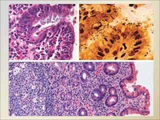
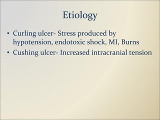
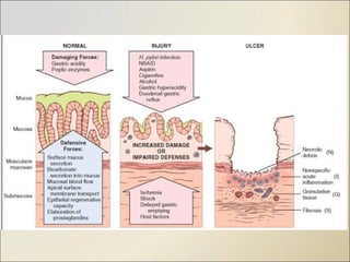
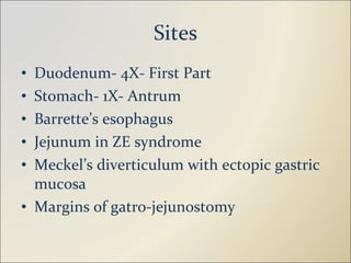
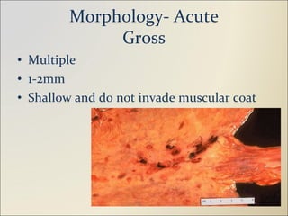
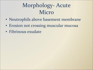
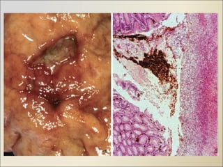
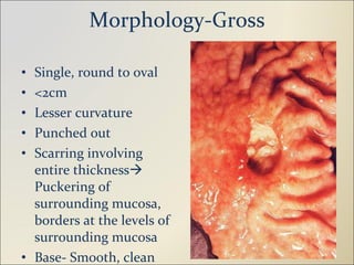
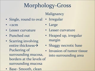
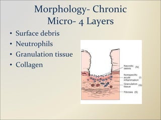
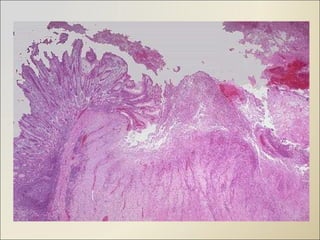
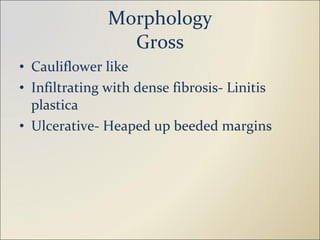
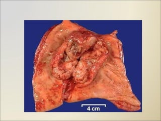
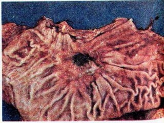
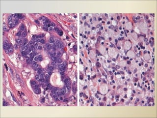
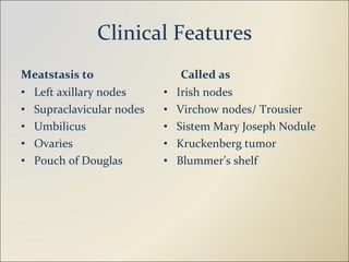
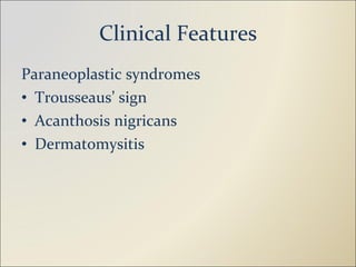
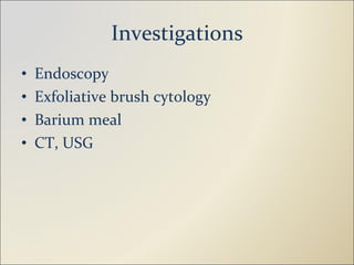
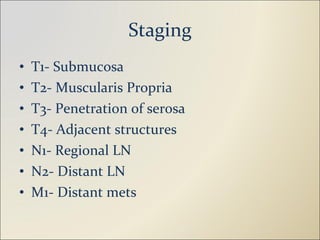
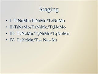
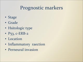
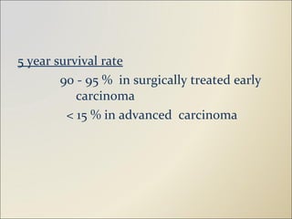
Stomach Dr. Girish Kamat
University Questions Peptic Ulcer Long Essay- 10M Describe the etiology, gross, and microscopy of gastric ulcer. List complications of gastric ulcer Define peptic ulcer. Mention the sites. Describe etiopathogenesis and morphology of chronic gastric ulcer Short Answers- 5M Helicobacter pylori Etiology of duodenal ulcer Short Notes- 3M Macroscopy of benign and malignant gastric ulcer
University Questions Tumors Short Answers- 5M Morphologic types of carcinoma of stomach Early gastric carcinoma Short Notes- 3M Morphology of gastric carcinoma Linitis plastica Signet ring cell Modes of spread of gastric cancer
Peptic Ulcer Definition Etiopathogenesis H.Pylori Sites Morphology- Acute, Chronic Difference between benign and malignant Clinical features Investigatuions Complications
Gastric Tumors Classification Carcinoma Etiology Morphology Clinical features Investigations Spread Staging Prognostic markers
Peptic Ulcer
Definition “ It is acid peptic digestion of alimentary mucosa, resulting in an ulcer, that extends through the muscularis mucosa into the submucosa or deeper.”
Epidemiology Men affected  more than Females M: F ratio  in  Duodenal ulcer is  3:1 M : F ratio  in  Gastric  ulcer  is 1.5 : 1 Women most affected at or after menopause
 
Etiology H.Pylori Infection 100%- Duodenal, 70%- Gastric 10-20% infected develop gastritis Non sporing, curvilinear, flagellated, Gram – neg Swims through mucus, urease, bacterialadhesins- Bab A, Produces cytotoxin (CagA gene), Vacuolating cytotoxin (VacA) Chronic gastritis   Atrophy   Intestinal metaplasia   Dysplasia   Carcinoma Also- MALToma
Etiology- H. Pylori Mechanism of production of gastritis Inflammatory cytokines- IL- 1,6,8, TNF Epithelial injury- Urease, protetease, Phospholipase Impairs duodenal bicarbonate production Thrombotic occlusion of surface capillaries- Bacterial PAF
Etiology- H. Pylori Tests Biopsy- H&E, Geimsa, Warthin Starry, Steiner silver, Aclian yellow tuleidine blue method, IHC Serology- Elisa Urea breath test- C13/C14 urea Culture and sensitivity Bacterial DNA detection by PCR
 
Etiology- NSAIDS & Steroids Inhibit cyclo-oxygenase   Reduced PG    Reduced cytoprotection   Mucosal injury
Etiology Emotions Diet Pylorodudenal reflux Endocrine- Zollinger Ellison, Cushing Trauma Smoking Hereditory- Blood Gr O
Etiology Curling ulcer- Stress produced by hypotension, endotoxic shock, MI, Burns Cushing ulcer- Increased intracranial tension
 
Sites Duodenum- 4X- First Part Stomach- 1X- Antrum Barrette’s esophagus Jejunum in ZE syndrome Meckel’s diverticulum with ectopic gastric mucosa Margins of gatro-jejunostomy
Morphology- Acute Gross Multiple 1-2mm Shallow and do not invade muscular coat
Morphology- Acute Micro Neutrophils above basement membrane Erosion not crossing muscular mucosa Fibrinous exudate
 
Morphology-Gross Single, round to oval <2cm Lesser curvature Punched out Scarring involving entire thickness   Puckering of surrounding mucosa, borders at the levels of surrounding mucosa Base- Smooth, clean
Morphology-Gross Single, round to oval <2cm Lesser curvature Punched out Scarring involving entire thickness   Puckering of surrounding mucosa, borders at the levels of surrounding mucosa Base- Smooth, clean Malignancy Irregular Large Lesser curvature Heaped up, irregular margin Shaggy necrotic base Invasion of tumor tissue into surrounding area
Morphology- Chronic Micr0- 4 Layers Surface debris Neutrophils Granulation tissue Collagen
 
Clinical Features Abdominal pain- Epigastric, burning type, vomiting (Relieves), episodic Food aggravates- Gastric Food relieves- Duodenal Hematemesis, malena
Investigations Blood- Anemia Stool-Occult blood Barium meal Endoscopy- H.Pylori, Malignancy H.Pylori Gatsric function tests- Obsolete
Complications Bleeding- 15-20% Perforation- 5% Obstruction due to edema and scarring
Other  gastritis Eosinophilic Lymphocytic Granulomatous Hemorrhagic CMV Radiation/ chemotherapy Autoimmune Xanthogranulomatous
Gastric Tumors
Classification Epithelial Adenoma Adenocarcinoma- Papillary, tubular, mucinous, signet ring, undifferentiated, adenosquamous Small cell carcinoma Carcinoid tumor Non Epithelial Leiomyoma Schwannoma GIST Lymphoma
Carcinoma of Stomach
Epidemiology Higher in Japan, China compared to US, UK More common in lower socio-economic groups Male to Female ratio is 2: 1 Steady decline in incidence & mortality for the past 6 decades
Etiology H.Pylori Diet- Salted/smoked food, nitrates Smoking and alcohol Decreased gastric acid secretion- Proton pump inhibitors, atrophic gastritis
Morphology Gross Sites-  Antrum- 50% Body- 30% Cardiac- 20% MC site- Lesser curvature
Morphology Gross Cauliflower like Infiltrating with dense fibrosis- Linitis plastica Ulcerative- Heaped up beeded margins
 
 
Morphology Micro- (DIO/ Lauren) Diffuse- Signet ring cells Intestinal- Similar to intestinal adenoca Others
 
 
Early Gastric Ca (Superficial/ microinvasive Ca) Ca confined to mucosa and submucosa regardless of status ofregional lymph nodes
Clinical Features Ulcer like pain Weight loss, anorexia Hematemesis Palpable epigastric mass
Clinical Features Meatstasis to Left axillary nodes Supraclavicular nodes Umbilicus Ovaries Pouch of Douglas Called as Irish nodes Virchow nodes/ Trousier Sistem Mary Joseph Nodule Kruckenberg tumor Blummer’s shelf
Clinical Features Paraneoplastic syndromes Trousseaus’ sign Acanthosis nigricans Dermatomysitis
Investigations Endoscopy Exfoliative brush cytology Barium meal CT, USG
Staging T1- Submucosa T2- Muscularis Propria T3- Penetration of serosa T4- Adjacent structures N1- Regional LN N2- Distant LN M1- Distant mets
Staging I- T1N0M0/T1N1M0/T2NoMo II-T1N2M0/T2N1Mo/T3N0M0 III- T2N2M0/T3N1M0/T4NoM0 IV- T4N2M0/T any  N any  M1
Prognostic markers Stage Grade Hsitologic type P53, c-ERB-2 Location Inflammatory  raection Perneural invasion
5 year survival rate 90 - 95 %  in surgically treated early  carcinoma < 15 % in advanced  carcinoma
Thank You…

2 stomach girish_10_11

  • 1.
  • 2.
    University Questions PepticUlcer Long Essay- 10M Describe the etiology, gross, and microscopy of gastric ulcer. List complications of gastric ulcer Define peptic ulcer. Mention the sites. Describe etiopathogenesis and morphology of chronic gastric ulcer Short Answers- 5M Helicobacter pylori Etiology of duodenal ulcer Short Notes- 3M Macroscopy of benign and malignant gastric ulcer
  • 3.
    University Questions TumorsShort Answers- 5M Morphologic types of carcinoma of stomach Early gastric carcinoma Short Notes- 3M Morphology of gastric carcinoma Linitis plastica Signet ring cell Modes of spread of gastric cancer
  • 4.
    Peptic Ulcer DefinitionEtiopathogenesis H.Pylori Sites Morphology- Acute, Chronic Difference between benign and malignant Clinical features Investigatuions Complications
  • 5.
    Gastric Tumors ClassificationCarcinoma Etiology Morphology Clinical features Investigations Spread Staging Prognostic markers
  • 6.
  • 7.
    Definition “ Itis acid peptic digestion of alimentary mucosa, resulting in an ulcer, that extends through the muscularis mucosa into the submucosa or deeper.”
  • 8.
    Epidemiology Men affected more than Females M: F ratio in Duodenal ulcer is 3:1 M : F ratio in Gastric ulcer is 1.5 : 1 Women most affected at or after menopause
  • 9.
  • 10.
    Etiology H.Pylori Infection100%- Duodenal, 70%- Gastric 10-20% infected develop gastritis Non sporing, curvilinear, flagellated, Gram – neg Swims through mucus, urease, bacterialadhesins- Bab A, Produces cytotoxin (CagA gene), Vacuolating cytotoxin (VacA) Chronic gastritis  Atrophy  Intestinal metaplasia  Dysplasia  Carcinoma Also- MALToma
  • 11.
    Etiology- H. PyloriMechanism of production of gastritis Inflammatory cytokines- IL- 1,6,8, TNF Epithelial injury- Urease, protetease, Phospholipase Impairs duodenal bicarbonate production Thrombotic occlusion of surface capillaries- Bacterial PAF
  • 12.
    Etiology- H. PyloriTests Biopsy- H&E, Geimsa, Warthin Starry, Steiner silver, Aclian yellow tuleidine blue method, IHC Serology- Elisa Urea breath test- C13/C14 urea Culture and sensitivity Bacterial DNA detection by PCR
  • 13.
  • 14.
    Etiology- NSAIDS &Steroids Inhibit cyclo-oxygenase  Reduced PG  Reduced cytoprotection  Mucosal injury
  • 15.
    Etiology Emotions DietPylorodudenal reflux Endocrine- Zollinger Ellison, Cushing Trauma Smoking Hereditory- Blood Gr O
  • 16.
    Etiology Curling ulcer-Stress produced by hypotension, endotoxic shock, MI, Burns Cushing ulcer- Increased intracranial tension
  • 17.
  • 18.
    Sites Duodenum- 4X-First Part Stomach- 1X- Antrum Barrette’s esophagus Jejunum in ZE syndrome Meckel’s diverticulum with ectopic gastric mucosa Margins of gatro-jejunostomy
  • 19.
    Morphology- Acute GrossMultiple 1-2mm Shallow and do not invade muscular coat
  • 20.
    Morphology- Acute MicroNeutrophils above basement membrane Erosion not crossing muscular mucosa Fibrinous exudate
  • 21.
  • 22.
    Morphology-Gross Single, roundto oval <2cm Lesser curvature Punched out Scarring involving entire thickness  Puckering of surrounding mucosa, borders at the levels of surrounding mucosa Base- Smooth, clean
  • 23.
    Morphology-Gross Single, roundto oval <2cm Lesser curvature Punched out Scarring involving entire thickness  Puckering of surrounding mucosa, borders at the levels of surrounding mucosa Base- Smooth, clean Malignancy Irregular Large Lesser curvature Heaped up, irregular margin Shaggy necrotic base Invasion of tumor tissue into surrounding area
  • 24.
    Morphology- Chronic Micr0-4 Layers Surface debris Neutrophils Granulation tissue Collagen
  • 25.
  • 26.
    Clinical Features Abdominalpain- Epigastric, burning type, vomiting (Relieves), episodic Food aggravates- Gastric Food relieves- Duodenal Hematemesis, malena
  • 27.
    Investigations Blood- AnemiaStool-Occult blood Barium meal Endoscopy- H.Pylori, Malignancy H.Pylori Gatsric function tests- Obsolete
  • 28.
    Complications Bleeding- 15-20%Perforation- 5% Obstruction due to edema and scarring
  • 29.
    Other gastritisEosinophilic Lymphocytic Granulomatous Hemorrhagic CMV Radiation/ chemotherapy Autoimmune Xanthogranulomatous
  • 30.
  • 31.
    Classification Epithelial AdenomaAdenocarcinoma- Papillary, tubular, mucinous, signet ring, undifferentiated, adenosquamous Small cell carcinoma Carcinoid tumor Non Epithelial Leiomyoma Schwannoma GIST Lymphoma
  • 32.
  • 33.
    Epidemiology Higher inJapan, China compared to US, UK More common in lower socio-economic groups Male to Female ratio is 2: 1 Steady decline in incidence & mortality for the past 6 decades
  • 34.
    Etiology H.Pylori Diet-Salted/smoked food, nitrates Smoking and alcohol Decreased gastric acid secretion- Proton pump inhibitors, atrophic gastritis
  • 35.
    Morphology Gross Sites- Antrum- 50% Body- 30% Cardiac- 20% MC site- Lesser curvature
  • 36.
    Morphology Gross Cauliflowerlike Infiltrating with dense fibrosis- Linitis plastica Ulcerative- Heaped up beeded margins
  • 37.
  • 38.
  • 39.
    Morphology Micro- (DIO/Lauren) Diffuse- Signet ring cells Intestinal- Similar to intestinal adenoca Others
  • 40.
  • 41.
  • 42.
    Early Gastric Ca(Superficial/ microinvasive Ca) Ca confined to mucosa and submucosa regardless of status ofregional lymph nodes
  • 43.
    Clinical Features Ulcerlike pain Weight loss, anorexia Hematemesis Palpable epigastric mass
  • 44.
    Clinical Features Meatstasisto Left axillary nodes Supraclavicular nodes Umbilicus Ovaries Pouch of Douglas Called as Irish nodes Virchow nodes/ Trousier Sistem Mary Joseph Nodule Kruckenberg tumor Blummer’s shelf
  • 45.
    Clinical Features Paraneoplasticsyndromes Trousseaus’ sign Acanthosis nigricans Dermatomysitis
  • 46.
    Investigations Endoscopy Exfoliativebrush cytology Barium meal CT, USG
  • 47.
    Staging T1- SubmucosaT2- Muscularis Propria T3- Penetration of serosa T4- Adjacent structures N1- Regional LN N2- Distant LN M1- Distant mets
  • 48.
    Staging I- T1N0M0/T1N1M0/T2NoMoII-T1N2M0/T2N1Mo/T3N0M0 III- T2N2M0/T3N1M0/T4NoM0 IV- T4N2M0/T any N any M1
  • 49.
    Prognostic markers StageGrade Hsitologic type P53, c-ERB-2 Location Inflammatory raection Perneural invasion
  • 50.
    5 year survivalrate 90 - 95 % in surgically treated early carcinoma < 15 % in advanced carcinoma
  • 51.