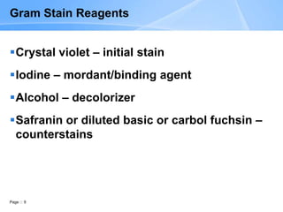
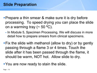
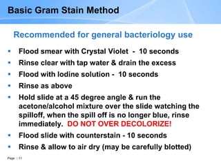
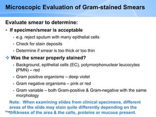
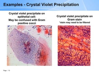
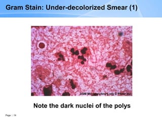
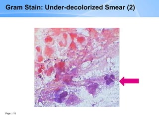
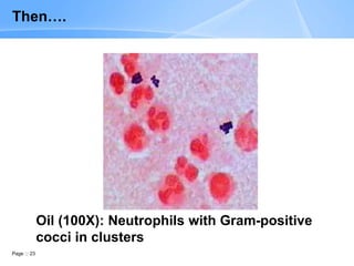
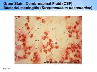
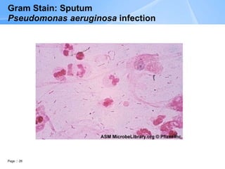
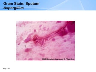
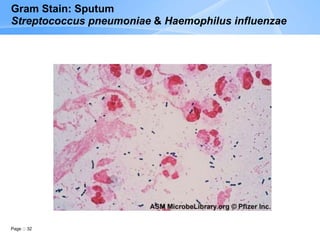
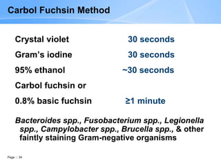
Module 4: Gram Stain Principle,
Procedure, & Interpretation
Page 2
Learning Objectives
At the end of this session participants should be able to :
 Describe the differences between gram-positive & gram-negative
bacteria that determine how they will stain with the Gram stain
 Properly prepare smears for Gram staining, both from clinical
specimens & cultures
 Properly perform the Gram stain procedure
 Interpret Gram stains based on sample type
 Recognize the morphological groups of organisms based on Gram
stain appearance
 Recognize the appearance of human cells associated with infectious
processes
 Prepare a set of slides to be used for Gram stain quality control (QC)
Page 3
Content Overview
Uses & advantages of Gram stains
Principle of the Gram stain
Specimens for Gram stain
Gram stain procedure
Examination & interpretation
Reporting of Gram stain results
QC of Gram stains
Page 4
 A critical test for a rapid, presumptive
diagnosis of infectious agents
 Used to classify bacteria on the basis of
their Gram reaction, morphology (shape),
& arrangement
 Serves to assess the quality of clinical
specimens
Gram Stain
 Originally developed by Christian Gram in 1884
 With minor modifications still used 126 years later
Page 5
Advantages of the Gram Stain
Rapid procedure
Cost effective
Accessible
Effective screening technique
Reliable
Semi-quantitative
Provides culture clues
Presumptive diagnosis of infection
Page 6
Gram positive Gram negative
Peptidoglycan Thick Thin
Teichoic acid Yes No
Outer membrane No Yes
Periplasm No Yes
Why do Bacteria Stain Differently?
It’s in the Cell Wall!
Page 7
Principle of the Gram Stain
 Bacteria stain either Gram-positive or Gram-
negative on the basis of differences in their cell wall
composition
 Gram positives have a thick peptidoglycan layer &
large amounts of teichoic acids
 Gram negatives have a thin peptidoglycan layer &
an outer membrane composed of a lipid bilayer
 The outer membrane of Gram-negative organisms
is damaged by the decolorizer (organic solvents)
Page 8
Specimens for Gram Stain
Clinical specimens (direct smears): wounds,
eye lesions, sterile fluids, body tissues,
discharges
Not done on: blood, throat swabs, nasal
swabs, stool (why?)
Young colonies on solid medium
Broth & blood cultures (turbid) - Arrangement
is best seen in broth
Page 9
Gram Stain Reagents
Crystal violet – initial stain
Iodine – mordant/binding agent
Alcohol – decolorizer
Safranin or diluted basic or carbol fuchsin –
counterstains
Page 10
Slide Preparation
Prepare a thin smear & make sure it is dry before
processing. To speed drying you can place the slide
on a warming tray (~ 50 ºC).
 In Module 5, Specimen Processing, We will discuss in more
detail how to prepare smears from clinical specimens.
Fix the slide with methanol (allow to dry) or by gently
passing through a flame 3 or 4 times. Touch the
slide after it has been passed through the flame, it
should be warm, NOT hot. Allow slide to dry.
You are now ready to stain the slide.
Page 11
Basic Gram Stain Method
 Flood smear with Crystal Violet - 10 seconds
 Rinse clear with tap water & drain the excess
 Flood with Iodine solution - 10 seconds
 Rinse as above
 Hold slide at a 45 degree angle & run the
acetone/alcohol mixture over the slide watching the
spilloff, when the spill off is no longer blue, rinse
immediately. DO NOT OVER DECOLORIZE!
 Flood slide with counterstain - 10 seconds
 Rinse & allow to air dry (may be carefully blotted)
Recommended for general bacteriology use
Page 12
Microscopic Evaluation of Gram-stained Smears
Evaluate smear to determine:
 If specimen/smear is acceptable
 e.g. reject sputum with many epithelial cells
 Check for stain deposits
 Determine if smear is too thick or too thin
 Was the smear properly stained?
 Background, epithelial cells (EC), polymorphonuclear leucocytes
(PMN) – red
 Gram positive organisms – deep violet
 Gram negative organisms – pink or red
 Gram variable – both Gram-positive & Gram-negative with the same
morphology
Note: When examining slides from clinical specimens, different
areas of the slide may stain quite differently depending on the
thickness of the area & the cells, proteins or mucous present.
Page 13
Crystal violet precipitate on
epithelial cell:
May be confused with Gram
positive cocci
Crystal violet precipitate on
Gram stain
*stain may need to be filtered
Examples - Crystal Violet Precipitation
Page 14
Gram Stain: Under-decolorized Smear (1)
Note the dark nuclei of the polys
Page 15
Gram Stain: Under-decolorized Smear (2)
Page 16
Gram Stain: Over-decolorized Smear
Page 17
Examination & Interpretation (1)
For smears prepared from clinical specimens
 105 organisms/ml of un-centrifuged fluid (104
centrifuged) organisms are required to be visible on slide
 At lower concentrations, organism will not be revealed
even if culture is positive
 Organisms seen on Gram stain, but fail to grow in culture
 fastidious (require specific media for growth)
 anaerobic bacteria
 patient has received antibiotics
Page 18
Examination & Interpretation (2)
 Cells are enumerated using the low power objective
(LPO)
 Squamous epithelial cells (SEC’s), PMN’s, red blood cells
(RBC’s)
 Examine multiple areas of the slide under both low power
& oil immersion magnification.
 Bacteria & yeasts are enumerated using the oil
immersion objective (OIO)
 Ignore one or two microorganisms on the entire slide (oil
objective field) unless results can be reproduced on a
second smear &, only if it is from an invasively collected
specimen
Page 19
Examination & Interpretation (3)
The presence of microorganisms from a
normally sterile site is likely to indicate
infections with the organism that is seen
The presence of large numbers of a single type
of organisms in a non-invasively collected
specimen, especially if associated with white
blood cells (WBC’s), is likely to indicate infection
Page 20
Interpretation & Reporting (1)
 Perform counts only in areas representative of
inflammation or necrosis, if present
 Specify gram-reaction, morphology (be as descriptive as
possible), arrangement & other information that can lead
to the suggestion of a particular pathogen
 Examples:
 Gram-positive diplococci consistent with Streptococcus
pneumoniae
 Small Gram-negative coccobacilli, consistent with
Haemophilus
 Gram-negative diplococci, consistent with Neisseria
species
Page 21
Interpretation & Reporting (2)
 Enumerate cells per low power field (LPF) Epis, PMNs,
RBCs)
1+ (rare or occasional): <1/LPF
2+ (few): 1– 9/LPF
3+ (moderate): 10-25/LPF
4+ (many): >25/LPF
 Enumerate bacteria & yeast per OIF
1+ (rare or occasional): <1/OIF
2+ (few): 1– 5/OIF
3+ (moderate): 6-25/OIF
4+ (many): >25/OIF
Page 22
10X: Many Neutrophils seen
Scan Slide on Low Power
Page 23
Oil (100X): Neutrophils with Gram-positive
cocci in clusters
Then….
Page 24
Gram Stain: Wound
Streptococcus pyogenes Infection
Page 25
Gram Stain: Cerebrospinal Fluid (CSF)
Bacterial meningitis (Streptococcus pneumoniae)
Page 26
Gram Stain: Sputum
Pseudomonas aeruginosa infection
Page 27
Page  27
Gram Stain: Sputum
Haemophilus influenzae
Page 28
Gram Stain: Urethral Discharge
Urethritis Neisseria gonorrhoaea
Page 29
Gram Stain: Sputum
Nocardia infection
Page 30
Gram Stain: Sputum
Aspergillus
Page 31
Gram Stain: Vaginal discharge:
Candida albicans
Page 32
Gram Stain: Sputum
Streptococcus pneumoniae & Haemophilus influenzae
Page 33
Do All Bacteria Stain with the Gram Stain?
Organisms that do not stain
Chlamydia & others - too small & intracellular
Spirochetes - many are too thin
Mycobacteria - waxy cell wall
Poorly staining
Some anaerobes
Legionella
Brucella & others
Gram-stain modifications used for poorly staining
organisms
Page 34
Carbol Fuchsin Method
Crystal violet 30 seconds
Gram’s iodine 30 seconds
95% ethanol ~30 seconds
Carbol fuchsin or
0.8% basic fuchsin ≥1 minute
Bacteroides spp., Fusobacterium spp., Legionella
spp., Campylobacter spp., Brucella spp., & other
faintly staining Gram-negative organisms
Page 35
Kopeloff’s Method
1. Alkaline crystal violet: flood with solution A;
add 5 drops of solution B 2–3 minutes
2. Kopeloff’s iodine ≥2 minutes
3. 3:7 acetone-alcohol rinse immediately after
applying
4. Kopeloff’s safranin 10-30 seconds
Anaerobes, diagnosis of bacterial vaginosis
Page 36
Summary

1_M4_Participant_Gram Stain.ppt

  • 1.
    Module 4: GramStain Principle, Procedure, & Interpretation
  • 2.
    Page 2 Learning Objectives Atthe end of this session participants should be able to :  Describe the differences between gram-positive & gram-negative bacteria that determine how they will stain with the Gram stain  Properly prepare smears for Gram staining, both from clinical specimens & cultures  Properly perform the Gram stain procedure  Interpret Gram stains based on sample type  Recognize the morphological groups of organisms based on Gram stain appearance  Recognize the appearance of human cells associated with infectious processes  Prepare a set of slides to be used for Gram stain quality control (QC)
  • 3.
    Page 3 Content Overview Uses& advantages of Gram stains Principle of the Gram stain Specimens for Gram stain Gram stain procedure Examination & interpretation Reporting of Gram stain results QC of Gram stains
  • 4.
    Page 4  Acritical test for a rapid, presumptive diagnosis of infectious agents  Used to classify bacteria on the basis of their Gram reaction, morphology (shape), & arrangement  Serves to assess the quality of clinical specimens Gram Stain  Originally developed by Christian Gram in 1884  With minor modifications still used 126 years later
  • 5.
    Page 5 Advantages ofthe Gram Stain Rapid procedure Cost effective Accessible Effective screening technique Reliable Semi-quantitative Provides culture clues Presumptive diagnosis of infection
  • 6.
    Page 6 Gram positiveGram negative Peptidoglycan Thick Thin Teichoic acid Yes No Outer membrane No Yes Periplasm No Yes Why do Bacteria Stain Differently? It’s in the Cell Wall!
  • 7.
    Page 7 Principle ofthe Gram Stain  Bacteria stain either Gram-positive or Gram- negative on the basis of differences in their cell wall composition  Gram positives have a thick peptidoglycan layer & large amounts of teichoic acids  Gram negatives have a thin peptidoglycan layer & an outer membrane composed of a lipid bilayer  The outer membrane of Gram-negative organisms is damaged by the decolorizer (organic solvents)
  • 8.
    Page 8 Specimens forGram Stain Clinical specimens (direct smears): wounds, eye lesions, sterile fluids, body tissues, discharges Not done on: blood, throat swabs, nasal swabs, stool (why?) Young colonies on solid medium Broth & blood cultures (turbid) - Arrangement is best seen in broth
  • 9.
    Page 9 Gram StainReagents Crystal violet – initial stain Iodine – mordant/binding agent Alcohol – decolorizer Safranin or diluted basic or carbol fuchsin – counterstains
  • 10.
    Page 10 Slide Preparation Preparea thin smear & make sure it is dry before processing. To speed drying you can place the slide on a warming tray (~ 50 ºC).  In Module 5, Specimen Processing, We will discuss in more detail how to prepare smears from clinical specimens. Fix the slide with methanol (allow to dry) or by gently passing through a flame 3 or 4 times. Touch the slide after it has been passed through the flame, it should be warm, NOT hot. Allow slide to dry. You are now ready to stain the slide.
  • 11.
    Page 11 Basic GramStain Method  Flood smear with Crystal Violet - 10 seconds  Rinse clear with tap water & drain the excess  Flood with Iodine solution - 10 seconds  Rinse as above  Hold slide at a 45 degree angle & run the acetone/alcohol mixture over the slide watching the spilloff, when the spill off is no longer blue, rinse immediately. DO NOT OVER DECOLORIZE!  Flood slide with counterstain - 10 seconds  Rinse & allow to air dry (may be carefully blotted) Recommended for general bacteriology use
  • 12.
    Page 12 Microscopic Evaluationof Gram-stained Smears Evaluate smear to determine:  If specimen/smear is acceptable  e.g. reject sputum with many epithelial cells  Check for stain deposits  Determine if smear is too thick or too thin  Was the smear properly stained?  Background, epithelial cells (EC), polymorphonuclear leucocytes (PMN) – red  Gram positive organisms – deep violet  Gram negative organisms – pink or red  Gram variable – both Gram-positive & Gram-negative with the same morphology Note: When examining slides from clinical specimens, different areas of the slide may stain quite differently depending on the thickness of the area & the cells, proteins or mucous present.
  • 13.
    Page 13 Crystal violetprecipitate on epithelial cell: May be confused with Gram positive cocci Crystal violet precipitate on Gram stain *stain may need to be filtered Examples - Crystal Violet Precipitation
  • 14.
    Page 14 Gram Stain:Under-decolorized Smear (1) Note the dark nuclei of the polys
  • 15.
    Page 15 Gram Stain:Under-decolorized Smear (2)
  • 16.
    Page 16 Gram Stain:Over-decolorized Smear
  • 17.
    Page 17 Examination &Interpretation (1) For smears prepared from clinical specimens  105 organisms/ml of un-centrifuged fluid (104 centrifuged) organisms are required to be visible on slide  At lower concentrations, organism will not be revealed even if culture is positive  Organisms seen on Gram stain, but fail to grow in culture  fastidious (require specific media for growth)  anaerobic bacteria  patient has received antibiotics
  • 18.
    Page 18 Examination &Interpretation (2)  Cells are enumerated using the low power objective (LPO)  Squamous epithelial cells (SEC’s), PMN’s, red blood cells (RBC’s)  Examine multiple areas of the slide under both low power & oil immersion magnification.  Bacteria & yeasts are enumerated using the oil immersion objective (OIO)  Ignore one or two microorganisms on the entire slide (oil objective field) unless results can be reproduced on a second smear &, only if it is from an invasively collected specimen
  • 19.
    Page 19 Examination &Interpretation (3) The presence of microorganisms from a normally sterile site is likely to indicate infections with the organism that is seen The presence of large numbers of a single type of organisms in a non-invasively collected specimen, especially if associated with white blood cells (WBC’s), is likely to indicate infection
  • 20.
    Page 20 Interpretation &Reporting (1)  Perform counts only in areas representative of inflammation or necrosis, if present  Specify gram-reaction, morphology (be as descriptive as possible), arrangement & other information that can lead to the suggestion of a particular pathogen  Examples:  Gram-positive diplococci consistent with Streptococcus pneumoniae  Small Gram-negative coccobacilli, consistent with Haemophilus  Gram-negative diplococci, consistent with Neisseria species
  • 21.
    Page 21 Interpretation &Reporting (2)  Enumerate cells per low power field (LPF) Epis, PMNs, RBCs) 1+ (rare or occasional): <1/LPF 2+ (few): 1– 9/LPF 3+ (moderate): 10-25/LPF 4+ (many): >25/LPF  Enumerate bacteria & yeast per OIF 1+ (rare or occasional): <1/OIF 2+ (few): 1– 5/OIF 3+ (moderate): 6-25/OIF 4+ (many): >25/OIF
  • 22.
    Page 22 10X: ManyNeutrophils seen Scan Slide on Low Power
  • 23.
    Page 23 Oil (100X):Neutrophils with Gram-positive cocci in clusters Then….
  • 24.
    Page 24 Gram Stain:Wound Streptococcus pyogenes Infection
  • 25.
    Page 25 Gram Stain:Cerebrospinal Fluid (CSF) Bacterial meningitis (Streptococcus pneumoniae)
  • 26.
    Page 26 Gram Stain:Sputum Pseudomonas aeruginosa infection
  • 27.
    Page 27 Page 27 Gram Stain: Sputum Haemophilus influenzae
  • 28.
    Page 28 Gram Stain:Urethral Discharge Urethritis Neisseria gonorrhoaea
  • 29.
    Page 29 Gram Stain:Sputum Nocardia infection
  • 30.
    Page 30 Gram Stain:Sputum Aspergillus
  • 31.
    Page 31 Gram Stain:Vaginal discharge: Candida albicans
  • 32.
    Page 32 Gram Stain:Sputum Streptococcus pneumoniae & Haemophilus influenzae
  • 33.
    Page 33 Do AllBacteria Stain with the Gram Stain? Organisms that do not stain Chlamydia & others - too small & intracellular Spirochetes - many are too thin Mycobacteria - waxy cell wall Poorly staining Some anaerobes Legionella Brucella & others Gram-stain modifications used for poorly staining organisms
  • 34.
    Page 34 Carbol FuchsinMethod Crystal violet 30 seconds Gram’s iodine 30 seconds 95% ethanol ~30 seconds Carbol fuchsin or 0.8% basic fuchsin ≥1 minute Bacteroides spp., Fusobacterium spp., Legionella spp., Campylobacter spp., Brucella spp., & other faintly staining Gram-negative organisms
  • 35.
    Page 35 Kopeloff’s Method 1.Alkaline crystal violet: flood with solution A; add 5 drops of solution B 2–3 minutes 2. Kopeloff’s iodine ≥2 minutes 3. 3:7 acetone-alcohol rinse immediately after applying 4. Kopeloff’s safranin 10-30 seconds Anaerobes, diagnosis of bacterial vaginosis
  • 36.