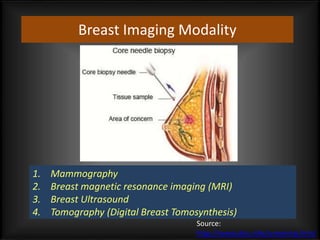
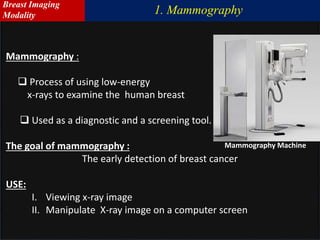
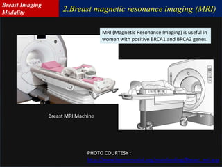
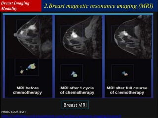
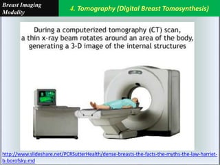
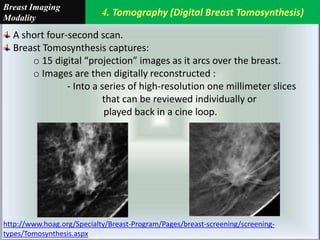
Department of Computer Science And Engineering
Shahjalal University of Science and Technology
Nashid Alam
Registration No: 2012321028
annanya_cse@yahoo.co.uk
Masters -2 Presentation
(Backup Slides# 1)
Breast Imaging Modality
1. Mammography
2. Breast magnetic resonance imaging (MRI)
3. Breast Ultrasound
4. Tomography (Digital Breast Tomosynthesis)
Source:
http://www.dcis.info/screening.html
1. Mammography
Breast Imaging
Modality
USE:
I. Viewing x-ray image
II. Manipulate X-ray image on a computer screen
Mammography :
 Process of using low-energy
x-rays to examine the human breast
 Used as a diagnostic and a screening tool.
The goal of mammography :
The early detection of breast cancer
Mammography Machine
1. Mammography
Breast Imaging
Modality
Mammogram:
A mammogram is an x-ray picture of the breast
Use:
To look for changes that are not normal.
Result Archive:
The results are recorded on x-ray film or directly into a computer
Types of mammograms:
I. Screening mammograms-Done for women who have no symptoms of breast
cancer.
II. Diagnostic mammograms -To check for breast cancer after a lump or other
symptom or sign of breast cancer has been found.
III. Digital mammogram-Uses x-rays to produce an image of the breast. The image
is stored directly on a computer.
mdb226.jpg
1. Mammography
Breast Imaging
Modality
http://www.dcis.info/screening.html
Records X-ray images in computer code instead of on film.
To view the images :
-The radiologist uses computer software
Magnification makes:
-Easier to spot a problem.
Can be sent electronically (called telemammography)
-For additional consultations.
Digital Mammography
1. Mammography
Breast Imaging
Modality
http://www.dcis.info/screening.html
The radiologist looks for :
Unusual shadows
Clusters of white specks (micro calcifications)
Distortions
Special patterns of tissue density
Any mass and its shape
Differences between the images of both breasts
The radiologist also looks at your previous
mammograms and compares them to your most recent
film(s)
What the radiologist looks for?
Mammograms are read by specialists called radiologists.
1. Mammography
Breast Imaging
Modality
2.Breast magnetic resonance imaging (MRI)
Breast Imaging
Modality
2.Breast magnetic resonance imaging (MRI)
Breast MRI Machine
PHOTO COURTESY :
http://www.leememorial.org/mainlanding/Breast_mri.asp
MRI (Magnetic Resonance Imaging) is useful in
women with positive BRCA1 and BRCA2 genes.
Breast Imaging
Modality
Breast MRI
PHOTO COURTESY :
http://radiology.ucsf.edu/patient-care/sections/womens-imaging/research
2.Breast magnetic resonance imaging (MRI)
Breast Imaging
Modality
3.Breast Ultrasound / Sonography
Breast Imaging
Modality
3.Breast Ultrasound / Sonography
The pictures produced during
an ultrasound are called:
Sonograms
Echograms or
Ultrasound Scans
An ultrasound of the breast:
- Shows All Areas Of The Breast
Including:
The area closest to the chest wall
(Which is hard to see with a mammogram.)
An Sonography/Ultrasound Machine
Breast Imaging
Modality
3.Breast Ultrasound / Sonography
ULTRASOUND TEST:
• Uses reflected sound waves to produce pictures of the inside of the body.
•It does not use X-rays or other types of possibly harmful radiation.
•A special kind of instrument called a transducer is rubbed back and forth over the area to
be examined.
It gives off high-pitched sound waves that show on a special screen.
Schematic of the Optoacoustic Plus UltraSound (OPUS) system
Breast Imaging
Modality
3.Breast Ultrasound / Sonography
An Sonography/Ultrasound image of the breast
PHOTO COURTESY :
http://www.ultrasoundschoolsinfo.com/breast-sonography/
Breast Imaging
Modality
3.Breast Ultrasound / Sonography
PHOTO COURTESY :
http://www.slideshare.net/PCRSutterHealth/dense-breasts-the-facts-the-myths-the-law-harriet-b-borofsky-md
Breast Ultrasound/Sonography
Breast Imaging
Modality
4. Tomography (Digital Breast Tomosynthesis)
Breast Imaging
Modality
4. Tomography (Digital Breast Tomosynthesis)
Breast Imaging
Modality
http://www.slideshare.net/PCRSutterHealth/dense-breasts-the-facts-the-myths-the-law-harriet-
b-borofsky-md
4. Tomography (Digital Breast Tomosynthesis)
Breast Imaging
Modality
A short four-second scan.
Breast Tomosynthesis captures:
o 15 digital “projection” images as it arcs over the breast.
o Images are then digitally reconstructed :
- Into a series of high-resolution one millimeter slices
that can be reviewed individually or
played back in a cine loop.
http://www.hoag.org/Specialty/Breast-Program/Pages/breast-screening/screening-
types/Tomosynthesis.aspx

1.breast cancer screening(backup slide-1)

  • 1.
    Department of ComputerScience And Engineering Shahjalal University of Science and Technology Nashid Alam Registration No: 2012321028 annanya_cse@yahoo.co.uk Masters -2 Presentation (Backup Slides# 1)
  • 3.
    Breast Imaging Modality 1.Mammography 2. Breast magnetic resonance imaging (MRI) 3. Breast Ultrasound 4. Tomography (Digital Breast Tomosynthesis) Source: http://www.dcis.info/screening.html
  • 4.
  • 5.
    USE: I. Viewing x-rayimage II. Manipulate X-ray image on a computer screen Mammography :  Process of using low-energy x-rays to examine the human breast  Used as a diagnostic and a screening tool. The goal of mammography : The early detection of breast cancer Mammography Machine 1. Mammography Breast Imaging Modality
  • 6.
    Mammogram: A mammogram isan x-ray picture of the breast Use: To look for changes that are not normal. Result Archive: The results are recorded on x-ray film or directly into a computer Types of mammograms: I. Screening mammograms-Done for women who have no symptoms of breast cancer. II. Diagnostic mammograms -To check for breast cancer after a lump or other symptom or sign of breast cancer has been found. III. Digital mammogram-Uses x-rays to produce an image of the breast. The image is stored directly on a computer. mdb226.jpg 1. Mammography Breast Imaging Modality
  • 7.
    http://www.dcis.info/screening.html Records X-ray imagesin computer code instead of on film. To view the images : -The radiologist uses computer software Magnification makes: -Easier to spot a problem. Can be sent electronically (called telemammography) -For additional consultations. Digital Mammography 1. Mammography Breast Imaging Modality
  • 8.
    http://www.dcis.info/screening.html The radiologist looksfor : Unusual shadows Clusters of white specks (micro calcifications) Distortions Special patterns of tissue density Any mass and its shape Differences between the images of both breasts The radiologist also looks at your previous mammograms and compares them to your most recent film(s) What the radiologist looks for? Mammograms are read by specialists called radiologists. 1. Mammography Breast Imaging Modality
  • 9.
    2.Breast magnetic resonanceimaging (MRI) Breast Imaging Modality
  • 10.
    2.Breast magnetic resonanceimaging (MRI) Breast MRI Machine PHOTO COURTESY : http://www.leememorial.org/mainlanding/Breast_mri.asp MRI (Magnetic Resonance Imaging) is useful in women with positive BRCA1 and BRCA2 genes. Breast Imaging Modality
  • 11.
    Breast MRI PHOTO COURTESY: http://radiology.ucsf.edu/patient-care/sections/womens-imaging/research 2.Breast magnetic resonance imaging (MRI) Breast Imaging Modality
  • 12.
    3.Breast Ultrasound /Sonography Breast Imaging Modality
  • 13.
    3.Breast Ultrasound /Sonography The pictures produced during an ultrasound are called: Sonograms Echograms or Ultrasound Scans An ultrasound of the breast: - Shows All Areas Of The Breast Including: The area closest to the chest wall (Which is hard to see with a mammogram.) An Sonography/Ultrasound Machine Breast Imaging Modality
  • 14.
    3.Breast Ultrasound /Sonography ULTRASOUND TEST: • Uses reflected sound waves to produce pictures of the inside of the body. •It does not use X-rays or other types of possibly harmful radiation. •A special kind of instrument called a transducer is rubbed back and forth over the area to be examined. It gives off high-pitched sound waves that show on a special screen. Schematic of the Optoacoustic Plus UltraSound (OPUS) system Breast Imaging Modality
  • 15.
    3.Breast Ultrasound /Sonography An Sonography/Ultrasound image of the breast PHOTO COURTESY : http://www.ultrasoundschoolsinfo.com/breast-sonography/ Breast Imaging Modality
  • 16.
    3.Breast Ultrasound /Sonography PHOTO COURTESY : http://www.slideshare.net/PCRSutterHealth/dense-breasts-the-facts-the-myths-the-law-harriet-b-borofsky-md Breast Ultrasound/Sonography Breast Imaging Modality
  • 17.
    4. Tomography (DigitalBreast Tomosynthesis) Breast Imaging Modality
  • 18.
    4. Tomography (DigitalBreast Tomosynthesis) Breast Imaging Modality http://www.slideshare.net/PCRSutterHealth/dense-breasts-the-facts-the-myths-the-law-harriet- b-borofsky-md
  • 19.
    4. Tomography (DigitalBreast Tomosynthesis) Breast Imaging Modality A short four-second scan. Breast Tomosynthesis captures: o 15 digital “projection” images as it arcs over the breast. o Images are then digitally reconstructed : - Into a series of high-resolution one millimeter slices that can be reviewed individually or played back in a cine loop. http://www.hoag.org/Specialty/Breast-Program/Pages/breast-screening/screening- types/Tomosynthesis.aspx