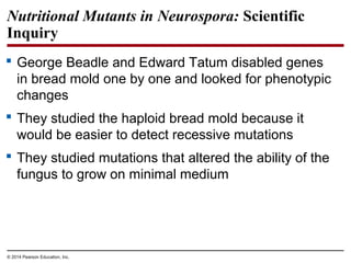
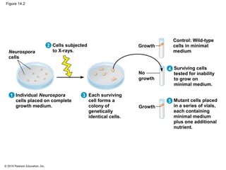
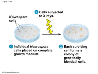
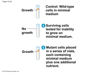
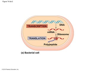
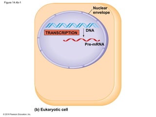
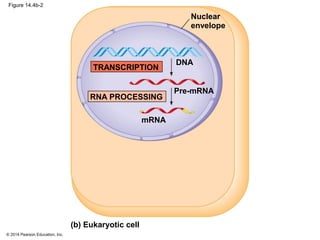
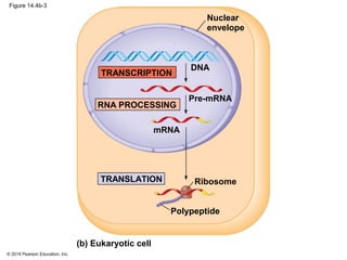
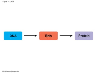
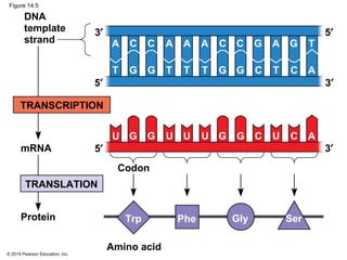
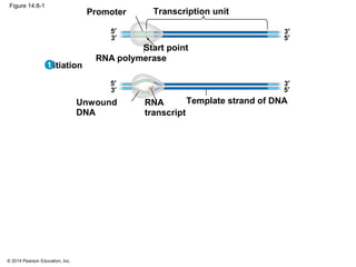
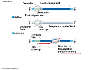
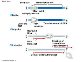
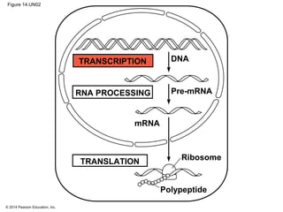
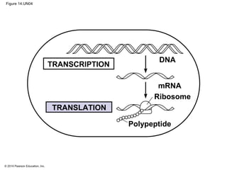
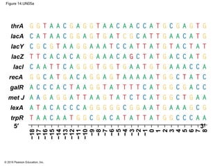
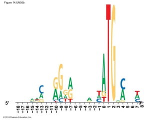
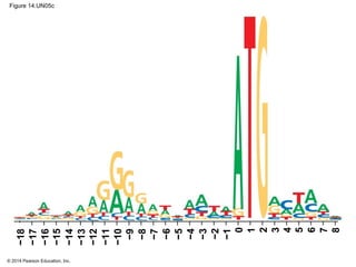
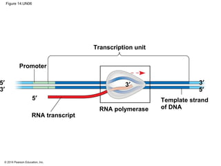
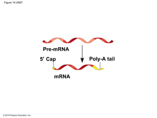
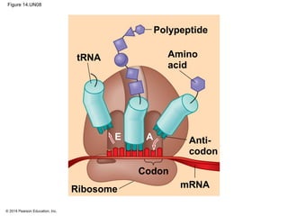
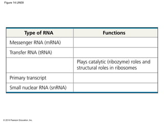
CAMPBELL BIOLOGY IN FOCUS
© 2014 Pearson Education, Inc.
Urry • Cain • Wasserman • Minorsky • Jackson • Reece
Lecture Presentations by
Kathleen Fitzpatrick and Nicole Tunbridge
14
Gene Expression:
From Gene to Protein
© 2014 Pearson Education, Inc.
Overview: The Flow of Genetic Information
 The information content of genes is in the form of
specific sequences of nucleotides in DNA
 The DNA inherited by an organism leads to specific
traits by dictating the synthesis of proteins
 Proteins are the links between genotype and
phenotype
 Gene expression, the process by which DNA
directs protein synthesis, includes two stages:
transcription and translation
© 2014 Pearson Education, Inc.
Figure 14.1
© 2014 Pearson Education, Inc.
Concept 14.1: Genes specify proteins via
transcription and translation
 How was the fundamental relationship between
genes and proteins discovered?
© 2014 Pearson Education, Inc.
Evidence from the Study of Metabolic Defects
 In 1902, British physician Archibald Garrod first
suggested that genes dictate phenotypes through
enzymes that catalyze specific chemical reactions
 He thought symptoms of an inherited disease reflect
an inability to synthesize a certain enzyme
 Cells synthesize and degrade molecules in a series
of steps, a metabolic pathway
© 2014 Pearson Education, Inc.
Nutritional Mutants in Neurospora: Scientific
Inquiry
 George Beadle and Edward Tatum disabled genes
in bread mold one by one and looked for phenotypic
changes
 They studied the haploid bread mold because it
would be easier to detect recessive mutations
 They studied mutations that altered the ability of the
fungus to grow on minimal medium
© 2014 Pearson Education, Inc.
Figure 14.2
Neurospora
cells
Each surviving
cell forms a
colony of
genetically
identical cells.
No
growth
Growth
Mutant cells placed
in a series of vials,
each containing
minimal medium
plus one additional
nutrient.
Surviving cells
tested for inability
to grow on
minimal medium.
Individual Neurospora
cells placed on complete
growth medium. Growth
Cells subjected
to X-rays.
Control: Wild-type
cells in minimal
medium
2
1 3
4
5
© 2014 Pearson Education, Inc.
Figure 14.2a
Neurospora
cells
Each surviving
cell forms a
colony of
genetically
identical cells.
Individual Neurospora
cells placed on complete
growth medium.
Cells subjected
to X-rays.
2
1 3
© 2014 Pearson Education, Inc.
Figure 14.2b
No
growth
Growth
Mutant cells placed
in a series of vials,
each containing
minimal medium
plus one additional
nutrient.
Surviving cells
tested for inability
to grow on
minimal medium.
Growth
Control: Wild-type
cells in minimal
medium
4
5
© 2014 Pearson Education, Inc.
 The researchers amassed a valuable collection of
Neurospora mutant strains, catalogued by their
defects
 For example, one set of mutants all required
arginine for growth
 It was determined that different classes of these
mutants were blocked at a different step in the
biochemical pathway for arginine biosynthesis
© 2014 Pearson Education, Inc.
Figure 14.3
Precursor
Gene A
Ornithine Citrulline Arginine
Gene B Gene C
Enzyme
A
Enzyme
B
Enzyme
C
© 2014 Pearson Education, Inc.
The Products of Gene Expression: A Developing
Story
 Some proteins are not enzymes, so researchers
later revised the one gene–one enzyme hypothesis:
one gene–one protein
 Many proteins are composed of several
polypeptides, each of which has its own gene
 Therefore, Beadle and Tatum’s hypothesis is now
restated as the one gene–one polypeptide
hypothesis
 It is common to refer to gene products as proteins
rather than polypeptides
© 2014 Pearson Education, Inc.
Basic Principles of Transcription and Translation
 RNA is the bridge between DNA and protein
synthesis
 RNA is chemically similar to DNA, but RNA has a
ribose sugar and the base uracil (U) rather than
thymine (T)
 RNA is usually single-stranded
 Getting from DNA to protein requires two stages:
transcription and translation
© 2014 Pearson Education, Inc.
 Transcription is the synthesis of RNA using
information in DNA
 Transcription produces messenger RNA
(mRNA)
 Translation is the synthesis of a polypeptide,
using information in the mRNA
 Ribosomes are the sites of translation
© 2014 Pearson Education, Inc.
 In prokaryotes, translation of mRNA can begin
before transcription has finished
 In eukaryotes, the nuclear envelope separates
transcription from translation
 Eukaryotic RNA transcripts are modified through
RNA processing to yield the finished mRNA
 Eukaryotic mRNA must be transported out of the
nucleus to be translated
© 2014 Pearson Education, Inc.
Figure 14.4
Nuclear
envelope
Pre-mRNA
mRNA
DNA
RNA PROCESSING
TRANSCRIPTION
TRANSLATION
Polypeptide
Ribosome
mRNA
DNA
TRANSCRIPTION
TRANSLATION
Polypeptide
Ribosome
(a) Bacterial cell (b) Eukaryotic cell
© 2014 Pearson Education, Inc.
Figure 14.4a-1
mRNA
DNA
TRANSCRIPTION
(a) Bacterial cell
© 2014 Pearson Education, Inc.
Figure 14.4a-2
mRNA
DNA
TRANSCRIPTION
TRANSLATION
Polypeptide
Ribosome
(a) Bacterial cell
© 2014 Pearson Education, Inc.
Figure 14.4b-1
Nuclear
envelope
Pre-mRNA
DNA
TRANSCRIPTION
(b) Eukaryotic cell
© 2014 Pearson Education, Inc.
Figure 14.4b-2
Nuclear
envelope
Pre-mRNA
mRNA
DNA
RNA PROCESSING
TRANSCRIPTION
(b) Eukaryotic cell
© 2014 Pearson Education, Inc.
Figure 14.4b-3
Nuclear
envelope
Pre-mRNA
mRNA
DNA
RNA PROCESSING
TRANSCRIPTION
TRANSLATION
Polypeptide
Ribosome
(b) Eukaryotic cell
© 2014 Pearson Education, Inc.
 A primary transcript is the initial RNA transcript
from any gene prior to processing
 The central dogma is the concept that cells are
governed by a cellular chain of command
© 2014 Pearson Education, Inc.
Figure 14.UN01
DNA RNA Protein
© 2014 Pearson Education, Inc.
The Genetic Code
 How are the instructions for assembling amino acids
into proteins encoded into DNA?
 There are 20 amino acids, but there are only four
nucleotide bases in DNA
 How many nucleotides correspond to an amino acid?
© 2014 Pearson Education, Inc.
Codons: Triplets of Nucleotides
 The flow of information from gene to protein is
based on a triplet code: a series of
nonoverlapping, three-nucleotide words
 The words of a gene are transcribed into
complementary nonoverlapping three-nucleotide
words of mRNA
 These words are then translated into a chain of
amino acids, forming a polypeptide
© 2014 Pearson Education, Inc.
Figure 14.5
DNA
template
strand
Protein
mRNA
3′
Trp
TRANSCRIPTION
TRANSLATION
Amino acid
Codon
5′
3′5′
3′
5′
Phe Gly Ser
GU G U UU G G UC C A
CA C A AA C C AG G T
GT G T TT G G TC C A
© 2014 Pearson Education, Inc.
 During transcription, one of the two DNA strands,
called the template strand, provides a template for
ordering the sequence of complementary
nucleotides in an RNA transcript
 The template strand is always the same strand for
any given gene
© 2014 Pearson Education, Inc.
 During translation, the mRNA base triplets, called
codons, are read in the 5′ to 3′ direction
 Each codon specifies the amino acid (one of 20)
to be placed at the corresponding position along
a polypeptide
© 2014 Pearson Education, Inc.
Cracking the Code
 All 64 codons were deciphered by the mid-1960s
 Of the 64 triplets, 61 code for amino acids; 3 triplets
are “stop” signals to end translation
 The genetic code is redundant: more than one
codon may specify a particular amino acid
 But it is not ambiguous: no codon specifies more
than one amino acid
© 2014 Pearson Education, Inc.
 Codons must be read in the correct reading frame
(correct groupings) in order for the specified
polypeptide to be produced
 Codons are read one at a time in a nonoverlapping
fashion
© 2014 Pearson Education, Inc.
Figure 14.6
UUU
Second mRNA base
UUC
UUA
UUG
UCU
UCC
UCA
UCG
UAU
UAC
UAA
UAG
UGU
UGC
UGA
UGG
CUU
CUC
CUA
CUG
CCU
CCC
CCA
CCG
CAU
CAC
CAA
CAG
CGU
CGC
CGA
CGG
AUU
AUC
AUA
AUG
ACU
ACC
ACA
ACG
AAU
AAC
AAA
AAG
AGU
AGC
AGA
AGG
GUU
GUC
GUA
GUG
GCU
GCC
GCA
GCG
GAU
GAC
GAA
GAG
GGU
GGC
GGA
GGG
FirstmRNAbase(5′endofcodon)
U
C
A
G
U
C
A
G
U
C
A
G
U
C
A
G
U
C
A
G
U C A G
Phe
Leu
Ser
Tyr Cys
Trp
Met or
start
Stop
Stop Stop
Arg
Gln
His
ProLeu
Val Ala
Asp
Glu
Gly
IIe
Thr
Lys
Asn
Arg
Ser
ThirdmRNAbase(3′endofcodon)
© 2014 Pearson Education, Inc.
Evolution of the Genetic Code
 The genetic code is nearly universal, shared by the
simplest bacteria and the most complex animals
 Genes can be transcribed and translated after being
transplanted from one species to another
© 2014 Pearson Education, Inc.
Figure 14.7
(a) Tobacco plant expressing
a firefly gene
(b) Pig expressing a jellyfish
gene
© 2014 Pearson Education, Inc.
Figure 14.7a
(a) Tobacco plant expressing
a firefly gene
© 2014 Pearson Education, Inc.
Figure 14.7b
(b) Pig expressing a jellyfish
gene
© 2014 Pearson Education, Inc.
Concept 14.2: Transcription is the DNA-directed
synthesis of RNA: a closer look
 Transcription is the first stage of gene expression
© 2014 Pearson Education, Inc.
Molecular Components of Transcription
 RNA synthesis is catalyzed by RNA polymerase,
which pries the DNA strands apart and joins
together the RNA nucleotides
 RNA polymerases assemble polynucleotides in the
5′ to 3′ direction
 However, RNA polymerases can start a chain
without a primer
Animation: Transcription Introduction
© 2014 Pearson Education, Inc.
Figure 14.8-1
Transcription unit
RNA polymerase
Promoter
Template strand of DNA
Start point
RNA
transcript
Unwound
DNA
Initiation
3′
5′
3′
5′ 3′
5′
3′
5′
1
© 2014 Pearson Education, Inc.
Figure 14.8-2
Transcription unit
RNA polymerase
Promoter
Template strand of DNA
Start point
RNA
transcript
Unwound
DNA
Rewound
DNA
RNA
transcript
Direction of
transcription
(“downstream”)
Initiation
Elongation
3′
5′
3′
5′
3′
5′ 3′
5′
3′
5′
3′
5′
3′
5′
2
1
© 2014 Pearson Education, Inc.
Figure 14.8-3
Transcription unit
RNA polymerase
Promoter
Template strand of DNA
Start point
Termination
Completed RNA transcript
RNA
transcript
Unwound
DNA
Rewound
DNA
RNA
transcript
Direction of
transcription
(“downstream”)
Initiation
Elongation
3′
5′
3′
5′
3′
5′ 3′
5′
3′
5′
3′5′
3′
5′
3′
5′
3′
5′
3′
5′
3
2
1
© 2014 Pearson Education, Inc.
 The DNA sequence where RNA polymerase
attaches is called the promoter; in bacteria, the
sequence signaling the end of transcription is called
the terminator
 The stretch of DNA that is transcribed is called a
transcription unit
© 2014 Pearson Education, Inc.
Synthesis of an RNA Transcript
 The three stages of transcription
 Initiation
 Elongation
 Termination
© 2014 Pearson Education, Inc.
RNA Polymerase Binding and Initiation of
Transcription
 Promoters signal the transcriptional start point and
usually extend several dozen nucleotide pairs
upstream of the start point
 Transcription factors mediate the binding of RNA
polymerase and the initiation of transcription
© 2014 Pearson Education, Inc.
 The completed assembly of transcription factors
and RNA polymerase II bound to a promoter is
called a transcription initiation complex
 A promoter called a TATA box is crucial in forming
the initiation complex in eukaryotes
© 2014 Pearson Education, Inc.
Figure 14.UN02
DNA
Pre-mRNA
mRNA
Ribosome
Polypeptide
TRANSLATION
TRANSCRIPTION
RNA PROCESSING
© 2014 Pearson Education, Inc.
Figure 14.9
Transcription
factors
TATA box
Promoter
Nontemplate strand
Start point
Transcription
initiation
complex forms.
Transcription initiation complex
DNA
RNA transcript
A eukaryotic
promoter
Several transcription
factors bind to DNA.
3′
5′
5′ 3′
3′
5′
3′
5′
3′
5′
3
2
1
Template
strand
Transcription
factors
RNA polymerase II
3′
5′
3′
5′ T A T A A A A
AT A T T T T
© 2014 Pearson Education, Inc.
Elongation of the RNA Strand
 As RNA polymerase moves along the DNA, it
untwists the double helix, 10 to 20 bases at a time
 Transcription progresses at a rate of 40 nucleotides
per second in eukaryotes
 A gene can be transcribed simultaneously by
several RNA polymerases
© 2014 Pearson Education, Inc.
Figure 14.10
Nontemplate
strand of DNA
Direction of transcription
RNA
polymerase
3′
5′3′
5′
RNA nucleotides
Template
strand of DNA
Newly made
RNA
3′ end
5′
U
C
U
G
A
A
A
A
AA
A
A
A
A
T T T
T
T
T
T
CC
C
CCC C
G
G
G
U
© 2014 Pearson Education, Inc.
Termination of Transcription
 The mechanisms of termination are different in
bacteria and eukaryotes
 In bacteria, the polymerase stops transcription at
the end of the terminator and the mRNA can be
translated without further modification
 In eukaryotes, RNA polymerase II transcribes the
polyadenylation signal sequence; the RNA
transcript is released 10–35 nucleotides past this
polyadenylation sequence
© 2014 Pearson Education, Inc.
Concept 14.3: Eukaryotic cells modify RNA after
transcription
 Enzymes in the eukaryotic nucleus modify pre-
mRNA (RNA processing) before the genetic
messages are dispatched to the cytoplasm
 During RNA processing, both ends of the primary
transcript are altered
 Also, usually some interior parts of the molecule
are cut out and the other parts spliced together
© 2014 Pearson Education, Inc.
Alteration of mRNA Ends
 Each end of a pre-mRNA molecule is modified in a
particular way
 The 5′ end receives a modified G nucleotide 5′ cap
 The 3′ end gets a poly-A tail
 These modifications share several functions
 Facilitating the export of mRNA to the cytoplasm
 Protecting mRNA from hydrolytic enzymes
 Helping ribosomes attach to the 5′ end
© 2014 Pearson Education, Inc.
Figure 14.UN03
DNA
Pre-mRNA
mRNA
Ribosome
Polypeptide
TRANSLATION
TRANSCRIPTION
RNA PROCESSING
© 2014 Pearson Education, Inc.
Figure 14.11
Protein-coding segment
Polyadenylation
signal
G P
A modified guanine
nucleotide added to
the 5′ end
50–250 adenine
nucleotides added
to the 3′ end
3′5′
5′ Cap 5′ UTR 3′ UTR Poly-A tail
Start
codon
Stop
codon
P P AAUAAA …AAA AAA
© 2014 Pearson Education, Inc.
Split Genes and RNA Splicing
 Most eukaryotic mRNAs have long noncoding
stretches of nucleotides that lie between coding
regions
 The noncoding regions are called intervening
sequences, or introns
 The other regions are called exons and are usually
translated into amino acid sequences
 RNA splicing removes introns and joins exons,
creating an mRNA molecule with a continuous
coding sequence
© 2014 Pearson Education, Inc.
Figure 14.12
Introns cut out and
exons spliced together
31–104
5′ Cap
5′ UTR 3′ UTR
Poly-A tail
Coding
segment
1–146
AAUAAA
105–
146
5′ Cap Poly-A tail
1–30
mRNA
Pre-mRNA
Intron Intron
© 2014 Pearson Education, Inc.
 Many genes can give rise to two or more different
polypeptides, depending on which segments are
used as exons
 This process is called alternative RNA splicing
 RNA splicing is carried out by spliceosomes
 Spliceosomes consist of proteins and small RNAs
© 2014 Pearson Education, Inc.
Figure 14.13
Spliceosome
components
5′
Pre-mRNA
5′
mRNA
Intron
Spliceosome
Exon 1
Small RNAs
Exon 2
Exon 2Exon 1
Cut-out
intron
© 2014 Pearson Education, Inc.
Ribozymes
 Ribozymes are RNA molecules that function as
enzymes
 RNA splicing can occur without proteins, or even
additional RNA molecules
 The introns can catalyze their own splicing
© 2014 Pearson Education, Inc.
Concept 14.4: Translation is the RNA-directed
synthesis of a polypeptide: a closer look
 Genetic information flows from mRNA to protein
through the process of translation
© 2014 Pearson Education, Inc.
Molecular Components of Translation
 A cell translates an mRNA message into protein with
the help of transfer RNA (tRNA)
 tRNAs transfer amino acids to the growing
polypeptide in a ribosome
 Translation is a complex process in terms of its
biochemistry and mechanics
© 2014 Pearson Education, Inc.
Figure 14.UN04
DNA
mRNA
Ribosome
Polypeptide
TRANSLATION
TRANSCRIPTION
© 2014 Pearson Education, Inc.
Figure 14.14
5′
tRNA
Polypeptide
Ribosome
Anticodon
mRNA
Codons 3′
tRNA with
amino acid
attached
Amino acids
Gly
Trp
Phe
A A A
A
C
C
C
C
G
U U U G G CU G G
© 2014 Pearson Education, Inc.
The Structure and Function of Transfer RNA
 Each tRNA can translate a particular mRNA codon
into a given amino acid
 The tRNA contains an amino acid at one end and at
the other end has a nucleotide triplet that can base-
pair with the complementary codon on mRNA
© 2014 Pearson Education, Inc.
 A tRNA molecule consists of a single RNA strand
that is only about 80 nucleotides long
 tRNA molecules can base-pair with themselves
 Flattened into one plane, a tRNA molecule looks like
a cloverleaf
 In three dimensions, tRNA is roughly L-shaped,
where one end of the L contains the anticodon that
base-pairs with an mRNA codon
Video: tRNA Model
© 2014 Pearson Education, Inc.
Figure 14.15
5′
Anticodon
3′
Amino acid
attachment site
Anticodon
Anticodon
A A G
5′3′
Hydrogen
bonds
5′
3′
Amino acid
attachment
site
Hydrogen
bonds
A
A
G
A
C
G
C
C
C
C
U
A
C G
U
U
A
A A
A
C
G U
A
C
G U
A
C
G
U
A
C
G
U
*
C
G
*
G
G
U
A
A
A
A
C C
C
C
C
G
G
GG
G
U
U
U
U
G
G
G
G
A
A
*
*
*
*
*
*
*
*
*
(b) Three-dimensional
structure
(c) Symbol used
in this book(a) Two-dimensional structure
*
© 2014 Pearson Education, Inc.
Figure 14.15a
5′
3′
Anticodon
Amino acid
attachment
site
Hydrogen
bonds
A
A
G
A
C
G
C
C
C
C
U
A
C G
U
U
A
A A
A
C
G U
A
C
G U
A
C
G
U
A
C
G
U
*
C
G
*
G
G
U
A
A
A
A
C C
C
C
C
G
G
GG
G
U
U
U
U
G
G
G
G
A
A
*
*
*
*
*
*
*
*
*
(a) Two-dimensional structure
*
© 2014 Pearson Education, Inc.
Figure 14.15b
Anticodon
Amino acid
attachment site
Anticodon
A A G
5′3′
Hydrogen
bonds
5′
3′
(b) Three-dimensional
structure
(c) Symbol used
in this book
© 2014 Pearson Education, Inc.
 Accurate translation requires two steps
 First: a correct match between a tRNA and an
amino acid, done by the enzyme aminoacyl-tRNA
synthetase
 Second: a correct match between the tRNA
anticodon and an mRNA codon
 Flexible pairing at the third base of a codon is
called wobble and allows some tRNAs to bind to
more than one codon
© 2014 Pearson Education, Inc.
Figure 14.16-1
Tyrosyl-tRNA
synthetase
Tyrosine (Tyr)
(amino acid)
Amino acid
and tRNA
enter active
site.
Tyr-tRNA
Complementary
tRNA anticodon
1
UA A
© 2014 Pearson Education, Inc.
Figure 14.16-2
Tyrosyl-tRNA
synthetase
Tyrosine (Tyr)
(amino acid)
Amino acid
and tRNA
enter active
site.
Tyr-tRNA
Complementary
tRNA anticodon
Using ATP,
synthetase
catalyzes
covalent
bonding.
AMP + 2
ATP
2
1
UA A
P i
© 2014 Pearson Education, Inc.
Figure 14.16-3
Tyrosyl-tRNA
synthetase
Tyrosine (Tyr)
(amino acid)
Amino acid
and tRNA
enter active
site.
Tyr-tRNA
Complementary
tRNA anticodon
Aminoacyl
tRNA
released.
Using ATP,
synthetase
catalyzes
covalent
bonding.
AMP + 2
ATP
2
3
1
UA A
P i
© 2014 Pearson Education, Inc.
Ribosomes
 Ribosomes facilitate specific coupling of tRNA
anticodons with mRNA codons during protein
synthesis
 The large and small ribosomal are made of
proteins and ribosomal RNAs (rRNAs)
 In bacterial and eukaryotic ribosomes the large and
small subunits join to form a ribosome only when
attached to an mRNA molecule
© 2014 Pearson Education, Inc.
Figure 14.17
PE A
tRNA
molecules
A
Large
subunit
Small
subunit
Growing polypeptide Exit tunnel
E P
mRNA
5′
3′
Growing polypeptide
(a) Computer model of functioning ribosome
tRNA
5′
3′
E
mRNA
(c) Schematic model with mRNA and tRNA
Codons
Amino end Next amino acid
to be added to
polypeptide
chain
Large
subunit
Small
subunit
A site (Aminoacyl-
tRNA binding site)
P site
(Peptidyl-tRNA
binding site)
Exit tunnel
E site
(Exit site)
mRNA
binding site
(b) Schematic model showing binding sites
© 2014 Pearson Education, Inc.
Figure 14.17a
tRNA
molecules
A
Large
subunit
Small
subunit
Growing polypeptide Exit tunnel
E P
mRNA
5′
3′
(a) Computer model of functioning ribosome
© 2014 Pearson Education, Inc.
Figure 14.17b
PE A Large
subunit
Small
subunit
P site
(Peptidyl-tRNA
binding site)
Exit tunnel
E site
(Exit site)
mRNA
binding site
(b) Schematic model showing binding sites
A site (Aminoacyl-
tRNA binding site)
© 2014 Pearson Education, Inc.
Figure 14.17c
Growing polypeptide
tRNA
5′
3′
E
mRNA
(c) Schematic model with mRNA and tRNA
Codons
Amino end Next amino acid
to be added to
polypeptide
chain
© 2014 Pearson Education, Inc.
 A ribosome has three binding sites for tRNA
 The P site holds the tRNA that carries the growing
polypeptide chain
 The A site holds the tRNA that carries the next
amino acid to be added to the chain
 The E site is the exit site, where discharged tRNAs
leave the ribosome
© 2014 Pearson Education, Inc.
Building a Polypeptide
 The three stages of translation
 Initiation
 Elongation
 Termination
 All three stages require protein “factors” that aid in
the translation process
© 2014 Pearson Education, Inc.
Ribosome Association and Initiation of Translation
 The initiation stage of translation brings together
mRNA, a tRNA with the first amino acid, and the two
ribosomal subunits
 A small ribosomal subunit binds with mRNA and a
special initiator tRNA
 Then the small subunit moves along the mRNA until
it reaches the start codon (AUG)
Animation: Translation Introduction
© 2014 Pearson Education, Inc.
Figure 14.18
A
Small ribosomal subunit binds
to mRNA.
GTP
P site
U A
mRNA
5′
3′
Met
P i
mRNA binding site
Start codon
Small
ribosomal
subunit
Initiator
tRNA
5′
3′
5′
3′
Large ribosomal subunit
completes the initiation complex.
U G
C
5′
3′
Translation initiation complex
Large
ribosomal
subunit
AE
Met
GDP
+
1 2
© 2014 Pearson Education, Inc.
 The start codon is important because it establishes
the reading frame for the mRNA
 The addition of the large ribosomal subunit is last
and completes the formation of the translation
initiation complex
 Proteins called initiation factors bring all these
components together
© 2014 Pearson Education, Inc.
Elongation of the Polypeptide Chain
 During elongation, amino acids are added one by
one to the previous amino acid at the C-terminus of
the growing chain
 Each addition involves proteins called elongation
factors and occurs in three steps: codon
recognition, peptide bond formation, and
translocation
 Translation proceeds along the mRNA in a 5′ to 3′
direction
© 2014 Pearson Education, Inc.
Figure 14.19-1
Amino end
of polypeptide
mRNA
P
site
P
i
5′
3′
E
GTP
+
A
site
GDP
Codon recognition
E
P A
1
© 2014 Pearson Education, Inc.
Figure 14.19-2
Amino end
of polypeptide
mRNA
P
site
P
i
5′
3′
E
GTP
+
A
site
GDP
Peptide bond
formation
Codon recognition
E
P A
E
P A
2
1
© 2014 Pearson Education, Inc.
Figure 14.19-3
Amino end
of polypeptide
mRNARibosome ready for
next aminoacyl tRNA P
site
P
i
5′
3′
E
GTP
+
A
site
GDP
Peptide bond
formation
Codon recognition
Translocation
E
P A
E
P A
P
i
GTP
+GDP
E
P A
3
2
1
© 2014 Pearson Education, Inc.
Termination of Translation
 Termination occurs when a stop codon in the mRNA
reaches the A site of the ribosome
 The A site accepts a protein called a release factor
 The release factor causes the addition of a water
molecule instead of an amino acid
 This reaction releases the polypeptide, and the
translation assembly then comes apart
© 2014 Pearson Education, Inc.
Figure 14.20-1
Ribosome reaches a
stop codon on mRNA.
5′
3′
Release
factor
Stop codon
(UAG, UAA, or UGA)
1
© 2014 Pearson Education, Inc.
Figure 14.20-2
Free
polypeptide
Ribosome reaches a
stop codon on mRNA.
5′
3′
Release
factor
Stop codon
(UAG, UAA, or UGA)
5′
3′
Release factor
promotes
hydrolysis.
21
© 2014 Pearson Education, Inc.
Figure 14.20-3
Free
polypeptide
Ribosome reaches a
stop codon on mRNA.
5′
3′
2 GTP
+2 GDP P
i
Release
factor
Stop codon
(UAG, UAA, or UGA)
5′
3′
5′
3′
Ribosomal
subunits and other
components
dissociate.
Release factor
promotes
hydrolysis.
2 31
© 2014 Pearson Education, Inc.
Completing and Targeting the Functional Protein
 Often translation is not sufficient to make a
functional protein
 Polypeptide chains are modified after translation or
targeted to specific sites in the cell
© 2014 Pearson Education, Inc.
Protein Folding and Post-Translational
Modifications
 During synthesis, a polypeptide chain spontaneously
coils and folds into its three-dimensional shape
 Proteins may also require post-translational
modifications before doing their jobs
© 2014 Pearson Education, Inc.
Targeting Polypeptides to Specific Locations
 Two populations of ribosomes are evident in cells:
free ribosomes (in the cytosol) and bound
ribosomes (attached to the ER)
 Free ribosomes mostly synthesize proteins that
function in the cytosol
 Bound ribosomes make proteins of the
endomembrane system and proteins that are
secreted from the cell
© 2014 Pearson Education, Inc.
 Polypeptide synthesis always begins in the cytosol
 Synthesis finishes in the cytosol unless the
polypeptide signals the ribosome to attach to the
ER
 Polypeptides destined for the ER or for secretion
are marked by a signal peptide
© 2014 Pearson Education, Inc.
 A signal-recognition particle (SRP) binds to the
signal peptide
 The SRP brings the signal peptide and its ribosome
to the ER
© 2014 Pearson Education, Inc.
Figure 14.21
Polypeptide
synthesis
begins.
SRP
binds to
signal
peptide.
SRP
binds to
receptor
protein.
SRP
detaches
and
polypeptide
synthesis
resumes.
Signal-
cleaving
enzyme cuts
off signal
peptide.
Completed
polypeptide
folds into
final
conformation.
Signal
peptide
removed
ER
membrane
Signal
peptide
Protein
SRP receptor
protein
Ribosome
mRNA
CYTOSOL
ER LUMEM
SRP
Translocation complex
1 2 3 4 5 6
© 2014 Pearson Education, Inc.
Making Multiple Polypeptides in Bacteria and
Eukaryotes
 In bacteria and eukaryotes multiple ribosomes
translate an mRNA at the same time
 Once a ribosome is far enough past the start codon,
another ribosome can attach to the mRNA
 Strings of ribosomes called polyribosomes (or
polysomes) can be seen with an electron
microscope
© 2014 Pearson Education, Inc.
Figure 14.22
Incoming
ribosomal
subunits
(b) A large polyribosome in a bacterial
cell (TEM)
Ribosomes
mRNA
(a) Several ribosomes simultaneously translating one
mRNA molecule
0.1 µm
Start of
mRNA
(5′ end)
End of mRNA
(3′ end)
Growing
polypeptides
Completed
polypeptide
Polyribosome
© 2014 Pearson Education, Inc.
Figure 14.22a
Incoming
ribosomal
subunits
(a) Several ribosomes simultaneously translating one
mRNA molecule
Start of
mRNA
(5′ end)
End of mRNA
(3′ end)
Growing
polypeptides
Completed
polypeptide
Polyribosome
© 2014 Pearson Education, Inc.
Figure 14.22b
(b) A large polyribosome in a bacterial
cell (TEM)
Ribosomes
mRNA
0.1 µm
© 2014 Pearson Education, Inc.
 Bacteria and eukaryotes can also transcribe
multiple mRNAs form the same gene
 In bacteria, the transcription and translation can
take place simultaneously
 In eukaryotes, the nuclear envelope separates
transcription and translation
© 2014 Pearson Education, Inc.
Figure 14.23
RNA polymerase
mRNA
0.25 µm
DNA
Polyribosome
RNA
polymerase DNA
Polyribosome
Direction of
transcription
Ribosome
mRNA (5′ end)
Polypeptide
(amino end)
© 2014 Pearson Education, Inc.
Figure 14.23a
RNA polymerase
mRNA
0.25 µm
DNA
Polyribosome
© 2014 Pearson Education, Inc.
Figure 14.24
A
U A
tRNA
5′ Cap
3′
mRNA
Ribosomal
subunits
Aminoacyl
(charged)
tRNA
P
E
G
5′
3′
Ribosome
Codon
Anticodon
TRANSLATION
Amino
acid
Aminoacyl-tRNA
synthetase
CYTOPLASM
NUCLEUS
AMINO ACID
ACTIVATION
Intron
5′ C
ap
Poly-A
TRANSCRIPTION
RNA
PROCESSING
RNA
transcript
RNA transcript
(pre-mRNA)
RNA
polymerase
Exon
DNA
Poly-A
Poly-A
U GUG U U
A A A
A
C
C U
A
C
AE
© 2014 Pearson Education, Inc.
Figure 14.24a
tRNA
mRNA
5′
3′
Amino
acid
Aminoacyl-tRNA
synthetase
CYTOPLASM
NUCLEUS
AMINO ACID
ACTIVATION
Intron
5′ C
ap
TRANSCRIPTION
RNA
PROCESSING
RNA
transcript
RNA transcript
(pre-mRNA)
RNA
polymerase
Exon
DNA
Poly-A
Poly-A
Aminoacyl
(charged)
tRNA
© 2014 Pearson Education, Inc.
Figure 14.24b
A
U A
5′ Cap
3′
mRNA
Ribosomal
subunits
Aminoacyl
(charged)
tRNA
P
E
G
Ribosome
Codon
Anticodon
TRANSLATION
5′ C
ap
Poly-A
U GUG U U
A A A
A
C
C U
A
C
AE
Growing polypeptide
© 2014 Pearson Education, Inc.
Concept 14.5: Mutations of one or a few
nucleotides can affect protein structure and
function
 Mutations are changes in the genetic material of a
cell or virus
 Point mutations are chemical changes in just one
or a few nucleotide pairs of a gene
 The change of a single nucleotide in a DNA
template strand can lead to the production of an
abnormal protein
© 2014 Pearson Education, Inc.
Figure 14.25
AG G
Wild-type hemoglobin
mRNA
5′
3′
mRNA
Wild-type hemoglobin DNA
5′
3′5′
3′
TC C
TG G
AC C 5′
3′
AG G5′ 5′3′ UG G 3′
Normal hemoglobin
Sickle-cell hemoglobin
Mutant hemoglobin DNA
Sickle-cell hemoglobin
ValGlu
© 2014 Pearson Education, Inc.
Types of Small-Scale Mutations
 Point mutations within a gene can be divided into
two general categories
 Nucleotide-pair substitutions
 One or more nucleotide-pair insertions or deletions
© 2014 Pearson Education, Inc.
Figure 14.26
mRNA
DNA template strand
Stop
Carboxyl end
Protein
Amino end
Phe GlyMet Lys
A G3′ T CC 5′T T T TA A A AC C
5′ 3′TA A A A AT T T TG G G G C
U5′ 3′G CA U U G GGA A A AU U
Wild type
Phe GlyMet Lys
A A3′ T CC 5′T T T TA A A AC C
5′ 3′TA A A A AT T T TG G G G T
U5′ 3′G UA U U G GGA A A AU U
Phe SerMet Lys
A G3′ T CC 5′T T T TA A A AC T
5′ 3′TA A A A AT T T TG G A G C
U5′ 3′G CA U U A GGA A A AU U
Met
A C3′ T CT 5′A T A TC A A GC A
5′ 3′TA T A T AG T T CG A T G G
U5′ 3′G GA G U U GAU A U AU U
Leu AlaMet Lys
A A3′ T GC 5′T T T
A
A A C TC C
5′ 3′TA A A AGT T AG G G C T
U5′ 3′G UA U G G GGA A A
U
U A
GlyMet Phe
A T3′ T TA 5′A A
T T
C C G
C
C A
5′ 3′TA T T
A
A
G G C
T
G T T A A
U5′ 3′G AA G C U AUU U
A G
G
A
Met
A G3′ T CC 5′A T T TA A A AC C
5′ 3′TA T A A AT T T TG G G G C
U5′ 3′G UA U U G GGU A A AU U
Stop Stop
Stop
Stop
Stop
A instead of G
Silent (no effect on amino acid sequence)
(a) Nucleotide-pair substitution
U instead of C
T instead of C
Missense
A instead of G
A instead of T
Nonsense
U instead of A
Extra A
Frameshift causing immediate nonsense
(1 nucleotide-pair insertion)
(b) Nucleotide-pair insertion or deletion
Extra U
Frameshift causing extensive missense
(1 nucleotide-pair deletion)
missing
missing
missing
missing
No frameshift, but one amino acid missing
(3 nucleotide-pair deletion)
© 2014 Pearson Education, Inc.
Substitutions
 A nucleotide-pair substitution replaces one
nucleotide and its partner with another pair of
nucleotides
 Silent mutations have no effect on the amino acid
produced by a codon because of redundancy in the
genetic code
© 2014 Pearson Education, Inc.
Figure 14.26a
DNA template strand
mRNA
Stop
Carboxyl end
Protein
Amino end
Phe GlyMet Lys
3′ 5′
5′ 3′
5′ 3′
Wild type
Phe GlyMet Lys
3′ 5′
5′ 3′
5′ 3′
Stop
A instead of G
Nucleotide-pair substitution: silent
U instead of C
T A T T A A A A T TC C C C G
TA T TA A AAT TG G G CG
UA U UA A AAU UG G G CG
T A T T A A A A T TC C C C A
TA T TA A AAT TG G G TG
UA U UA A AAU UG G G UG
© 2014 Pearson Education, Inc.
 Missense mutations still code for an amino acid,
but not the correct amino acid
 Substitution mutations are usually missense
mutations
 Nonsense mutations change an amino acid codon
into a stop codon, nearly always leading to a
nonfunctional protein
Animation: Protein Synthesis
© 2014 Pearson Education, Inc.
Figure 14.26b
DNA template strand
mRNA
Stop
Carboxyl end
Protein
Amino end
Phe GlyMet Lys
3′ 5′
5′ 3′
5′ 3′
Wild type
Phe SerMet Lys
3′ 5′
5′ 3′
5′ 3′
Stop
T instead of C
A instead of G
Nucleotide-pair substitution: missense
T A T T A A A A T TC C C C G
TA T TA A AAT TG G G CG
UA U UA A AAU UG G G CG
T A T T A A A A T TC C T C G
TA T TA A AAT TG A G CG
UA U UA A AAU UG A G CG
© 2014 Pearson Education, Inc.
Figure 14.26c
DNA template strand
mRNA
Stop
Carboxyl end
Protein
Amino end
Phe GlyMet Lys
3′ 5′
5′ 3′
5′ 3′
Wild type
Nucleotide-pair substitution: nonsense
3′ 5′
5′ 3′
5′ 3′
Met
Stop
A instead of T
U instead of A
T A T T A A A A T TC C C C G
TA T TA A AAT TG G G CG
UA U UA A AAU UG G G CG
T A A T A A A A T TC C C C G
TA T TT A AAT TG G G CG
UA U UU A AAU UG G G CG
© 2014 Pearson Education, Inc.
Insertions and Deletions
 Insertions and deletions are additions or losses of
nucleotide pairs in a gene
 These mutations have a disastrous effect on the
resulting protein more often than substitutions do
 Insertion or deletion of nucleotides may alter the
reading frame of the genetic message, producing a
frameshift mutation
© 2014 Pearson Education, Inc.
Figure 14.26d
DNA template strand
mRNA
Stop
Carboxyl end
Protein
Amino end
Phe GlyMet Lys
3′ 5′
5′ 3′
5′ 3′
Wild type
Nucleotide-pair insertion: frameshift causing immediate nonsense
Met
3′ 5′
5′ 3′
5′ 3′
Stop
Extra A
Extra U
T A T T A A A A T TC C C C G
TA T TA A AAT TG G G CG
UA U UA A AAU UG G G CG
T A T T A A A A T TC C C C G
TA T TA A AAT TG G G CG
UA U UA A AAU UG G G CG
A
T
U
© 2014 Pearson Education, Inc.
Figure 14.26e
DNA template strand
mRNA
Stop
Carboxyl end
Protein
Amino end
Phe GlyMet Lys
3′ 5′
5′ 3′
5′ 3′
Wild type
Nucleotide-pair deletion: frameshift causing extensive missense
Leu AlaMet Lys
3′ 5′
A
5′ 3′
5′ 3′
U
missing
missing
T A T T A A A A T TC C C C G
TA T TA A AAT TG G G CG
UA U UA A AAU UG G G CG
T A T T A A
G
A T TC C C C G
TA T TA A AATG G CG
UA U UA A AAG UG G CG
© 2014 Pearson Education, Inc.
Figure 14.26f
DNA template strand
mRNA
Stop
Carboxyl end
Protein
Amino end
Phe GlyMet Lys
3′ 5′
5′ 3′
5′ 3′
Wild type
3 nucleotide-pair deletion: no frameshift, but one amino acid
missing
GlyMet Phe
3′ 5′
T T C
5′ 3′
5′ 3′
A GA
Stop
missing
missing
T A T T A A A A T TC C C C G
TA T TA A AAT TG G G CG
UA U UA A AAU UG G G CG
T A A A C C GC A A T T
TA G GT T CT T A AG
UA U U UG G G C U A A
© 2014 Pearson Education, Inc.
Mutagens
 Spontaneous mutations can occur during DNA
replication, recombination, or repair
 Mutagens are physical or chemical agents that can
cause mutations
 Researchers have developed methods to test the
mutagenic activity of chemicals
 Most cancer-causing chemicals (carcinogens) are
mutagenic, and the converse is also true
© 2014 Pearson Education, Inc.
What Is a Gene? Revisiting the Question
 The definition of a gene has evolved through the
history of genetics
 We have considered a gene as
 A discrete unit of inheritance
 A region of specific nucleotide sequence in a
chromosome
 A DNA sequence that codes for a specific polypeptide
chain
© 2014 Pearson Education, Inc.
 A gene can be defined as a region of DNA that can
be expressed to produce a final functional product,
either a polypeptide or an RNA molecule
© 2014 Pearson Education, Inc.
Figure 14.UN05a
thrA
−18
lacA
lacY
lacZ
lacl
recA
lexA
galR
met J
trpR
3′5′
−17
−16
−15
−14
−13
−12
−11
−10
0
−1
−2
−3
−4
−5
−6
−7
−8
−9
1
2
3
4
5
6
7
8
© 2014 Pearson Education, Inc.
Figure 14.UN05b
3′5′
−18
−17
−16
−15
−14
−13
−12
−11
−10
0
−1
−2
−3
−4
−5
−6
−7
−8
−9
1
2
3
4
5
6
7
8
© 2014 Pearson Education, Inc.
Figure 14.UN05c
−18
−17
−16
−15
−14
−13
−12
−11
−10
0
−1
−2
−3
−4
−5
−6
−7
−8
−9
1
2
3
4
5
6
7
8
© 2014 Pearson Education, Inc.
Figure 14.UN06
3′
5′
3′
5′ 3′
5′
RNA transcript
RNA polymerase
Transcription unit
Promoter
Template strand
of DNA
© 2014 Pearson Education, Inc.
Figure 14.UN07
5′ Cap
Pre-mRNA
mRNA
Poly-A tail
© 2014 Pearson Education, Inc.
Figure 14.UN08
tRNA
Polypeptide
Ribosome
Amino
acid
Anti-
codon
Codon
mRNA
E A
© 2014 Pearson Education, Inc.
Figure 14.UN09

Biology in Focus - Chapter 14

  • 1.
    CAMPBELL BIOLOGY INFOCUS © 2014 Pearson Education, Inc. Urry • Cain • Wasserman • Minorsky • Jackson • Reece Lecture Presentations by Kathleen Fitzpatrick and Nicole Tunbridge 14 Gene Expression: From Gene to Protein
  • 2.
    © 2014 PearsonEducation, Inc. Overview: The Flow of Genetic Information  The information content of genes is in the form of specific sequences of nucleotides in DNA  The DNA inherited by an organism leads to specific traits by dictating the synthesis of proteins  Proteins are the links between genotype and phenotype  Gene expression, the process by which DNA directs protein synthesis, includes two stages: transcription and translation
  • 3.
    © 2014 PearsonEducation, Inc. Figure 14.1
  • 4.
    © 2014 PearsonEducation, Inc. Concept 14.1: Genes specify proteins via transcription and translation  How was the fundamental relationship between genes and proteins discovered?
  • 5.
    © 2014 PearsonEducation, Inc. Evidence from the Study of Metabolic Defects  In 1902, British physician Archibald Garrod first suggested that genes dictate phenotypes through enzymes that catalyze specific chemical reactions  He thought symptoms of an inherited disease reflect an inability to synthesize a certain enzyme  Cells synthesize and degrade molecules in a series of steps, a metabolic pathway
  • 6.
    © 2014 PearsonEducation, Inc. Nutritional Mutants in Neurospora: Scientific Inquiry  George Beadle and Edward Tatum disabled genes in bread mold one by one and looked for phenotypic changes  They studied the haploid bread mold because it would be easier to detect recessive mutations  They studied mutations that altered the ability of the fungus to grow on minimal medium
  • 7.
    © 2014 PearsonEducation, Inc. Figure 14.2 Neurospora cells Each surviving cell forms a colony of genetically identical cells. No growth Growth Mutant cells placed in a series of vials, each containing minimal medium plus one additional nutrient. Surviving cells tested for inability to grow on minimal medium. Individual Neurospora cells placed on complete growth medium. Growth Cells subjected to X-rays. Control: Wild-type cells in minimal medium 2 1 3 4 5
  • 8.
    © 2014 PearsonEducation, Inc. Figure 14.2a Neurospora cells Each surviving cell forms a colony of genetically identical cells. Individual Neurospora cells placed on complete growth medium. Cells subjected to X-rays. 2 1 3
  • 9.
    © 2014 PearsonEducation, Inc. Figure 14.2b No growth Growth Mutant cells placed in a series of vials, each containing minimal medium plus one additional nutrient. Surviving cells tested for inability to grow on minimal medium. Growth Control: Wild-type cells in minimal medium 4 5
  • 10.
    © 2014 PearsonEducation, Inc.  The researchers amassed a valuable collection of Neurospora mutant strains, catalogued by their defects  For example, one set of mutants all required arginine for growth  It was determined that different classes of these mutants were blocked at a different step in the biochemical pathway for arginine biosynthesis
  • 11.
    © 2014 PearsonEducation, Inc. Figure 14.3 Precursor Gene A Ornithine Citrulline Arginine Gene B Gene C Enzyme A Enzyme B Enzyme C
  • 12.
    © 2014 PearsonEducation, Inc. The Products of Gene Expression: A Developing Story  Some proteins are not enzymes, so researchers later revised the one gene–one enzyme hypothesis: one gene–one protein  Many proteins are composed of several polypeptides, each of which has its own gene  Therefore, Beadle and Tatum’s hypothesis is now restated as the one gene–one polypeptide hypothesis  It is common to refer to gene products as proteins rather than polypeptides
  • 13.
    © 2014 PearsonEducation, Inc. Basic Principles of Transcription and Translation  RNA is the bridge between DNA and protein synthesis  RNA is chemically similar to DNA, but RNA has a ribose sugar and the base uracil (U) rather than thymine (T)  RNA is usually single-stranded  Getting from DNA to protein requires two stages: transcription and translation
  • 14.
    © 2014 PearsonEducation, Inc.  Transcription is the synthesis of RNA using information in DNA  Transcription produces messenger RNA (mRNA)  Translation is the synthesis of a polypeptide, using information in the mRNA  Ribosomes are the sites of translation
  • 15.
    © 2014 PearsonEducation, Inc.  In prokaryotes, translation of mRNA can begin before transcription has finished  In eukaryotes, the nuclear envelope separates transcription from translation  Eukaryotic RNA transcripts are modified through RNA processing to yield the finished mRNA  Eukaryotic mRNA must be transported out of the nucleus to be translated
  • 16.
    © 2014 PearsonEducation, Inc. Figure 14.4 Nuclear envelope Pre-mRNA mRNA DNA RNA PROCESSING TRANSCRIPTION TRANSLATION Polypeptide Ribosome mRNA DNA TRANSCRIPTION TRANSLATION Polypeptide Ribosome (a) Bacterial cell (b) Eukaryotic cell
  • 17.
    © 2014 PearsonEducation, Inc. Figure 14.4a-1 mRNA DNA TRANSCRIPTION (a) Bacterial cell
  • 18.
    © 2014 PearsonEducation, Inc. Figure 14.4a-2 mRNA DNA TRANSCRIPTION TRANSLATION Polypeptide Ribosome (a) Bacterial cell
  • 19.
    © 2014 PearsonEducation, Inc. Figure 14.4b-1 Nuclear envelope Pre-mRNA DNA TRANSCRIPTION (b) Eukaryotic cell
  • 20.
    © 2014 PearsonEducation, Inc. Figure 14.4b-2 Nuclear envelope Pre-mRNA mRNA DNA RNA PROCESSING TRANSCRIPTION (b) Eukaryotic cell
  • 21.
    © 2014 PearsonEducation, Inc. Figure 14.4b-3 Nuclear envelope Pre-mRNA mRNA DNA RNA PROCESSING TRANSCRIPTION TRANSLATION Polypeptide Ribosome (b) Eukaryotic cell
  • 22.
    © 2014 PearsonEducation, Inc.  A primary transcript is the initial RNA transcript from any gene prior to processing  The central dogma is the concept that cells are governed by a cellular chain of command
  • 23.
    © 2014 PearsonEducation, Inc. Figure 14.UN01 DNA RNA Protein
  • 24.
    © 2014 PearsonEducation, Inc. The Genetic Code  How are the instructions for assembling amino acids into proteins encoded into DNA?  There are 20 amino acids, but there are only four nucleotide bases in DNA  How many nucleotides correspond to an amino acid?
  • 25.
    © 2014 PearsonEducation, Inc. Codons: Triplets of Nucleotides  The flow of information from gene to protein is based on a triplet code: a series of nonoverlapping, three-nucleotide words  The words of a gene are transcribed into complementary nonoverlapping three-nucleotide words of mRNA  These words are then translated into a chain of amino acids, forming a polypeptide
  • 26.
    © 2014 PearsonEducation, Inc. Figure 14.5 DNA template strand Protein mRNA 3′ Trp TRANSCRIPTION TRANSLATION Amino acid Codon 5′ 3′5′ 3′ 5′ Phe Gly Ser GU G U UU G G UC C A CA C A AA C C AG G T GT G T TT G G TC C A
  • 27.
    © 2014 PearsonEducation, Inc.  During transcription, one of the two DNA strands, called the template strand, provides a template for ordering the sequence of complementary nucleotides in an RNA transcript  The template strand is always the same strand for any given gene
  • 28.
    © 2014 PearsonEducation, Inc.  During translation, the mRNA base triplets, called codons, are read in the 5′ to 3′ direction  Each codon specifies the amino acid (one of 20) to be placed at the corresponding position along a polypeptide
  • 29.
    © 2014 PearsonEducation, Inc. Cracking the Code  All 64 codons were deciphered by the mid-1960s  Of the 64 triplets, 61 code for amino acids; 3 triplets are “stop” signals to end translation  The genetic code is redundant: more than one codon may specify a particular amino acid  But it is not ambiguous: no codon specifies more than one amino acid
  • 30.
    © 2014 PearsonEducation, Inc.  Codons must be read in the correct reading frame (correct groupings) in order for the specified polypeptide to be produced  Codons are read one at a time in a nonoverlapping fashion
  • 31.
    © 2014 PearsonEducation, Inc. Figure 14.6 UUU Second mRNA base UUC UUA UUG UCU UCC UCA UCG UAU UAC UAA UAG UGU UGC UGA UGG CUU CUC CUA CUG CCU CCC CCA CCG CAU CAC CAA CAG CGU CGC CGA CGG AUU AUC AUA AUG ACU ACC ACA ACG AAU AAC AAA AAG AGU AGC AGA AGG GUU GUC GUA GUG GCU GCC GCA GCG GAU GAC GAA GAG GGU GGC GGA GGG FirstmRNAbase(5′endofcodon) U C A G U C A G U C A G U C A G U C A G U C A G Phe Leu Ser Tyr Cys Trp Met or start Stop Stop Stop Arg Gln His ProLeu Val Ala Asp Glu Gly IIe Thr Lys Asn Arg Ser ThirdmRNAbase(3′endofcodon)
  • 32.
    © 2014 PearsonEducation, Inc. Evolution of the Genetic Code  The genetic code is nearly universal, shared by the simplest bacteria and the most complex animals  Genes can be transcribed and translated after being transplanted from one species to another
  • 33.
    © 2014 PearsonEducation, Inc. Figure 14.7 (a) Tobacco plant expressing a firefly gene (b) Pig expressing a jellyfish gene
  • 34.
    © 2014 PearsonEducation, Inc. Figure 14.7a (a) Tobacco plant expressing a firefly gene
  • 35.
    © 2014 PearsonEducation, Inc. Figure 14.7b (b) Pig expressing a jellyfish gene
  • 36.
    © 2014 PearsonEducation, Inc. Concept 14.2: Transcription is the DNA-directed synthesis of RNA: a closer look  Transcription is the first stage of gene expression
  • 37.
    © 2014 PearsonEducation, Inc. Molecular Components of Transcription  RNA synthesis is catalyzed by RNA polymerase, which pries the DNA strands apart and joins together the RNA nucleotides  RNA polymerases assemble polynucleotides in the 5′ to 3′ direction  However, RNA polymerases can start a chain without a primer Animation: Transcription Introduction
  • 38.
    © 2014 PearsonEducation, Inc. Figure 14.8-1 Transcription unit RNA polymerase Promoter Template strand of DNA Start point RNA transcript Unwound DNA Initiation 3′ 5′ 3′ 5′ 3′ 5′ 3′ 5′ 1
  • 39.
    © 2014 PearsonEducation, Inc. Figure 14.8-2 Transcription unit RNA polymerase Promoter Template strand of DNA Start point RNA transcript Unwound DNA Rewound DNA RNA transcript Direction of transcription (“downstream”) Initiation Elongation 3′ 5′ 3′ 5′ 3′ 5′ 3′ 5′ 3′ 5′ 3′ 5′ 3′ 5′ 2 1
  • 40.
    © 2014 PearsonEducation, Inc. Figure 14.8-3 Transcription unit RNA polymerase Promoter Template strand of DNA Start point Termination Completed RNA transcript RNA transcript Unwound DNA Rewound DNA RNA transcript Direction of transcription (“downstream”) Initiation Elongation 3′ 5′ 3′ 5′ 3′ 5′ 3′ 5′ 3′ 5′ 3′5′ 3′ 5′ 3′ 5′ 3′ 5′ 3′ 5′ 3 2 1
  • 41.
    © 2014 PearsonEducation, Inc.  The DNA sequence where RNA polymerase attaches is called the promoter; in bacteria, the sequence signaling the end of transcription is called the terminator  The stretch of DNA that is transcribed is called a transcription unit
  • 42.
    © 2014 PearsonEducation, Inc. Synthesis of an RNA Transcript  The three stages of transcription  Initiation  Elongation  Termination
  • 43.
    © 2014 PearsonEducation, Inc. RNA Polymerase Binding and Initiation of Transcription  Promoters signal the transcriptional start point and usually extend several dozen nucleotide pairs upstream of the start point  Transcription factors mediate the binding of RNA polymerase and the initiation of transcription
  • 44.
    © 2014 PearsonEducation, Inc.  The completed assembly of transcription factors and RNA polymerase II bound to a promoter is called a transcription initiation complex  A promoter called a TATA box is crucial in forming the initiation complex in eukaryotes
  • 45.
    © 2014 PearsonEducation, Inc. Figure 14.UN02 DNA Pre-mRNA mRNA Ribosome Polypeptide TRANSLATION TRANSCRIPTION RNA PROCESSING
  • 46.
    © 2014 PearsonEducation, Inc. Figure 14.9 Transcription factors TATA box Promoter Nontemplate strand Start point Transcription initiation complex forms. Transcription initiation complex DNA RNA transcript A eukaryotic promoter Several transcription factors bind to DNA. 3′ 5′ 5′ 3′ 3′ 5′ 3′ 5′ 3′ 5′ 3 2 1 Template strand Transcription factors RNA polymerase II 3′ 5′ 3′ 5′ T A T A A A A AT A T T T T
  • 47.
    © 2014 PearsonEducation, Inc. Elongation of the RNA Strand  As RNA polymerase moves along the DNA, it untwists the double helix, 10 to 20 bases at a time  Transcription progresses at a rate of 40 nucleotides per second in eukaryotes  A gene can be transcribed simultaneously by several RNA polymerases
  • 48.
    © 2014 PearsonEducation, Inc. Figure 14.10 Nontemplate strand of DNA Direction of transcription RNA polymerase 3′ 5′3′ 5′ RNA nucleotides Template strand of DNA Newly made RNA 3′ end 5′ U C U G A A A A AA A A A A T T T T T T T CC C CCC C G G G U
  • 49.
    © 2014 PearsonEducation, Inc. Termination of Transcription  The mechanisms of termination are different in bacteria and eukaryotes  In bacteria, the polymerase stops transcription at the end of the terminator and the mRNA can be translated without further modification  In eukaryotes, RNA polymerase II transcribes the polyadenylation signal sequence; the RNA transcript is released 10–35 nucleotides past this polyadenylation sequence
  • 50.
    © 2014 PearsonEducation, Inc. Concept 14.3: Eukaryotic cells modify RNA after transcription  Enzymes in the eukaryotic nucleus modify pre- mRNA (RNA processing) before the genetic messages are dispatched to the cytoplasm  During RNA processing, both ends of the primary transcript are altered  Also, usually some interior parts of the molecule are cut out and the other parts spliced together
  • 51.
    © 2014 PearsonEducation, Inc. Alteration of mRNA Ends  Each end of a pre-mRNA molecule is modified in a particular way  The 5′ end receives a modified G nucleotide 5′ cap  The 3′ end gets a poly-A tail  These modifications share several functions  Facilitating the export of mRNA to the cytoplasm  Protecting mRNA from hydrolytic enzymes  Helping ribosomes attach to the 5′ end
  • 52.
    © 2014 PearsonEducation, Inc. Figure 14.UN03 DNA Pre-mRNA mRNA Ribosome Polypeptide TRANSLATION TRANSCRIPTION RNA PROCESSING
  • 53.
    © 2014 PearsonEducation, Inc. Figure 14.11 Protein-coding segment Polyadenylation signal G P A modified guanine nucleotide added to the 5′ end 50–250 adenine nucleotides added to the 3′ end 3′5′ 5′ Cap 5′ UTR 3′ UTR Poly-A tail Start codon Stop codon P P AAUAAA …AAA AAA
  • 54.
    © 2014 PearsonEducation, Inc. Split Genes and RNA Splicing  Most eukaryotic mRNAs have long noncoding stretches of nucleotides that lie between coding regions  The noncoding regions are called intervening sequences, or introns  The other regions are called exons and are usually translated into amino acid sequences  RNA splicing removes introns and joins exons, creating an mRNA molecule with a continuous coding sequence
  • 55.
    © 2014 PearsonEducation, Inc. Figure 14.12 Introns cut out and exons spliced together 31–104 5′ Cap 5′ UTR 3′ UTR Poly-A tail Coding segment 1–146 AAUAAA 105– 146 5′ Cap Poly-A tail 1–30 mRNA Pre-mRNA Intron Intron
  • 56.
    © 2014 PearsonEducation, Inc.  Many genes can give rise to two or more different polypeptides, depending on which segments are used as exons  This process is called alternative RNA splicing  RNA splicing is carried out by spliceosomes  Spliceosomes consist of proteins and small RNAs
  • 57.
    © 2014 PearsonEducation, Inc. Figure 14.13 Spliceosome components 5′ Pre-mRNA 5′ mRNA Intron Spliceosome Exon 1 Small RNAs Exon 2 Exon 2Exon 1 Cut-out intron
  • 58.
    © 2014 PearsonEducation, Inc. Ribozymes  Ribozymes are RNA molecules that function as enzymes  RNA splicing can occur without proteins, or even additional RNA molecules  The introns can catalyze their own splicing
  • 59.
    © 2014 PearsonEducation, Inc. Concept 14.4: Translation is the RNA-directed synthesis of a polypeptide: a closer look  Genetic information flows from mRNA to protein through the process of translation
  • 60.
    © 2014 PearsonEducation, Inc. Molecular Components of Translation  A cell translates an mRNA message into protein with the help of transfer RNA (tRNA)  tRNAs transfer amino acids to the growing polypeptide in a ribosome  Translation is a complex process in terms of its biochemistry and mechanics
  • 61.
    © 2014 PearsonEducation, Inc. Figure 14.UN04 DNA mRNA Ribosome Polypeptide TRANSLATION TRANSCRIPTION
  • 62.
    © 2014 PearsonEducation, Inc. Figure 14.14 5′ tRNA Polypeptide Ribosome Anticodon mRNA Codons 3′ tRNA with amino acid attached Amino acids Gly Trp Phe A A A A C C C C G U U U G G CU G G
  • 63.
    © 2014 PearsonEducation, Inc. The Structure and Function of Transfer RNA  Each tRNA can translate a particular mRNA codon into a given amino acid  The tRNA contains an amino acid at one end and at the other end has a nucleotide triplet that can base- pair with the complementary codon on mRNA
  • 64.
    © 2014 PearsonEducation, Inc.  A tRNA molecule consists of a single RNA strand that is only about 80 nucleotides long  tRNA molecules can base-pair with themselves  Flattened into one plane, a tRNA molecule looks like a cloverleaf  In three dimensions, tRNA is roughly L-shaped, where one end of the L contains the anticodon that base-pairs with an mRNA codon Video: tRNA Model
  • 65.
    © 2014 PearsonEducation, Inc. Figure 14.15 5′ Anticodon 3′ Amino acid attachment site Anticodon Anticodon A A G 5′3′ Hydrogen bonds 5′ 3′ Amino acid attachment site Hydrogen bonds A A G A C G C C C C U A C G U U A A A A C G U A C G U A C G U A C G U * C G * G G U A A A A C C C C C G G GG G U U U U G G G G A A * * * * * * * * * (b) Three-dimensional structure (c) Symbol used in this book(a) Two-dimensional structure *
  • 66.
    © 2014 PearsonEducation, Inc. Figure 14.15a 5′ 3′ Anticodon Amino acid attachment site Hydrogen bonds A A G A C G C C C C U A C G U U A A A A C G U A C G U A C G U A C G U * C G * G G U A A A A C C C C C G G GG G U U U U G G G G A A * * * * * * * * * (a) Two-dimensional structure *
  • 67.
    © 2014 PearsonEducation, Inc. Figure 14.15b Anticodon Amino acid attachment site Anticodon A A G 5′3′ Hydrogen bonds 5′ 3′ (b) Three-dimensional structure (c) Symbol used in this book
  • 68.
    © 2014 PearsonEducation, Inc.  Accurate translation requires two steps  First: a correct match between a tRNA and an amino acid, done by the enzyme aminoacyl-tRNA synthetase  Second: a correct match between the tRNA anticodon and an mRNA codon  Flexible pairing at the third base of a codon is called wobble and allows some tRNAs to bind to more than one codon
  • 69.
    © 2014 PearsonEducation, Inc. Figure 14.16-1 Tyrosyl-tRNA synthetase Tyrosine (Tyr) (amino acid) Amino acid and tRNA enter active site. Tyr-tRNA Complementary tRNA anticodon 1 UA A
  • 70.
    © 2014 PearsonEducation, Inc. Figure 14.16-2 Tyrosyl-tRNA synthetase Tyrosine (Tyr) (amino acid) Amino acid and tRNA enter active site. Tyr-tRNA Complementary tRNA anticodon Using ATP, synthetase catalyzes covalent bonding. AMP + 2 ATP 2 1 UA A P i
  • 71.
    © 2014 PearsonEducation, Inc. Figure 14.16-3 Tyrosyl-tRNA synthetase Tyrosine (Tyr) (amino acid) Amino acid and tRNA enter active site. Tyr-tRNA Complementary tRNA anticodon Aminoacyl tRNA released. Using ATP, synthetase catalyzes covalent bonding. AMP + 2 ATP 2 3 1 UA A P i
  • 72.
    © 2014 PearsonEducation, Inc. Ribosomes  Ribosomes facilitate specific coupling of tRNA anticodons with mRNA codons during protein synthesis  The large and small ribosomal are made of proteins and ribosomal RNAs (rRNAs)  In bacterial and eukaryotic ribosomes the large and small subunits join to form a ribosome only when attached to an mRNA molecule
  • 73.
    © 2014 PearsonEducation, Inc. Figure 14.17 PE A tRNA molecules A Large subunit Small subunit Growing polypeptide Exit tunnel E P mRNA 5′ 3′ Growing polypeptide (a) Computer model of functioning ribosome tRNA 5′ 3′ E mRNA (c) Schematic model with mRNA and tRNA Codons Amino end Next amino acid to be added to polypeptide chain Large subunit Small subunit A site (Aminoacyl- tRNA binding site) P site (Peptidyl-tRNA binding site) Exit tunnel E site (Exit site) mRNA binding site (b) Schematic model showing binding sites
  • 74.
    © 2014 PearsonEducation, Inc. Figure 14.17a tRNA molecules A Large subunit Small subunit Growing polypeptide Exit tunnel E P mRNA 5′ 3′ (a) Computer model of functioning ribosome
  • 75.
    © 2014 PearsonEducation, Inc. Figure 14.17b PE A Large subunit Small subunit P site (Peptidyl-tRNA binding site) Exit tunnel E site (Exit site) mRNA binding site (b) Schematic model showing binding sites A site (Aminoacyl- tRNA binding site)
  • 76.
    © 2014 PearsonEducation, Inc. Figure 14.17c Growing polypeptide tRNA 5′ 3′ E mRNA (c) Schematic model with mRNA and tRNA Codons Amino end Next amino acid to be added to polypeptide chain
  • 77.
    © 2014 PearsonEducation, Inc.  A ribosome has three binding sites for tRNA  The P site holds the tRNA that carries the growing polypeptide chain  The A site holds the tRNA that carries the next amino acid to be added to the chain  The E site is the exit site, where discharged tRNAs leave the ribosome
  • 78.
    © 2014 PearsonEducation, Inc. Building a Polypeptide  The three stages of translation  Initiation  Elongation  Termination  All three stages require protein “factors” that aid in the translation process
  • 79.
    © 2014 PearsonEducation, Inc. Ribosome Association and Initiation of Translation  The initiation stage of translation brings together mRNA, a tRNA with the first amino acid, and the two ribosomal subunits  A small ribosomal subunit binds with mRNA and a special initiator tRNA  Then the small subunit moves along the mRNA until it reaches the start codon (AUG) Animation: Translation Introduction
  • 80.
    © 2014 PearsonEducation, Inc. Figure 14.18 A Small ribosomal subunit binds to mRNA. GTP P site U A mRNA 5′ 3′ Met P i mRNA binding site Start codon Small ribosomal subunit Initiator tRNA 5′ 3′ 5′ 3′ Large ribosomal subunit completes the initiation complex. U G C 5′ 3′ Translation initiation complex Large ribosomal subunit AE Met GDP + 1 2
  • 81.
    © 2014 PearsonEducation, Inc.  The start codon is important because it establishes the reading frame for the mRNA  The addition of the large ribosomal subunit is last and completes the formation of the translation initiation complex  Proteins called initiation factors bring all these components together
  • 82.
    © 2014 PearsonEducation, Inc. Elongation of the Polypeptide Chain  During elongation, amino acids are added one by one to the previous amino acid at the C-terminus of the growing chain  Each addition involves proteins called elongation factors and occurs in three steps: codon recognition, peptide bond formation, and translocation  Translation proceeds along the mRNA in a 5′ to 3′ direction
  • 83.
    © 2014 PearsonEducation, Inc. Figure 14.19-1 Amino end of polypeptide mRNA P site P i 5′ 3′ E GTP + A site GDP Codon recognition E P A 1
  • 84.
    © 2014 PearsonEducation, Inc. Figure 14.19-2 Amino end of polypeptide mRNA P site P i 5′ 3′ E GTP + A site GDP Peptide bond formation Codon recognition E P A E P A 2 1
  • 85.
    © 2014 PearsonEducation, Inc. Figure 14.19-3 Amino end of polypeptide mRNARibosome ready for next aminoacyl tRNA P site P i 5′ 3′ E GTP + A site GDP Peptide bond formation Codon recognition Translocation E P A E P A P i GTP +GDP E P A 3 2 1
  • 86.
    © 2014 PearsonEducation, Inc. Termination of Translation  Termination occurs when a stop codon in the mRNA reaches the A site of the ribosome  The A site accepts a protein called a release factor  The release factor causes the addition of a water molecule instead of an amino acid  This reaction releases the polypeptide, and the translation assembly then comes apart
  • 87.
    © 2014 PearsonEducation, Inc. Figure 14.20-1 Ribosome reaches a stop codon on mRNA. 5′ 3′ Release factor Stop codon (UAG, UAA, or UGA) 1
  • 88.
    © 2014 PearsonEducation, Inc. Figure 14.20-2 Free polypeptide Ribosome reaches a stop codon on mRNA. 5′ 3′ Release factor Stop codon (UAG, UAA, or UGA) 5′ 3′ Release factor promotes hydrolysis. 21
  • 89.
    © 2014 PearsonEducation, Inc. Figure 14.20-3 Free polypeptide Ribosome reaches a stop codon on mRNA. 5′ 3′ 2 GTP +2 GDP P i Release factor Stop codon (UAG, UAA, or UGA) 5′ 3′ 5′ 3′ Ribosomal subunits and other components dissociate. Release factor promotes hydrolysis. 2 31
  • 90.
    © 2014 PearsonEducation, Inc. Completing and Targeting the Functional Protein  Often translation is not sufficient to make a functional protein  Polypeptide chains are modified after translation or targeted to specific sites in the cell
  • 91.
    © 2014 PearsonEducation, Inc. Protein Folding and Post-Translational Modifications  During synthesis, a polypeptide chain spontaneously coils and folds into its three-dimensional shape  Proteins may also require post-translational modifications before doing their jobs
  • 92.
    © 2014 PearsonEducation, Inc. Targeting Polypeptides to Specific Locations  Two populations of ribosomes are evident in cells: free ribosomes (in the cytosol) and bound ribosomes (attached to the ER)  Free ribosomes mostly synthesize proteins that function in the cytosol  Bound ribosomes make proteins of the endomembrane system and proteins that are secreted from the cell
  • 93.
    © 2014 PearsonEducation, Inc.  Polypeptide synthesis always begins in the cytosol  Synthesis finishes in the cytosol unless the polypeptide signals the ribosome to attach to the ER  Polypeptides destined for the ER or for secretion are marked by a signal peptide
  • 94.
    © 2014 PearsonEducation, Inc.  A signal-recognition particle (SRP) binds to the signal peptide  The SRP brings the signal peptide and its ribosome to the ER
  • 95.
    © 2014 PearsonEducation, Inc. Figure 14.21 Polypeptide synthesis begins. SRP binds to signal peptide. SRP binds to receptor protein. SRP detaches and polypeptide synthesis resumes. Signal- cleaving enzyme cuts off signal peptide. Completed polypeptide folds into final conformation. Signal peptide removed ER membrane Signal peptide Protein SRP receptor protein Ribosome mRNA CYTOSOL ER LUMEM SRP Translocation complex 1 2 3 4 5 6
  • 96.
    © 2014 PearsonEducation, Inc. Making Multiple Polypeptides in Bacteria and Eukaryotes  In bacteria and eukaryotes multiple ribosomes translate an mRNA at the same time  Once a ribosome is far enough past the start codon, another ribosome can attach to the mRNA  Strings of ribosomes called polyribosomes (or polysomes) can be seen with an electron microscope
  • 97.
    © 2014 PearsonEducation, Inc. Figure 14.22 Incoming ribosomal subunits (b) A large polyribosome in a bacterial cell (TEM) Ribosomes mRNA (a) Several ribosomes simultaneously translating one mRNA molecule 0.1 µm Start of mRNA (5′ end) End of mRNA (3′ end) Growing polypeptides Completed polypeptide Polyribosome
  • 98.
    © 2014 PearsonEducation, Inc. Figure 14.22a Incoming ribosomal subunits (a) Several ribosomes simultaneously translating one mRNA molecule Start of mRNA (5′ end) End of mRNA (3′ end) Growing polypeptides Completed polypeptide Polyribosome
  • 99.
    © 2014 PearsonEducation, Inc. Figure 14.22b (b) A large polyribosome in a bacterial cell (TEM) Ribosomes mRNA 0.1 µm
  • 100.
    © 2014 PearsonEducation, Inc.  Bacteria and eukaryotes can also transcribe multiple mRNAs form the same gene  In bacteria, the transcription and translation can take place simultaneously  In eukaryotes, the nuclear envelope separates transcription and translation
  • 101.
    © 2014 PearsonEducation, Inc. Figure 14.23 RNA polymerase mRNA 0.25 µm DNA Polyribosome RNA polymerase DNA Polyribosome Direction of transcription Ribosome mRNA (5′ end) Polypeptide (amino end)
  • 102.
    © 2014 PearsonEducation, Inc. Figure 14.23a RNA polymerase mRNA 0.25 µm DNA Polyribosome
  • 103.
    © 2014 PearsonEducation, Inc. Figure 14.24 A U A tRNA 5′ Cap 3′ mRNA Ribosomal subunits Aminoacyl (charged) tRNA P E G 5′ 3′ Ribosome Codon Anticodon TRANSLATION Amino acid Aminoacyl-tRNA synthetase CYTOPLASM NUCLEUS AMINO ACID ACTIVATION Intron 5′ C ap Poly-A TRANSCRIPTION RNA PROCESSING RNA transcript RNA transcript (pre-mRNA) RNA polymerase Exon DNA Poly-A Poly-A U GUG U U A A A A C C U A C AE
  • 104.
    © 2014 PearsonEducation, Inc. Figure 14.24a tRNA mRNA 5′ 3′ Amino acid Aminoacyl-tRNA synthetase CYTOPLASM NUCLEUS AMINO ACID ACTIVATION Intron 5′ C ap TRANSCRIPTION RNA PROCESSING RNA transcript RNA transcript (pre-mRNA) RNA polymerase Exon DNA Poly-A Poly-A Aminoacyl (charged) tRNA
  • 105.
    © 2014 PearsonEducation, Inc. Figure 14.24b A U A 5′ Cap 3′ mRNA Ribosomal subunits Aminoacyl (charged) tRNA P E G Ribosome Codon Anticodon TRANSLATION 5′ C ap Poly-A U GUG U U A A A A C C U A C AE Growing polypeptide
  • 106.
    © 2014 PearsonEducation, Inc. Concept 14.5: Mutations of one or a few nucleotides can affect protein structure and function  Mutations are changes in the genetic material of a cell or virus  Point mutations are chemical changes in just one or a few nucleotide pairs of a gene  The change of a single nucleotide in a DNA template strand can lead to the production of an abnormal protein
  • 107.
    © 2014 PearsonEducation, Inc. Figure 14.25 AG G Wild-type hemoglobin mRNA 5′ 3′ mRNA Wild-type hemoglobin DNA 5′ 3′5′ 3′ TC C TG G AC C 5′ 3′ AG G5′ 5′3′ UG G 3′ Normal hemoglobin Sickle-cell hemoglobin Mutant hemoglobin DNA Sickle-cell hemoglobin ValGlu
  • 108.
    © 2014 PearsonEducation, Inc. Types of Small-Scale Mutations  Point mutations within a gene can be divided into two general categories  Nucleotide-pair substitutions  One or more nucleotide-pair insertions or deletions
  • 109.
    © 2014 PearsonEducation, Inc. Figure 14.26 mRNA DNA template strand Stop Carboxyl end Protein Amino end Phe GlyMet Lys A G3′ T CC 5′T T T TA A A AC C 5′ 3′TA A A A AT T T TG G G G C U5′ 3′G CA U U G GGA A A AU U Wild type Phe GlyMet Lys A A3′ T CC 5′T T T TA A A AC C 5′ 3′TA A A A AT T T TG G G G T U5′ 3′G UA U U G GGA A A AU U Phe SerMet Lys A G3′ T CC 5′T T T TA A A AC T 5′ 3′TA A A A AT T T TG G A G C U5′ 3′G CA U U A GGA A A AU U Met A C3′ T CT 5′A T A TC A A GC A 5′ 3′TA T A T AG T T CG A T G G U5′ 3′G GA G U U GAU A U AU U Leu AlaMet Lys A A3′ T GC 5′T T T A A A C TC C 5′ 3′TA A A AGT T AG G G C T U5′ 3′G UA U G G GGA A A U U A GlyMet Phe A T3′ T TA 5′A A T T C C G C C A 5′ 3′TA T T A A G G C T G T T A A U5′ 3′G AA G C U AUU U A G G A Met A G3′ T CC 5′A T T TA A A AC C 5′ 3′TA T A A AT T T TG G G G C U5′ 3′G UA U U G GGU A A AU U Stop Stop Stop Stop Stop A instead of G Silent (no effect on amino acid sequence) (a) Nucleotide-pair substitution U instead of C T instead of C Missense A instead of G A instead of T Nonsense U instead of A Extra A Frameshift causing immediate nonsense (1 nucleotide-pair insertion) (b) Nucleotide-pair insertion or deletion Extra U Frameshift causing extensive missense (1 nucleotide-pair deletion) missing missing missing missing No frameshift, but one amino acid missing (3 nucleotide-pair deletion)
  • 110.
    © 2014 PearsonEducation, Inc. Substitutions  A nucleotide-pair substitution replaces one nucleotide and its partner with another pair of nucleotides  Silent mutations have no effect on the amino acid produced by a codon because of redundancy in the genetic code
  • 111.
    © 2014 PearsonEducation, Inc. Figure 14.26a DNA template strand mRNA Stop Carboxyl end Protein Amino end Phe GlyMet Lys 3′ 5′ 5′ 3′ 5′ 3′ Wild type Phe GlyMet Lys 3′ 5′ 5′ 3′ 5′ 3′ Stop A instead of G Nucleotide-pair substitution: silent U instead of C T A T T A A A A T TC C C C G TA T TA A AAT TG G G CG UA U UA A AAU UG G G CG T A T T A A A A T TC C C C A TA T TA A AAT TG G G TG UA U UA A AAU UG G G UG
  • 112.
    © 2014 PearsonEducation, Inc.  Missense mutations still code for an amino acid, but not the correct amino acid  Substitution mutations are usually missense mutations  Nonsense mutations change an amino acid codon into a stop codon, nearly always leading to a nonfunctional protein Animation: Protein Synthesis
  • 113.
    © 2014 PearsonEducation, Inc. Figure 14.26b DNA template strand mRNA Stop Carboxyl end Protein Amino end Phe GlyMet Lys 3′ 5′ 5′ 3′ 5′ 3′ Wild type Phe SerMet Lys 3′ 5′ 5′ 3′ 5′ 3′ Stop T instead of C A instead of G Nucleotide-pair substitution: missense T A T T A A A A T TC C C C G TA T TA A AAT TG G G CG UA U UA A AAU UG G G CG T A T T A A A A T TC C T C G TA T TA A AAT TG A G CG UA U UA A AAU UG A G CG
  • 114.
    © 2014 PearsonEducation, Inc. Figure 14.26c DNA template strand mRNA Stop Carboxyl end Protein Amino end Phe GlyMet Lys 3′ 5′ 5′ 3′ 5′ 3′ Wild type Nucleotide-pair substitution: nonsense 3′ 5′ 5′ 3′ 5′ 3′ Met Stop A instead of T U instead of A T A T T A A A A T TC C C C G TA T TA A AAT TG G G CG UA U UA A AAU UG G G CG T A A T A A A A T TC C C C G TA T TT A AAT TG G G CG UA U UU A AAU UG G G CG
  • 115.
    © 2014 PearsonEducation, Inc. Insertions and Deletions  Insertions and deletions are additions or losses of nucleotide pairs in a gene  These mutations have a disastrous effect on the resulting protein more often than substitutions do  Insertion or deletion of nucleotides may alter the reading frame of the genetic message, producing a frameshift mutation
  • 116.
    © 2014 PearsonEducation, Inc. Figure 14.26d DNA template strand mRNA Stop Carboxyl end Protein Amino end Phe GlyMet Lys 3′ 5′ 5′ 3′ 5′ 3′ Wild type Nucleotide-pair insertion: frameshift causing immediate nonsense Met 3′ 5′ 5′ 3′ 5′ 3′ Stop Extra A Extra U T A T T A A A A T TC C C C G TA T TA A AAT TG G G CG UA U UA A AAU UG G G CG T A T T A A A A T TC C C C G TA T TA A AAT TG G G CG UA U UA A AAU UG G G CG A T U
  • 117.
    © 2014 PearsonEducation, Inc. Figure 14.26e DNA template strand mRNA Stop Carboxyl end Protein Amino end Phe GlyMet Lys 3′ 5′ 5′ 3′ 5′ 3′ Wild type Nucleotide-pair deletion: frameshift causing extensive missense Leu AlaMet Lys 3′ 5′ A 5′ 3′ 5′ 3′ U missing missing T A T T A A A A T TC C C C G TA T TA A AAT TG G G CG UA U UA A AAU UG G G CG T A T T A A G A T TC C C C G TA T TA A AATG G CG UA U UA A AAG UG G CG
  • 118.
    © 2014 PearsonEducation, Inc. Figure 14.26f DNA template strand mRNA Stop Carboxyl end Protein Amino end Phe GlyMet Lys 3′ 5′ 5′ 3′ 5′ 3′ Wild type 3 nucleotide-pair deletion: no frameshift, but one amino acid missing GlyMet Phe 3′ 5′ T T C 5′ 3′ 5′ 3′ A GA Stop missing missing T A T T A A A A T TC C C C G TA T TA A AAT TG G G CG UA U UA A AAU UG G G CG T A A A C C GC A A T T TA G GT T CT T A AG UA U U UG G G C U A A
  • 119.
    © 2014 PearsonEducation, Inc. Mutagens  Spontaneous mutations can occur during DNA replication, recombination, or repair  Mutagens are physical or chemical agents that can cause mutations  Researchers have developed methods to test the mutagenic activity of chemicals  Most cancer-causing chemicals (carcinogens) are mutagenic, and the converse is also true
  • 120.
    © 2014 PearsonEducation, Inc. What Is a Gene? Revisiting the Question  The definition of a gene has evolved through the history of genetics  We have considered a gene as  A discrete unit of inheritance  A region of specific nucleotide sequence in a chromosome  A DNA sequence that codes for a specific polypeptide chain
  • 121.
    © 2014 PearsonEducation, Inc.  A gene can be defined as a region of DNA that can be expressed to produce a final functional product, either a polypeptide or an RNA molecule
  • 122.
    © 2014 PearsonEducation, Inc. Figure 14.UN05a thrA −18 lacA lacY lacZ lacl recA lexA galR met J trpR 3′5′ −17 −16 −15 −14 −13 −12 −11 −10 0 −1 −2 −3 −4 −5 −6 −7 −8 −9 1 2 3 4 5 6 7 8
  • 123.
    © 2014 PearsonEducation, Inc. Figure 14.UN05b 3′5′ −18 −17 −16 −15 −14 −13 −12 −11 −10 0 −1 −2 −3 −4 −5 −6 −7 −8 −9 1 2 3 4 5 6 7 8
  • 124.
    © 2014 PearsonEducation, Inc. Figure 14.UN05c −18 −17 −16 −15 −14 −13 −12 −11 −10 0 −1 −2 −3 −4 −5 −6 −7 −8 −9 1 2 3 4 5 6 7 8
  • 125.
    © 2014 PearsonEducation, Inc. Figure 14.UN06 3′ 5′ 3′ 5′ 3′ 5′ RNA transcript RNA polymerase Transcription unit Promoter Template strand of DNA
  • 126.
    © 2014 PearsonEducation, Inc. Figure 14.UN07 5′ Cap Pre-mRNA mRNA Poly-A tail
  • 127.
    © 2014 PearsonEducation, Inc. Figure 14.UN08 tRNA Polypeptide Ribosome Amino acid Anti- codon Codon mRNA E A
  • 128.
    © 2014 PearsonEducation, Inc. Figure 14.UN09

Editor's Notes

  • #4 Figure 14.1 How does a single faulty gene result in the dramatic appearance of an albino deer?
  • #8 Figure 14.2 The experimental approach of Beadle and Tatum
  • #9 Figure 14.2a The experimental approach of Beadle and Tatum (part 1)
  • #10 Figure 14.2b The experimental approach of Beadle and Tatum (part 2)
  • #12 Figure 14.3 The one gene–one protein hypothesis
  • #17 Figure 14.4 Overview: the roles of transcription and translation in the flow of genetic information
  • #18 Figure 14.4a-1 Overview: the roles of transcription and translation in the flow of genetic information (part 1, step 1)
  • #19 Figure 14.4a-2 Overview: the roles of transcription and translation in the flow of genetic information (part 1, step 2)
  • #20 Figure 14.4b-1 Overview: the roles of transcription and translation in the flow of genetic information (part 2, step 1)
  • #21 Figure 14.4b-2 Overview: the roles of transcription and translation in the flow of genetic information (part 2, step 2)
  • #22 Figure 14.4b-3 Overview: the roles of transcription and translation in the flow of genetic information (part 2, step 3)
  • #23 NOTE: I’ve just copy-pasted this out of the text file - I like it here. Obviously, feel free to move it to a separate slide or delete if you want to.
  • #24 Figure 14.UN01 In-text figure, central dogma, p. 272
  • #27 Figure 14.5 The triplet code
  • #32 Figure 14.6 The codon table for mRNA
  • #34 Figure 14.7 Expression of genes from different species
  • #35 Figure 14.7a Expression of genes from different species (part 1: tobacco plant)
  • #36 Figure 14.7b Expression of genes from different species (part 2: pig)
  • #39 Figure 14.8-1 The stages of transcription: initiation, elongation, and termination (step 1)
  • #40 Figure 14.8-2 The stages of transcription: initiation, elongation, and termination (step 2)
  • #41 Figure 14.8-3 The stages of transcription: initiation, elongation, and termination (step 3)
  • #46 Figure 14.UN02 In-text figure, transcription, p. 275
  • #47 Figure 14.9 The initiation of transcription at a eukaryotic promoter
  • #49 Figure 14.10 Transcription elongation
  • #53 Figure 14.UN03 In-text figure, RNA processing, p. 277
  • #54 Figure 14.11 RNA processing: addition of the 5' cap and poly-A tail
  • #56 Figure 14.12 RNA processing: RNA splicing
  • #58 Figure 14.13 A spliceosome splicing a pre-mRNA
  • #62 Figure 14.UN04 In-text figure, translation, p. 279
  • #63 Figure 14.14 Translation: the basic concept
  • #66 Figure 14.15 The structure of transfer RNA (tRNA)
  • #67 Figure 14.15a The structure of transfer RNA (tRNA) (part 1: two-dimensional structure)
  • #68 Figure 14.15b The structure of transfer RNA (tRNA) (part 2: three-dimensional structure)
  • #70 Figure 14.16-1 Aminoacyl-tRNA synthetases provide specificity in joining amino acids to their tRNAs (step 1)
  • #71 Figure 14.16-2 Aminoacyl-tRNA synthetases provide specificity in joining amino acids to their tRNAs (step 2)
  • #72 Figure 14.16-3 Aminoacyl-tRNA synthetases provide specificity in joining amino acids to their tRNAs (step 3)
  • #74 Figure 14.17 The anatomy of a functioning ribosome
  • #75 Figure 14.17a The anatomy of a functioning ribosome (part 1: computer model)
  • #76 Figure 14.17b The anatomy of a functioning ribosome (part 2: binding sites)
  • #77 Figure 14.17c The anatomy of a functioning ribosome (part 3: mRNA and tRNA)
  • #81 Figure 14.18 The initiation of translation
  • #84 Figure 14.19-1 The elongation cycle of translation (step 1)
  • #85 Figure 14.19-2 The elongation cycle of translation (step 2)
  • #86 Figure 14.19-3 The elongation cycle of translation (step 3)
  • #88 Figure 14.20-1 The termination of translation (step 1)
  • #89 Figure 14.20-2 The termination of translation (step 2)
  • #90 Figure 14.20-3 The termination of translation (step 3)
  • #96 Figure 14.21 The signal mechanism for targeting proteins to the ER
  • #98 Figure 14.22 Polyribosomes
  • #99 Figure 14.22a Polyribosomes (part 1: art)
  • #100 Figure 14.22b Polyribosomes (part 2: TEM)
  • #102 Figure 14.23 Coupled transcription and translation in bacteria
  • #103 Figure 14.23a Coupled transcription and translation in bacteria (part 1: TEM)
  • #104 Figure 14.24 A summary of transcription and translation in a eukaryotic cell
  • #105 Figure 14.24a A summary of transcription and translation in a eukaryotic cell (part 1)
  • #106 Figure 14.24b A summary of transcription and translation in a eukaryotic cell (part 2)
  • #108 Figure 14.25 The molecular basis of sickle-cell disease: a point mutation
  • #110 Figure 14.26 Types of small-scale mutations that affect mRNA sequence
  • #112 Figure 14.26a Types of small-scale mutations that affect mRNA sequence (part 1: silent)
  • #114 Figure 14.26b Types of small-scale mutations that affect mRNA sequence (part 2: missense)
  • #115 Figure 14.26c Types of small-scale mutations that affect mRNA sequence (part 3: nonsense)
  • #117 Figure 14.26d Types of small-scale mutations that affect mRNA sequence (part 4: frameshift, nonsense)
  • #118 Figure 14.26e Types of small-scale mutations that affect mRNA sequence (part 5: frameshift, missense)
  • #119 Figure 14.26f Types of small-scale mutations that affect mRNA sequence (part 6: missing amino acid)
  • #123 Figure 14.UN05a Skills exercise: interpreting a sequence logo (part 1)
  • #124 Figure 14.UN05b Skills exercise: interpreting a sequence logo (part 2)
  • #125 Figure 14.UN05c Skills exercise: interpreting a sequence logo (part 3)
  • #126 Figure 14.UN06 Summary of key concepts: transcription
  • #127 Figure 14.UN07 Summary of key concepts: RNA processing
  • #128 Figure 14.UN08 Summary of key concepts: translation
  • #129 Figure 14.UN09 Test your understanding, question 8 (types of RNA)