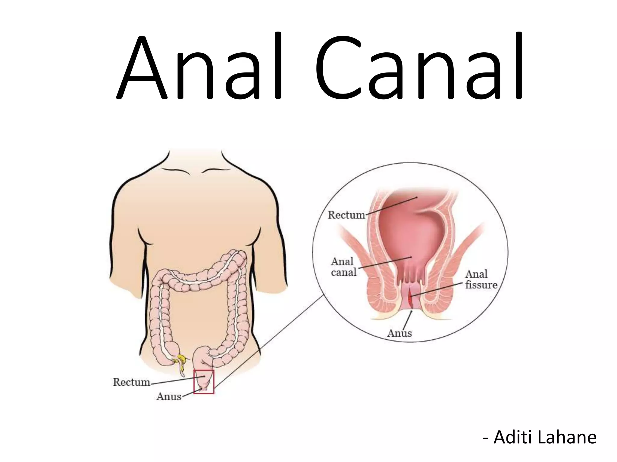
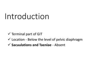
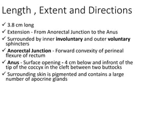
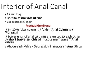
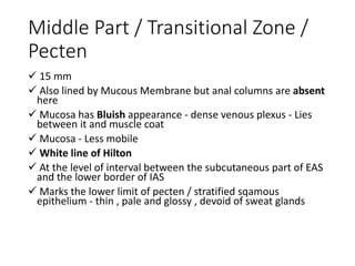
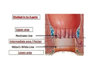
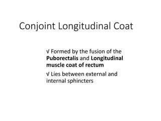
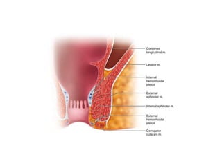
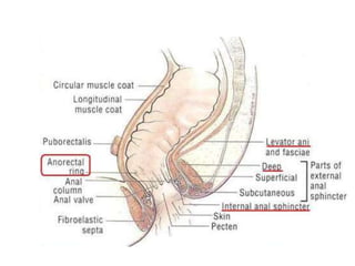
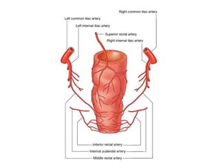
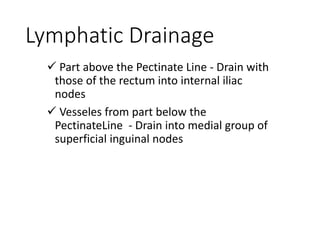
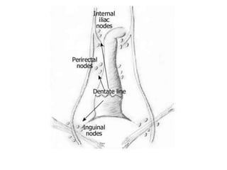
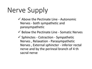
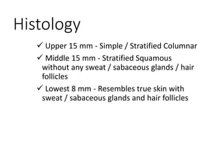
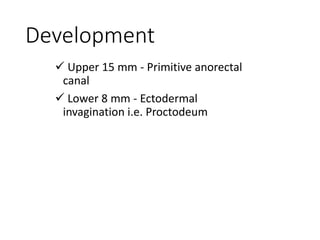
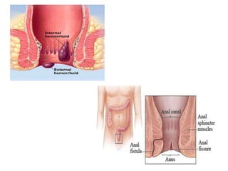
Anal Canal
- Aditi Lahane
Introduction
 Terminal part of GIT
 Location - Below the level of pelvic diaphragm
 Sacuulations and Taeniae - Absent
Length , Extent and Directions
 3.8 cm long
 Extension - From Anorectal Junction to the Anus
 Surrounded by inner involuntary and outer voluntary
sphincters
 Anorectal Junction - Forward convexity of perineal
flexure of rectum
 Anus - Surface opening - 4 cm below and infront of the
tip of the coccyx in the cleft between two buttocks
 Surrounding skin is pigmented and contains a large
number of apocrine glands
Relations of the Anal Canal
Anteriorly
 In both sexes - Perineal
body
 In Males -
Membranous urethra
and bulb of Penis
 In Female - Lower end
of Vagina
Posteriorly
 Anococcygeal ligament
 Tip of the coccyx
 Laterally - Ischioanal
Fossa
 All round -Surrounded
by sphincter muscles
Interior of Anal Canal
Divided into 3 parts -
• Upper Mucous Part
• Middle Part / Transistional Zone / Pecten
• Lower Cutaneous Part
Interior of Anal Canal
• 15 mm long
• Lined by Mucous Membrane
• Endodermal in origin
Mucous Membrane
√ 6 - 10 vertical columns / folds ~ Anal Columns /
Morgagni
√ Lower ends of anal columns are united to each other
by short trasverse folds of mucous membrane ~ Anal
Valves
√ Above each Valve - Depression in mucosa ~ Anal Sinus
√ Anal Valves - Together - Trasverse Line - Runs all round
the anal canal ~ Pectinate / Dentate Line
|
Situated opposite to middle of
intetnal Anal Sphincter - The junction of ectodermal and
endodermal parts
√ Anal Valves ocaasionally show Epithelial Projections -
Anal Papillae
√ Anal sinuses contain Anal Glands
Middle Part / Transitional Zone /
Pecten
 15 mm
 Also lined by Mucous Membrane but anal columns are absent
here
 Mucosa has Bluish appearance - dense venous plexus - Lies
between it and muscle coat
 Mucosa - Less mobile
 White line of Hilton
 At the level of interval between the subcutaneous part of EAS
and the lower border of IAS
 Marks the lower limit of pecten / stratified sqamous
epithelium - thin , pale and glossy , devoid of sweat glands
Lower Cutaneous Part
 8 mm long , lined by true skin , contaning
sabceous glands
 Epithelium of lowest part resembles that of
pigmented skin - Sabaceous glands , sweat glands
and hair
Musculature of Anal Canal
Anal Sphincters
1. Internal Anal Sphincter
2. External Anal Sphincter
1. Internal Anal Sphincter
 Involuntary
 Formed by thickned circular muscle coat
 Surrounds the upper 3/4 ths - 30 mm of the anal
canal to the white line of Hilton
2 . External Anal Sphincter
 Voluntary
 Made up of striated muscles
 Surrounds the whole length of the anal canal
 3 parts - Subcutaneous , Superficial and Deep
Conjoint Longitudinal Coat
√ Formed by the fusion of the
Puborectalis and Longitudinal
muscle coat of rectum
√ Lies between external and
internal sphincters
Anorectal Ring
 Muscular ring
 At the Anorectal Junction
 Formation - Puborectalis ,uppermost fibers
of EAS and the internal sphincter
Surgical Spaces Related to Anal
Canal
 Ischioanal Space - Lies on each side of the anal canal
 Perineal Space - Surrounds the anal canal below the
white line - Contains - Lower fibers of EAS , External
rectal venous plexus ,terminal branches of the inferior
rectal venous vesseles and nerves
 Submucous Space -Lies above the white line between
the mucuous membrane and intetnal sphincter -
Contain - Internal rectal venous plexus , Lymphatics
Arterial Supply
 Above the Pectinate Line -
Superior Rectal Artery
 Below the Pectinate Line - Inferior
Rectal Artery
Venous Drainage
 Internal rectal venous plexus / Haemorrhoidal plexus
- Submucosa of the anal canal- Drains mainly into
superior rectal vein
 External rectal plexus - Outside the muscular coat of
rectum and anal canal - lower part - drained by the
inferior rectal vein into internal pudendal vein
 Anal veins - Arranged radially around the anal margin
Lymphatic Drainage
 Part above the Pectinate Line - Drain with
those of the rectum into internal iliac
nodes
 Vesseles from part below the
PectinateLine - Drain into medial group of
superficial inguinal nodes
Nerve Supply
 Above the Pectinate Line - Autonomic
Nerves - both sympathetic and
parasympathetic
 Below the Pectinate Line - Somatic Nerves
 Sphinctes - Cotraction - Sympathetic
Nerves , Relaxation - Parasympathetic
Nerves , External sphincter - inferior rectal
nerve and by the perineal branch of 4 th
sacral nerve
Histology
 Upper 15 mm - Simple / Stratified Columnar
 Middle 15 mm - Stratified Squamous
without any sweat / sabaceous glands / hair
follicles
 Lowest 8 mm - Resembles true skin with
sweat / sabaceous glands and hair follicles
Development
 Upper 15 mm - Primitive anorectal
canal
 Lower 8 mm - Ectodermal
invagination i.e. Proctodeum
Clinical Anatomy
 Piles / Haemorrhoids
 Fistula / Fissure in Ano
 Anal Stenosis
 Anal Agencies with or without Fistula
Anal Canal-WPS Office.pptx

Anal Canal-WPS Office.pptx

  • 1.
  • 2.
    Introduction  Terminal partof GIT  Location - Below the level of pelvic diaphragm  Sacuulations and Taeniae - Absent
  • 3.
    Length , Extentand Directions  3.8 cm long  Extension - From Anorectal Junction to the Anus  Surrounded by inner involuntary and outer voluntary sphincters  Anorectal Junction - Forward convexity of perineal flexure of rectum  Anus - Surface opening - 4 cm below and infront of the tip of the coccyx in the cleft between two buttocks  Surrounding skin is pigmented and contains a large number of apocrine glands
  • 4.
    Relations of theAnal Canal Anteriorly  In both sexes - Perineal body  In Males - Membranous urethra and bulb of Penis  In Female - Lower end of Vagina Posteriorly  Anococcygeal ligament  Tip of the coccyx  Laterally - Ischioanal Fossa  All round -Surrounded by sphincter muscles
  • 5.
    Interior of AnalCanal Divided into 3 parts - • Upper Mucous Part • Middle Part / Transistional Zone / Pecten • Lower Cutaneous Part
  • 6.
    Interior of AnalCanal • 15 mm long • Lined by Mucous Membrane • Endodermal in origin Mucous Membrane √ 6 - 10 vertical columns / folds ~ Anal Columns / Morgagni √ Lower ends of anal columns are united to each other by short trasverse folds of mucous membrane ~ Anal Valves √ Above each Valve - Depression in mucosa ~ Anal Sinus
  • 7.
    √ Anal Valves- Together - Trasverse Line - Runs all round the anal canal ~ Pectinate / Dentate Line | Situated opposite to middle of intetnal Anal Sphincter - The junction of ectodermal and endodermal parts √ Anal Valves ocaasionally show Epithelial Projections - Anal Papillae √ Anal sinuses contain Anal Glands
  • 8.
    Middle Part /Transitional Zone / Pecten  15 mm  Also lined by Mucous Membrane but anal columns are absent here  Mucosa has Bluish appearance - dense venous plexus - Lies between it and muscle coat  Mucosa - Less mobile  White line of Hilton  At the level of interval between the subcutaneous part of EAS and the lower border of IAS  Marks the lower limit of pecten / stratified sqamous epithelium - thin , pale and glossy , devoid of sweat glands
  • 9.
    Lower Cutaneous Part 8 mm long , lined by true skin , contaning sabceous glands  Epithelium of lowest part resembles that of pigmented skin - Sabaceous glands , sweat glands and hair
  • 11.
    Musculature of AnalCanal Anal Sphincters 1. Internal Anal Sphincter 2. External Anal Sphincter
  • 12.
    1. Internal AnalSphincter  Involuntary  Formed by thickned circular muscle coat  Surrounds the upper 3/4 ths - 30 mm of the anal canal to the white line of Hilton
  • 13.
    2 . ExternalAnal Sphincter  Voluntary  Made up of striated muscles  Surrounds the whole length of the anal canal  3 parts - Subcutaneous , Superficial and Deep
  • 15.
    Conjoint Longitudinal Coat √Formed by the fusion of the Puborectalis and Longitudinal muscle coat of rectum √ Lies between external and internal sphincters
  • 17.
    Anorectal Ring  Muscularring  At the Anorectal Junction  Formation - Puborectalis ,uppermost fibers of EAS and the internal sphincter
  • 19.
    Surgical Spaces Relatedto Anal Canal  Ischioanal Space - Lies on each side of the anal canal  Perineal Space - Surrounds the anal canal below the white line - Contains - Lower fibers of EAS , External rectal venous plexus ,terminal branches of the inferior rectal venous vesseles and nerves  Submucous Space -Lies above the white line between the mucuous membrane and intetnal sphincter - Contain - Internal rectal venous plexus , Lymphatics
  • 20.
    Arterial Supply  Abovethe Pectinate Line - Superior Rectal Artery  Below the Pectinate Line - Inferior Rectal Artery
  • 22.
    Venous Drainage  Internalrectal venous plexus / Haemorrhoidal plexus - Submucosa of the anal canal- Drains mainly into superior rectal vein  External rectal plexus - Outside the muscular coat of rectum and anal canal - lower part - drained by the inferior rectal vein into internal pudendal vein  Anal veins - Arranged radially around the anal margin
  • 25.
    Lymphatic Drainage  Partabove the Pectinate Line - Drain with those of the rectum into internal iliac nodes  Vesseles from part below the PectinateLine - Drain into medial group of superficial inguinal nodes
  • 27.
    Nerve Supply  Abovethe Pectinate Line - Autonomic Nerves - both sympathetic and parasympathetic  Below the Pectinate Line - Somatic Nerves  Sphinctes - Cotraction - Sympathetic Nerves , Relaxation - Parasympathetic Nerves , External sphincter - inferior rectal nerve and by the perineal branch of 4 th sacral nerve
  • 28.
    Histology  Upper 15mm - Simple / Stratified Columnar  Middle 15 mm - Stratified Squamous without any sweat / sabaceous glands / hair follicles  Lowest 8 mm - Resembles true skin with sweat / sabaceous glands and hair follicles
  • 29.
    Development  Upper 15mm - Primitive anorectal canal  Lower 8 mm - Ectodermal invagination i.e. Proctodeum
  • 30.
    Clinical Anatomy  Piles/ Haemorrhoids  Fistula / Fissure in Ano  Anal Stenosis  Anal Agencies with or without Fistula