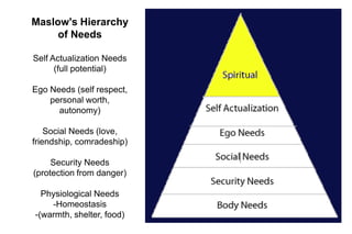
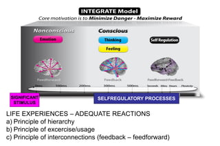
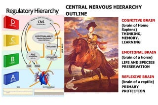
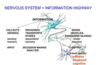
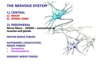
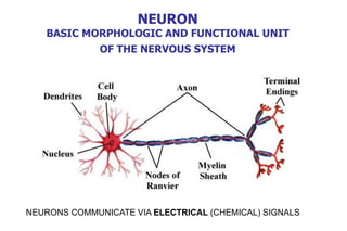
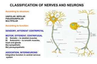
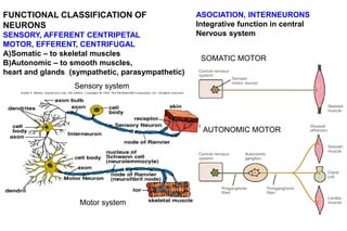
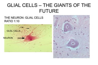
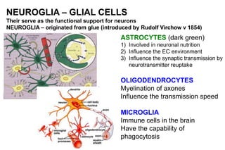
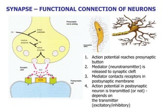
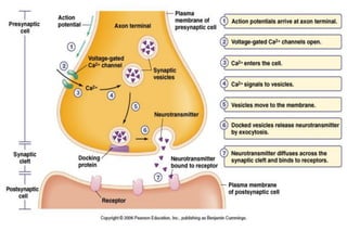
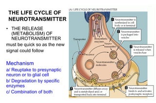
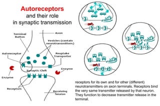
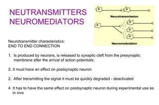
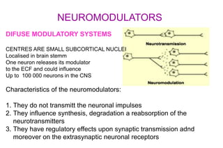
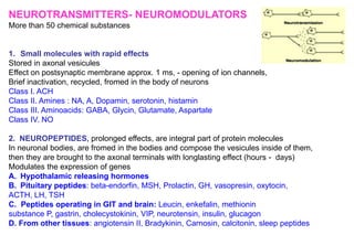
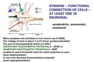
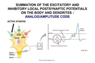
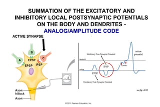
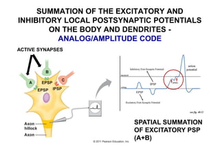
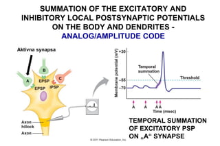
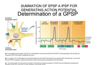
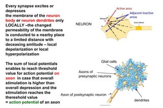
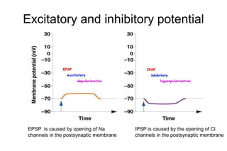
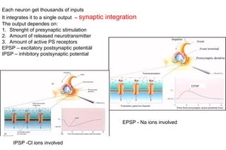
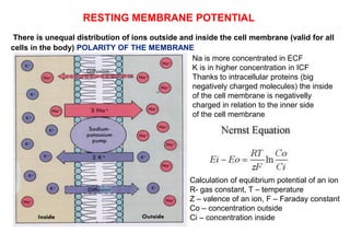
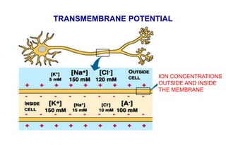
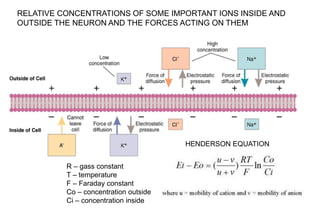
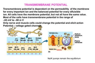
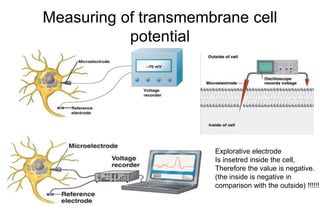
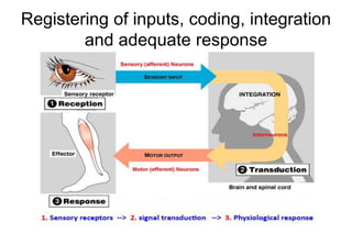
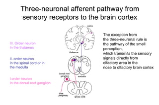
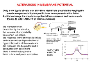
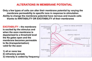
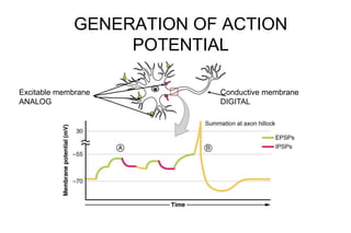
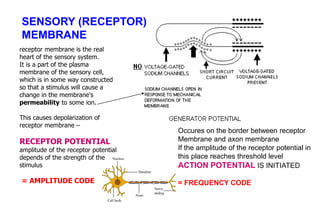
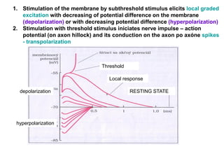
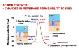
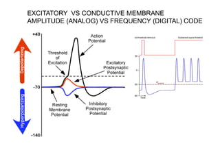
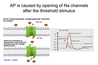
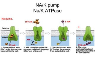
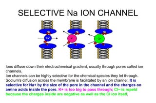
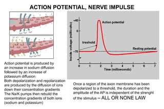
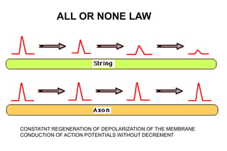
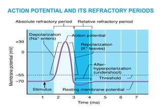
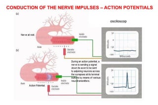
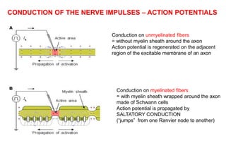
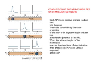
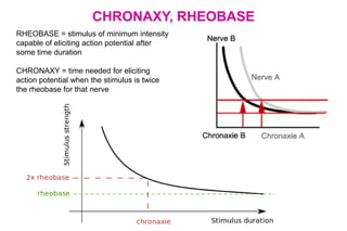
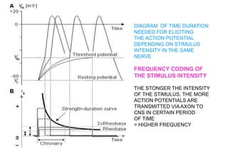
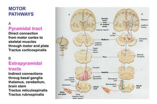
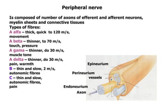
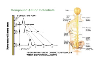
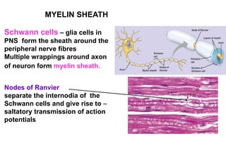
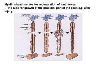
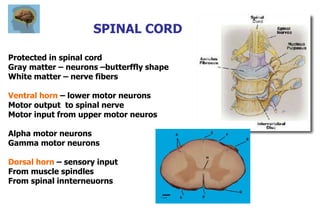
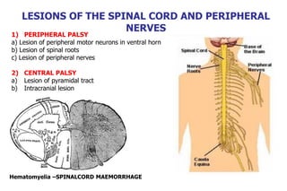
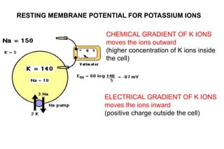
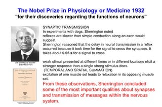
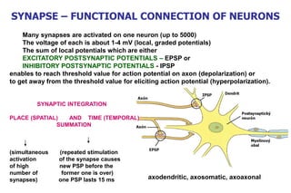
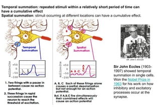
NEUROPHYSIOLOGY
Prof. MUDr. Daniela Ostatníková, PhD
Institute of Physiology, Faculty of Medicine
Comenius University
PHYSIOLOGY –
LOGIC OF LIFE
Rembrandt van Rijn –
1606 – 1669
Anatomy lecture
MOTIVATION TO LEARN
Maslow's Hierarchy
of Needs
Self Actualization Needs
(full potential)
Ego Needs (self respect,
personal worth,
autonomy)
Social Needs (love,
friendship, comradeship)
Security Needs
(protection from danger)
Physiological Needs
-Homeostasis
-(warmth, shelter, food)
SIGNIFICANT
STIMULUS
SELFREGULATORY PROCESSES
LIFE EXPERIENCES – ADEQUATE REACTIONS
a) Principle of hierarchy
b) Principle of excercise/usage
c) Principle of interconnections (feedback – feedforward)
•
TRAIN YOUR BRAIN
COGNITIVE BRAIN
(brain of Homo
Sapiens)
THINKING,
MEMORY,
LEARNING
EMOTIONAL BRAIN
(brain of a horse)
LIFE AND SPECIES
PRESERVATION
REFLEXIVE BRAIN
(brain of a reptile)
PRIMARY
PROTECTION
CENTRAL NERVOUS HIERARCHY
OUTLINE
LIFE = ADAPTATION
Regulatory mechanisms for life preservation = regulatory
mechanisms for preservation of inner environment stability -
HOMEOSTASIS in spite of changes in external environment
IMMUNE SYSTEM
PROTECTION, an ability to discriminate between self and non self, protection against
microorganisms´ reproduction
Systems of innate and adaptive immune reactions, fast and slow immune systems
ENDOCRINNE SYSTEM
Regulation of metabolic and functional processes in human body, ensuring the
biorhythms, life stereotypes and responses to stress
Secretion of minimum doses of hormones (nanomoles, picomoles) to blood or to
extracellular space. Slow but very effective systems
NERVOUS SYSTEM
Very fast regulation system, communication via nerve impulses – action potentials that
are spreading via nerve fibers to distant tissues (muscles, glands, brain tissue)
HOMEOSTASIS & HOMEORHESIS
the principles for life preservation
For a dynamical system,
homeostasis can be viewed
as sustaining internal
stability.
For a dynamical system,
homeorhesis is viewed as
maintenance of order and
normalcy in the state
trajectory under internal
and external disturbances
by avoiding a potential
chaos.
Homeorhesis, derived from the Greek for "similar flow", is a concept
encompassing dynamical systems which return to a trajectory, as
opposed to systems which return to a particular state, which is termed
homeostasis
OUTLINE OF THE LECTURES
• Physiology of the nerve cell
• Resting transmembrane potential, receptor
potential
• Depolarisation, hyperpolarisation, action
potential of the nerve cell
• Conduction of action potentials
• Physiology of senses (vision, hearing, pain
perception)
• Physiology of skeletal and smooth muscles
• Physiology of autonomous nervous system
THE NERVOUS SYSTEM
NERVOUS SYSTEM = INFORMATION HIGHWAY
INFORMATION
COLLECTS ORGANISES SENDS
(SENSES) TRANSPORTS (MUSCLES,
STORES ENDOCRINE GLANDS)
sensory association motor
neurons neurons neurons
INPUT DECISSION MAKING OUTPUT
ANALYSIS
3-level system:
reflexive
emotional
cognitive
THE NERVOUS SYSTEM
1) CENTRAL
a) BRAIN
b) SPINAL CORD
2) PERIPHERAL
Nerve fibers - AXONS – connections to
muscles and glands
MOTOR NERVE FIBERS
AUTONOMIC (VEGETATIVE)
NERVE FIBERS
• Sympaticus
• Parasympaticus
SENSORY NERVE FIBERS
NEURON
BASIC MORPHOLOGIC AND FUNCTIONAL UNIT
OF THE NERVOUS SYSTEM
NEURONS COMMUNICATE VIA ELECTRICAL (CHEMICAL) SIGNALS
According to structure:
UNIPOLAR, BIPOLAR
PSEUDOUNIPOLAR
MULTIPOLAR
According to function:
SENSORY, AFFERENT CENTRIPETAL
MOTOR, EFFERENT, CENTRIFUGAL
A) Somatic – to skeletal muscles
B) Autonomic – to smooth muscles,
heart and glands
Ba) sympathetic
Bb) parasympathetic
ASOCIATION, INTERNEURONS
Integrative function in central nervous
system
CLASSIFICATION OF NERVES AND NEURONS
Motor system
Sensory system
FUNCTIONAL CLASSIFICATION OF
NEURONS
SENSORY, AFFERENT CENTRIPETAL
MOTOR, EFFERENT, CENTRIFUGAL
A)Somatic – to skeletal muscles
B)Autonomic – to smooth muscles,
heart and glands (sympathetic, parasympathetic)
ASOCIATION, INTERNEURONS
Integrative function in central
Nervous system
SOMATIC MOTOR
AUTONOMIC MOTOR
Registering of inputs, coding, integration
and adequate response
GLIAL CELLS – THE GIANTS OF THE
FUTURE
NEURON
GLIAL CELLS
THE NEURON: GLIAL CELLS
RATIO 1:10
NEUROGLIA – GLIAL CELLS
Their serve as the functional support for neurons
NEUROGLIA – originated from glue (introduced by Rudolf Virchow v 1854)
ASTROCYTES (dark green)
1) Involved in neuronal nutrition
2) Influence the EC environment
3) Influence the synaptic transmission by
neurotransmitter reuptake
OLIGODENDROCYTES
Myelination of axones
Influence the transmission speed
MICROGLIA
Immune cells in the brain
Have the capability of
phagocytosis
SYNAPSE – FUNCTIONAL CONNECTION OF NEURONS
1. Action potential reaches presynaptic
button
2. Mediator (neurotransmitter) is
released to synaptic cleft
3. Mediator contacts receptors in
postsynaptic membrane
4. Action potential in postsynaptic
neuron is transmitted (or not) -
depends on
the transmitter
(excitatory/inhibitory)
THE LIFE CYCLE OF
NEUROTRANSMITTER
• THE RELEASE
(METABOLISM) OF
NEUROTRANSMITTER
must be quick so as the new
signal could follow
Mechanism
a/ Reuptake to presynaptic
neuron or to glial cell
b/ Degradation by specific
enzymes
c/ Combination of both
Autoreceptors
and their role
in synaptic transmission
receptors for its own and for other (different)
neurotransmitters on axon terminals. Receptors bind
the very same transmitter released by that neuron.
They function to decrease transmitter release in the
terminal.
NEUTRANSMITTERS
NEUROMEDIATORS
Neurotransmitter characteristics:
END TO END CONNECTION
1. Is produced by neurons, is released to synaptic cleft from the presynaptic
membrane after the arrival of action potentials.
2. It must have an effect on postsynaptic neuron
2. After trensmitting the signal it must be quickly degraded - deactivated
4. It has to have the same effect on postsynaptic neuron during experimental use as
in vivo
NEUROMODULATORS
DIFUSE MODULATORY SYSTEMS
CENTRES ARE SMALL SUBCORTICAL NUCLEI
Localised in brain stemm
One neuron releases its modulator
to the ECF and could influence
Up to 100 000 neurons in the CNS
Characteristics of the neuromodulators:
1. They do not transmitt the neuronal impulses
2. They influence synthesis, degradation a reabsorption of the
neurotransmitters
3. They have regulatory effects upon synaptic transmission adnd
moreover on the extrasynaptic neuronal receptors
NEUROTRANSMITTERS- NEUROMODULATORS
More than 50 chemical substances
1. Small molecules with rapid effects
Stored in axonal vesicules
Effect on postsynaptic membrane approx. 1 ms, - opening of ion channels,
Brief inactivation, recycled, fromed in the body of neurons
Class I. ACH
Class II. Amines : NA, A, Dopamin, serotonin, histamin
Class III. Aminoacids: GABA, Glycin, Glutamate, Aspartate
Class IV. NO
2. NEUROPEPTIDES, prolonged effects, are integral part of protein molecules
In neuronal bodies, are fromed in the bodies and compose the vesicules inside of them,
then they are brought to the axonal terminals with longlasting effect (hours - days)
Modulates the expression of genes
A. Hypothalamic releasing hormones
B. Pituitary peptides: beta-endorfin, MSH, Prolactin, GH, vasopresin, oxytocin,
ACTH, LH, TSH
C. Peptides operating in GIT and brain: Leucin, enkefalin, methionin
substance P, gastrin, cholecystokinin, VIP, neurotensin, insulin, glucagon
D. From other tissues: angiotensin II, Bradykinin, Carnosin, calcitonin, sleep peptides
Many synapses are activated on one neuron (up to 5000)
The voltage of each is about 1-4 mV (local, graded potentials)
The sum of local potentials which are either
EXCITATORY POSTSYNAPTIC POTENTIALS – EPSP or
INHIBITORY POSTSYNAPTIC POTENTIALS - IPSP
enables to reach threshold value for action potential on axon
(axon depolarization)
or can even decrease transmembrane potential
(axon hyperpolarization).
axodendritic, axosomatic,
axoaxonal
SYNAPSE – FUNCTIONAL
CONNECTION OF CELLS –
AT LEAST ONE IS
NEURONAL
SUMMATION OF THE EXCITATORY AND
INHIBITORY LOCAL POSTSYNAPTIC POTENTIALS
ON THE BODY AND DENDRITES -
ANALOG/AMPLITUDE CODE
ACTIVE SYNAPSE
ACTIVE SYNAPSE
SUMMATION OF THE EXCITATORY AND
INHIBITORY LOCAL POSTSYNAPTIC POTENTIALS
ON THE BODY AND DENDRITES -
ANALOG/AMPLITUDE CODE
SPATIAL SUMMATION
OF EXCITATORY PSP
(A+B)
ACTIVE SYNAPSES
SUMMATION OF THE EXCITATORY AND
INHIBITORY LOCAL POSTSYNAPTIC POTENTIALS
ON THE BODY AND DENDRITES -
ANALOG/AMPLITUDE CODE
TEMPORAL SUMMATION
OF EXCITATORY PSP
ON „A“ SYNAPSE
Aktívna synapsa
SUMMATION OF THE EXCITATORY AND
INHIBITORY LOCAL POSTSYNAPTIC POTENTIALS
ON THE BODY AND DENDRITES -
ANALOG/AMPLITUDE CODE
SUMMATION OF EPSP A IPSP FOR
GENERATING ACTION POTENTIAL
Every synapse excites or
depresses
the membrane of the neuron
body or neuron dendrites only
LOCALLY –the changed
permeability of the membrane
is conducted to a nearby place
to a limited distance with
deceasing amlitude – local
depolarization or local
hyperpolarization
The sum of local potentials
enables to reach threshold
value for action potential on
axon in case that overall
stimulation is higher than
overall depression and the
stimulation reaches the
thereshold value
= action potential of an axon
NEURON
dendrites
Glial cells
Axon of postsynaptic neuron
Axons of
presynaptic neurons
Excitatory and inhibitory potential
EPSP is caused by opening of Na
channels in the postsynaptic membrane
IPSP is caused by the opening of Cl
channels in the postsynaptic membrane
Each neuron get thousands of inputs
It integrates it to a single output – synaptic integration
The output dependes on:
1. Strenght of presynaptic stimulation
2. Amount of released neurotransmitter
3. Amount of active PS receptors
EPSP – excitatory postsynaptic potentiál
IPSP – inhibitory postsynaptic potential
IPSP -Cl ions involved
EPSP - Na ions involved
RESTING MEMBRANE
POTENTIAL
RESTING MEMBRANE POTENTIAL
There is unequal distribution of ions outside and inside the cell membrane (valid for all
cells in the body) POLARITY OF THE MEMBRANE
Na is more concentrated in ECF
K is in higher concentration in ICF
Thanks to intracellular proteins (big
negatively charged molecules) the inside
of the cell membrane is negativelly
charged in relation to the inner side
of the cell membrane
Calculation of equlibrium potential of an ion
R- gas constant, T – temperature
Z – valence of an ion, F – Faraday constant
Co – concentration outside
Ci – concentration inside
TRANSMEMBRANE POTENTIAL
ION CONCENTRATIONS
OUTSIDE AND INSIDE
THE MEMBRANE
RELATIVE CONCENTRATIONS OF SOME IMPORTANT IONS INSIDE AND
OUTSIDE THE NEURON AND THE FORCES ACTING ON THEM
HENDERSON EQUATION
R – gass constant
T – temperature
F – Faraday constant
Co – concentration outside
Ci – concentration inside
TRANSMEMBRANE POTENTIAL
Transmembrane potential is dependant on the permeability of the membrane
for every important ion and the balanced potential for every difuisible
ion. All cells have the membrane potential, but not all have the same value.
Most of the cells have transmembrane potential in the range of
–65 mV to –90 m V
Only nerve and muscle cells could change the potential and elicit action
Potential – voltage gated channels
Na/K pumps remain the equilibrium
Measuring of transmembrane cell
potential
Explorative electrode
Is insetred inside the cell,
Therefore the value is negative.
(the inside is negative in
comparison with the outside) !!!!!!
ALTERATIONS OF AXON
MEMBRANE
POTENTIAL
ACTION POTENTIAL
Registering of inputs, coding, integration
and adequate response
Three-neuronal afferent pathway from
sensory receptors to the brain cortex
I.order neuron
In the dorsal root ganglion
II. order neuron
In the spinal cord or in
the medulla
III. Order neuron
In the thalamus
The exception from
the three-neuronal rule is
the pathway of the smell
perception,
which transmits the sensory
signals directly from
olfactory area in the
nose to olfactory brain cortex
Only a few types of cells can alter their membrane potential by varying the
membrane permeability to specific ions in response to stimulation
Ability to change the membrane potential have nervous and muscle cells
thanks to EXCITABILITY of their membranes
the membrane can
be excited by the stimulus,
the increase of premeability
to a certain ion occurs,
the response to the stimulus is limited
and causes either depolarization or
hyperpolarization of the membrane,
the response can be graded and is
conducted with decrement
there is no refractory phase
there is time and place summation
ALTERATIONS IN MEMBRANE POTENTIAL
AMPLITUDE
ANALOG
CODE
Only a few types of cells can alter their membrane potential by varying the
membrane permeability to specific ions in response to stimulation
Ability to change the membrane potential have nervous and muscle cells
thanks to IRRITABILITY OR EXCITABILITY of their membranes
EXCITABILITY – the membrane
is excited by the stimulus and
when the axon membrane is
depolarized to a threshold level
the Na gates open and the
membrane becomes permeable
to Na (transpolarization)
valid for the axon
1) all or none law
2) refractory periods
3) intensity is coded by frequency
ALTERATIONS IN MEMBRANE POTENTIAL
GENERATION OF ACTION
POTENTIAL
Excitable membrane
ANALOG
Conductive membrane
DIGITAL
receptor membrane is the real
heart of the sensory system.
It is a part of the plasma
membrane of the sensory cell,
which is in some way constructed
so that a stimulus will cause a
change in the membrane's
permeability to some ion.
This causes depolarization of
receptor membrane –
RECEPTOR POTENTIAL
amplitude of the receptor potential
depends of the strength of the
stimulus
= AMPLITUDE CODE
SENSORY (RECEPTOR)
MEMBRANE
Occures on the border between receptor
Membrane and axon membrane
If the amplitude of the receptor potential in
this place reaches threshold level
ACTION POTENTIAL IS INITIATED
= FREQUENCY CODE
1. Stimulation of the membrane by subthreshold stimulus elicits local graded
excitation with decreasing of potential difference on the membrane
(depolarization) or with decreasing potential difference (hyperpolarization)
2. Stimulation with threshold stimulus iniciates nerve impulse – action
potential (on axon hillock) and its conduction on the axon po axóne spikes
- transpolarization
RESTING STATE
depolarization
hyperpolarization
Threshold
Local response
ACTION POTENTIAL
– CHANGES IN MEMBRANE PERMEABILITY TO IONS
AP time duration 4ms
EXCITATORY VS CONDUCTIVE MEMBRANE
AMPLITUDE (ANALOG) VS FREQUENCY (DIGITAL) CODE
AP is caused by opening of Na channels
after the threshold stimulus
NA/K pump
Na/K ATPase
SELECTIVE Na ION CHANNEL
Ions diffuse down their electrochemical gradient, usually through pores called ion
channels.
Ion channels can be highly selective for the chemical species they let through.
Sodium's diffusion across the membrane is facilitated by an ion channel. It is
selective for Na+ by the size of the pore in the channel and the charges on
amino acids inside the pore. K+ is too big to pass through; Cl− is repeld
because the charges inside are negative as well as the Cl ion itself.
Action potential is produced by
an increase in sodium diffusion
followed by an increase of
potassium diffusion
Both depolarization and repolarization
are produced by the diffusion of ions
down their concentration gradients
The Na/K pumps then rebuild the
concentration gradients of both ions
(sodium and potassium)
ACTION POTENTIAL, NERVE IMPULSE
treshold
Once a region of the axon membrane has been
depolarized to a threshold, the duration and the
amplitude of the AP is independent of the strenght
of the stimulus – ALL OR NONE LAW
ALL OR NONE LAW
CONSTATNT REGENERATION OF DEPOLARIZATION OF THE MEMBRANE
CONDUCTION OF ACTION POTENTIALS WITHOUT DECREMENT
ACTION POTENTIAL AND ITS REFRACTORY PERIODS
CONDUCTION OF
ACTION POTENTIALS
CONDUCTION OF THE NERVE IMPULSES – ACTION POTENTIALS
osciloscop
CONDUCTION OF THE NERVE IMPULSES – ACTION POTENTIALS
Conduction on unmyelinated fibers
= without myelin sheath around the axon
Action potential is regenerated on the adjacent
region of the excitable membrane of an axon
Conduction on myelinated fibers
= with myelin sheath wrapped around the axon
made of Schwann cells
Action potential is propagated by
SALTATORY CONDUCTION
(“jumps” from one Ranvier node to another)
CONDUCTION OF THE NERVE IMPULSES
ON UNMYELINATED FIBERS
Each AP injects positive charges (sodium
ions)
Into the axon
These are conducted by the cable
properties
of the axon to an adjacent region that still
has
a membrane potential of –65 mV.
When this adjacent region of the
membrane
reaches threshold level of depolarization
It too produces an AP as its voltage
regulated
gates open
CHRONAXY, RHEOBASE
RHEOBASE = stimulus of minimum intensity
capable of eliciting action potential after
some time duration
CHRONAXY = time needed for eliciting
action potential when the stimulus is twice
the rheobase for that nerve
DIAGRAM OF TIME DURATION
NEEDED FOR ELICITING
THE ACTION POTENTIAL
DEPENDING ON STIMULUS
INTENSITY IN THE SAME
NERVE
FREQUENCY CODING OF
THE STIMULUS INTENSITY
THE STONGER THE INTENSITY
OF THE STIMULUS, THE MORE
ACTION POTENTIALS ARE
TRANSMITTED VIA AXON TO
CNS IN CERTAIN PERIOD
OF TIME
= HIGHER FREQUENCY
PERIPHERAL NERVOUS
SYSTEM
MOTOR
PATHWAYS
A
Pyramidal tract
Direct connection
from motor cortex to
skeletal muscles
through motor end plate
Tractus corticospinalis
B
Extrapyramidal
tracts
Indirect connections
throug basal ganglia
thalamus, cerebellum,
brain stem
Tractus reticulospinalis
Tractus rubrospinalis
Epineurium
Endoneurium
Axon
Peripheral nerve
Is composed of number of axons of efferent and afferent neurons,
myelin sheets and connective tissues
Types of fibres:
A alfa – thick, quick to 120 m/s,
movement
A beta – thinner, to 70 m/s,
touch, pressure
A gama – thinner, do 30 m/s,
muscle tone
A delta – thinner, do 30 m/s,
pain, warmth
B – thin and slow, 2 m/s,
autonomic fibres
C – thin and slow,
autonomic fibres,
pain
Perineurium
vessels
LATENCY
STIMULATION POINT
FIBERS OF DIFFERENT CONDUCTION VELOCITY
WITHIN ON PERIPHERAL NERVE
Schwann cells – glia cells in
PNS form the sheath around the
peripheral nerve fibres
Multiple wrappings around axon
of neuron form myelin sheath.
Nodes of Ranvier
separate the internodia of the
Schwann cells and give rise to –
saltatory transmission of action
potentials
MYELIN SHEATH
Myelin sheath serves for regeneration of cut nerves
– the tube for growth of the proximal part of the axon e.g. after
injury.
Protected in spinal cord
Gray matter – neurons –butterffly shape
White matter – nerve fibers
Ventral horn – lower motor neurons
Motor output to spinal nerve
Motor input from upper motor neuros
Alpha motor neurons
Gamma motor neurons
Dorsal horn – sensory input
From muscle spindles
From spinal innterneuorns
SPINAL CORD
Hematomyelia –SPINALCORD MAEMORRHAGE
LESIONS OF THE SPINAL CORD AND PERIPHERAL
NERVES
1) PERIPHERAL PALSY
a) Lesion of peripheral motor neurons in ventral horn
b) Lesion of spinal roots
c) Lesion of peripheral nerves
2) CENTRAL PALSY
a) Lesion of pyramidal tract
b) Intracranial lesion
RESTING MEMBRANE POTENTIAL FOR POTASSIUM IONS
CHEMICAL GRADIENT OF K IONS
moves the ions outward
(higher concentration of K ions inside
the cell)
ELECTRICAL GRADIENT OF K IONS
moves the ions inward
(positive charge outside the cell)
The Nobel Prize in Physiology or Medicine 1932
"for their discoveries regarding the functions of neurons"
SYNAPTIC TRANSMISSION
In experiments with dogs, Sherrington noted
reflexes are slower than simple conduction along an axon would
suggest.
Sherrington reasoned that the delay in neural transmission in a reflex
occurred because it took time for the signal to cross the synapses. It
takes about 0.05 s for a signal to cross.
weak stimuli presented at different times or in different locations elicit a
stronger response than a single strong stimulus does.
(TEMPORAL AND SPATIAL SUMMATION)
excitation of one muscle set leads to relaxation in its opposing muscle
set.
From these observations, Sherrington concluded
some of the most important qualities about synapses
and transmission of messages within the nervous
system.
Many synapses are activated on one neuron (up to 5000)
The voltage of each is about 1-4 mV (local, graded potentials)
The sum of local potentials which are either
EXCITATORY POSTSYNAPTIC POTENTIALS – EPSP or
INHIBITORY POSTSYNAPTIC POTENTIALS - IPSP
enables to reach threshold value for action potential on axon (depolarization) or
to get away from the threshold value for eliciting action potential (hyperpolarization).
SYNAPTIC INTEGRATION
PLACE (SPATIAL) AND TIME (TEMPORAL)
SUMMATION
(simultaneous (repeated stimulation
activation of the synapse causes
of high new PSP before the
number of former one is over)
synapses) one PSP lasts 15 ms axodendritic, axosomatic, axoaxonal
SYNAPSE – FUNCTIONAL CONNECTION OF NEURONS
Temporal summation: repeated stimuli within a relatively short period of time can
have a cumulative effect
Spatial summation: stimuli occurring at different locations can have a cumulative effect.
Sir John Eccles (1903-
1997) showed temporal
summation in single cells.
Won the Nobel Prize in
1963 for his work on how
inhibitory and excitatory
processes occur at the
synapse.

NEUROPHYSIOLOGY_GM_1_2018.pdf

  • 1.
    NEUROPHYSIOLOGY Prof. MUDr. DanielaOstatníková, PhD Institute of Physiology, Faculty of Medicine Comenius University
  • 2.
  • 3.
    Rembrandt van Rijn– 1606 – 1669 Anatomy lecture MOTIVATION TO LEARN
  • 4.
    Maslow's Hierarchy of Needs SelfActualization Needs (full potential) Ego Needs (self respect, personal worth, autonomy) Social Needs (love, friendship, comradeship) Security Needs (protection from danger) Physiological Needs -Homeostasis -(warmth, shelter, food)
  • 5.
    SIGNIFICANT STIMULUS SELFREGULATORY PROCESSES LIFE EXPERIENCES– ADEQUATE REACTIONS a) Principle of hierarchy b) Principle of excercise/usage c) Principle of interconnections (feedback – feedforward)
  • 6.
  • 7.
    COGNITIVE BRAIN (brain ofHomo Sapiens) THINKING, MEMORY, LEARNING EMOTIONAL BRAIN (brain of a horse) LIFE AND SPECIES PRESERVATION REFLEXIVE BRAIN (brain of a reptile) PRIMARY PROTECTION CENTRAL NERVOUS HIERARCHY OUTLINE
  • 8.
    LIFE = ADAPTATION Regulatorymechanisms for life preservation = regulatory mechanisms for preservation of inner environment stability - HOMEOSTASIS in spite of changes in external environment IMMUNE SYSTEM PROTECTION, an ability to discriminate between self and non self, protection against microorganisms´ reproduction Systems of innate and adaptive immune reactions, fast and slow immune systems ENDOCRINNE SYSTEM Regulation of metabolic and functional processes in human body, ensuring the biorhythms, life stereotypes and responses to stress Secretion of minimum doses of hormones (nanomoles, picomoles) to blood or to extracellular space. Slow but very effective systems NERVOUS SYSTEM Very fast regulation system, communication via nerve impulses – action potentials that are spreading via nerve fibers to distant tissues (muscles, glands, brain tissue)
  • 9.
    HOMEOSTASIS & HOMEORHESIS theprinciples for life preservation For a dynamical system, homeostasis can be viewed as sustaining internal stability. For a dynamical system, homeorhesis is viewed as maintenance of order and normalcy in the state trajectory under internal and external disturbances by avoiding a potential chaos. Homeorhesis, derived from the Greek for "similar flow", is a concept encompassing dynamical systems which return to a trajectory, as opposed to systems which return to a particular state, which is termed homeostasis
  • 10.
    OUTLINE OF THELECTURES • Physiology of the nerve cell • Resting transmembrane potential, receptor potential • Depolarisation, hyperpolarisation, action potential of the nerve cell • Conduction of action potentials • Physiology of senses (vision, hearing, pain perception) • Physiology of skeletal and smooth muscles • Physiology of autonomous nervous system
  • 11.
  • 12.
    NERVOUS SYSTEM =INFORMATION HIGHWAY INFORMATION COLLECTS ORGANISES SENDS (SENSES) TRANSPORTS (MUSCLES, STORES ENDOCRINE GLANDS) sensory association motor neurons neurons neurons INPUT DECISSION MAKING OUTPUT ANALYSIS 3-level system: reflexive emotional cognitive
  • 13.
    THE NERVOUS SYSTEM 1)CENTRAL a) BRAIN b) SPINAL CORD 2) PERIPHERAL Nerve fibers - AXONS – connections to muscles and glands MOTOR NERVE FIBERS AUTONOMIC (VEGETATIVE) NERVE FIBERS • Sympaticus • Parasympaticus SENSORY NERVE FIBERS
  • 14.
    NEURON BASIC MORPHOLOGIC ANDFUNCTIONAL UNIT OF THE NERVOUS SYSTEM NEURONS COMMUNICATE VIA ELECTRICAL (CHEMICAL) SIGNALS
  • 15.
    According to structure: UNIPOLAR,BIPOLAR PSEUDOUNIPOLAR MULTIPOLAR According to function: SENSORY, AFFERENT CENTRIPETAL MOTOR, EFFERENT, CENTRIFUGAL A) Somatic – to skeletal muscles B) Autonomic – to smooth muscles, heart and glands Ba) sympathetic Bb) parasympathetic ASOCIATION, INTERNEURONS Integrative function in central nervous system CLASSIFICATION OF NERVES AND NEURONS
  • 16.
    Motor system Sensory system FUNCTIONALCLASSIFICATION OF NEURONS SENSORY, AFFERENT CENTRIPETAL MOTOR, EFFERENT, CENTRIFUGAL A)Somatic – to skeletal muscles B)Autonomic – to smooth muscles, heart and glands (sympathetic, parasympathetic) ASOCIATION, INTERNEURONS Integrative function in central Nervous system SOMATIC MOTOR AUTONOMIC MOTOR
  • 17.
    Registering of inputs,coding, integration and adequate response
  • 18.
    GLIAL CELLS –THE GIANTS OF THE FUTURE NEURON GLIAL CELLS THE NEURON: GLIAL CELLS RATIO 1:10
  • 19.
    NEUROGLIA – GLIALCELLS Their serve as the functional support for neurons NEUROGLIA – originated from glue (introduced by Rudolf Virchow v 1854) ASTROCYTES (dark green) 1) Involved in neuronal nutrition 2) Influence the EC environment 3) Influence the synaptic transmission by neurotransmitter reuptake OLIGODENDROCYTES Myelination of axones Influence the transmission speed MICROGLIA Immune cells in the brain Have the capability of phagocytosis
  • 20.
    SYNAPSE – FUNCTIONALCONNECTION OF NEURONS 1. Action potential reaches presynaptic button 2. Mediator (neurotransmitter) is released to synaptic cleft 3. Mediator contacts receptors in postsynaptic membrane 4. Action potential in postsynaptic neuron is transmitted (or not) - depends on the transmitter (excitatory/inhibitory)
  • 22.
    THE LIFE CYCLEOF NEUROTRANSMITTER • THE RELEASE (METABOLISM) OF NEUROTRANSMITTER must be quick so as the new signal could follow Mechanism a/ Reuptake to presynaptic neuron or to glial cell b/ Degradation by specific enzymes c/ Combination of both
  • 23.
    Autoreceptors and their role insynaptic transmission receptors for its own and for other (different) neurotransmitters on axon terminals. Receptors bind the very same transmitter released by that neuron. They function to decrease transmitter release in the terminal.
  • 24.
    NEUTRANSMITTERS NEUROMEDIATORS Neurotransmitter characteristics: END TOEND CONNECTION 1. Is produced by neurons, is released to synaptic cleft from the presynaptic membrane after the arrival of action potentials. 2. It must have an effect on postsynaptic neuron 2. After trensmitting the signal it must be quickly degraded - deactivated 4. It has to have the same effect on postsynaptic neuron during experimental use as in vivo
  • 25.
    NEUROMODULATORS DIFUSE MODULATORY SYSTEMS CENTRESARE SMALL SUBCORTICAL NUCLEI Localised in brain stemm One neuron releases its modulator to the ECF and could influence Up to 100 000 neurons in the CNS Characteristics of the neuromodulators: 1. They do not transmitt the neuronal impulses 2. They influence synthesis, degradation a reabsorption of the neurotransmitters 3. They have regulatory effects upon synaptic transmission adnd moreover on the extrasynaptic neuronal receptors
  • 26.
    NEUROTRANSMITTERS- NEUROMODULATORS More than50 chemical substances 1. Small molecules with rapid effects Stored in axonal vesicules Effect on postsynaptic membrane approx. 1 ms, - opening of ion channels, Brief inactivation, recycled, fromed in the body of neurons Class I. ACH Class II. Amines : NA, A, Dopamin, serotonin, histamin Class III. Aminoacids: GABA, Glycin, Glutamate, Aspartate Class IV. NO 2. NEUROPEPTIDES, prolonged effects, are integral part of protein molecules In neuronal bodies, are fromed in the bodies and compose the vesicules inside of them, then they are brought to the axonal terminals with longlasting effect (hours - days) Modulates the expression of genes A. Hypothalamic releasing hormones B. Pituitary peptides: beta-endorfin, MSH, Prolactin, GH, vasopresin, oxytocin, ACTH, LH, TSH C. Peptides operating in GIT and brain: Leucin, enkefalin, methionin substance P, gastrin, cholecystokinin, VIP, neurotensin, insulin, glucagon D. From other tissues: angiotensin II, Bradykinin, Carnosin, calcitonin, sleep peptides
  • 27.
    Many synapses areactivated on one neuron (up to 5000) The voltage of each is about 1-4 mV (local, graded potentials) The sum of local potentials which are either EXCITATORY POSTSYNAPTIC POTENTIALS – EPSP or INHIBITORY POSTSYNAPTIC POTENTIALS - IPSP enables to reach threshold value for action potential on axon (axon depolarization) or can even decrease transmembrane potential (axon hyperpolarization). axodendritic, axosomatic, axoaxonal SYNAPSE – FUNCTIONAL CONNECTION OF CELLS – AT LEAST ONE IS NEURONAL
  • 28.
    SUMMATION OF THEEXCITATORY AND INHIBITORY LOCAL POSTSYNAPTIC POTENTIALS ON THE BODY AND DENDRITES - ANALOG/AMPLITUDE CODE ACTIVE SYNAPSE
  • 29.
    ACTIVE SYNAPSE SUMMATION OFTHE EXCITATORY AND INHIBITORY LOCAL POSTSYNAPTIC POTENTIALS ON THE BODY AND DENDRITES - ANALOG/AMPLITUDE CODE
  • 30.
    SPATIAL SUMMATION OF EXCITATORYPSP (A+B) ACTIVE SYNAPSES SUMMATION OF THE EXCITATORY AND INHIBITORY LOCAL POSTSYNAPTIC POTENTIALS ON THE BODY AND DENDRITES - ANALOG/AMPLITUDE CODE
  • 31.
    TEMPORAL SUMMATION OF EXCITATORYPSP ON „A“ SYNAPSE Aktívna synapsa SUMMATION OF THE EXCITATORY AND INHIBITORY LOCAL POSTSYNAPTIC POTENTIALS ON THE BODY AND DENDRITES - ANALOG/AMPLITUDE CODE
  • 32.
    SUMMATION OF EPSPA IPSP FOR GENERATING ACTION POTENTIAL
  • 33.
    Every synapse excitesor depresses the membrane of the neuron body or neuron dendrites only LOCALLY –the changed permeability of the membrane is conducted to a nearby place to a limited distance with deceasing amlitude – local depolarization or local hyperpolarization The sum of local potentials enables to reach threshold value for action potential on axon in case that overall stimulation is higher than overall depression and the stimulation reaches the thereshold value = action potential of an axon NEURON dendrites Glial cells Axon of postsynaptic neuron Axons of presynaptic neurons
  • 34.
    Excitatory and inhibitorypotential EPSP is caused by opening of Na channels in the postsynaptic membrane IPSP is caused by the opening of Cl channels in the postsynaptic membrane
  • 35.
    Each neuron getthousands of inputs It integrates it to a single output – synaptic integration The output dependes on: 1. Strenght of presynaptic stimulation 2. Amount of released neurotransmitter 3. Amount of active PS receptors EPSP – excitatory postsynaptic potentiál IPSP – inhibitory postsynaptic potential IPSP -Cl ions involved EPSP - Na ions involved
  • 36.
  • 37.
    RESTING MEMBRANE POTENTIAL Thereis unequal distribution of ions outside and inside the cell membrane (valid for all cells in the body) POLARITY OF THE MEMBRANE Na is more concentrated in ECF K is in higher concentration in ICF Thanks to intracellular proteins (big negatively charged molecules) the inside of the cell membrane is negativelly charged in relation to the inner side of the cell membrane Calculation of equlibrium potential of an ion R- gas constant, T – temperature Z – valence of an ion, F – Faraday constant Co – concentration outside Ci – concentration inside
  • 38.
  • 39.
    RELATIVE CONCENTRATIONS OFSOME IMPORTANT IONS INSIDE AND OUTSIDE THE NEURON AND THE FORCES ACTING ON THEM HENDERSON EQUATION R – gass constant T – temperature F – Faraday constant Co – concentration outside Ci – concentration inside
  • 40.
    TRANSMEMBRANE POTENTIAL Transmembrane potentialis dependant on the permeability of the membrane for every important ion and the balanced potential for every difuisible ion. All cells have the membrane potential, but not all have the same value. Most of the cells have transmembrane potential in the range of –65 mV to –90 m V Only nerve and muscle cells could change the potential and elicit action Potential – voltage gated channels Na/K pumps remain the equilibrium
  • 41.
    Measuring of transmembranecell potential Explorative electrode Is insetred inside the cell, Therefore the value is negative. (the inside is negative in comparison with the outside) !!!!!!
  • 42.
  • 43.
    Registering of inputs,coding, integration and adequate response
  • 44.
    Three-neuronal afferent pathwayfrom sensory receptors to the brain cortex I.order neuron In the dorsal root ganglion II. order neuron In the spinal cord or in the medulla III. Order neuron In the thalamus The exception from the three-neuronal rule is the pathway of the smell perception, which transmits the sensory signals directly from olfactory area in the nose to olfactory brain cortex
  • 45.
    Only a fewtypes of cells can alter their membrane potential by varying the membrane permeability to specific ions in response to stimulation Ability to change the membrane potential have nervous and muscle cells thanks to EXCITABILITY of their membranes the membrane can be excited by the stimulus, the increase of premeability to a certain ion occurs, the response to the stimulus is limited and causes either depolarization or hyperpolarization of the membrane, the response can be graded and is conducted with decrement there is no refractory phase there is time and place summation ALTERATIONS IN MEMBRANE POTENTIAL AMPLITUDE ANALOG CODE
  • 46.
    Only a fewtypes of cells can alter their membrane potential by varying the membrane permeability to specific ions in response to stimulation Ability to change the membrane potential have nervous and muscle cells thanks to IRRITABILITY OR EXCITABILITY of their membranes EXCITABILITY – the membrane is excited by the stimulus and when the axon membrane is depolarized to a threshold level the Na gates open and the membrane becomes permeable to Na (transpolarization) valid for the axon 1) all or none law 2) refractory periods 3) intensity is coded by frequency ALTERATIONS IN MEMBRANE POTENTIAL
  • 47.
    GENERATION OF ACTION POTENTIAL Excitablemembrane ANALOG Conductive membrane DIGITAL
  • 48.
    receptor membrane isthe real heart of the sensory system. It is a part of the plasma membrane of the sensory cell, which is in some way constructed so that a stimulus will cause a change in the membrane's permeability to some ion. This causes depolarization of receptor membrane – RECEPTOR POTENTIAL amplitude of the receptor potential depends of the strength of the stimulus = AMPLITUDE CODE SENSORY (RECEPTOR) MEMBRANE Occures on the border between receptor Membrane and axon membrane If the amplitude of the receptor potential in this place reaches threshold level ACTION POTENTIAL IS INITIATED = FREQUENCY CODE
  • 49.
    1. Stimulation ofthe membrane by subthreshold stimulus elicits local graded excitation with decreasing of potential difference on the membrane (depolarization) or with decreasing potential difference (hyperpolarization) 2. Stimulation with threshold stimulus iniciates nerve impulse – action potential (on axon hillock) and its conduction on the axon po axóne spikes - transpolarization RESTING STATE depolarization hyperpolarization Threshold Local response
  • 50.
    ACTION POTENTIAL – CHANGESIN MEMBRANE PERMEABILITY TO IONS AP time duration 4ms
  • 51.
    EXCITATORY VS CONDUCTIVEMEMBRANE AMPLITUDE (ANALOG) VS FREQUENCY (DIGITAL) CODE
  • 52.
    AP is causedby opening of Na channels after the threshold stimulus
  • 53.
  • 54.
    SELECTIVE Na IONCHANNEL Ions diffuse down their electrochemical gradient, usually through pores called ion channels. Ion channels can be highly selective for the chemical species they let through. Sodium's diffusion across the membrane is facilitated by an ion channel. It is selective for Na+ by the size of the pore in the channel and the charges on amino acids inside the pore. K+ is too big to pass through; Cl− is repeld because the charges inside are negative as well as the Cl ion itself.
  • 55.
    Action potential isproduced by an increase in sodium diffusion followed by an increase of potassium diffusion Both depolarization and repolarization are produced by the diffusion of ions down their concentration gradients The Na/K pumps then rebuild the concentration gradients of both ions (sodium and potassium) ACTION POTENTIAL, NERVE IMPULSE treshold Once a region of the axon membrane has been depolarized to a threshold, the duration and the amplitude of the AP is independent of the strenght of the stimulus – ALL OR NONE LAW
  • 56.
    ALL OR NONELAW CONSTATNT REGENERATION OF DEPOLARIZATION OF THE MEMBRANE CONDUCTION OF ACTION POTENTIALS WITHOUT DECREMENT
  • 57.
    ACTION POTENTIAL ANDITS REFRACTORY PERIODS
  • 58.
  • 59.
    CONDUCTION OF THENERVE IMPULSES – ACTION POTENTIALS osciloscop
  • 60.
    CONDUCTION OF THENERVE IMPULSES – ACTION POTENTIALS Conduction on unmyelinated fibers = without myelin sheath around the axon Action potential is regenerated on the adjacent region of the excitable membrane of an axon Conduction on myelinated fibers = with myelin sheath wrapped around the axon made of Schwann cells Action potential is propagated by SALTATORY CONDUCTION (“jumps” from one Ranvier node to another)
  • 61.
    CONDUCTION OF THENERVE IMPULSES ON UNMYELINATED FIBERS Each AP injects positive charges (sodium ions) Into the axon These are conducted by the cable properties of the axon to an adjacent region that still has a membrane potential of –65 mV. When this adjacent region of the membrane reaches threshold level of depolarization It too produces an AP as its voltage regulated gates open
  • 62.
    CHRONAXY, RHEOBASE RHEOBASE =stimulus of minimum intensity capable of eliciting action potential after some time duration CHRONAXY = time needed for eliciting action potential when the stimulus is twice the rheobase for that nerve
  • 63.
    DIAGRAM OF TIMEDURATION NEEDED FOR ELICITING THE ACTION POTENTIAL DEPENDING ON STIMULUS INTENSITY IN THE SAME NERVE FREQUENCY CODING OF THE STIMULUS INTENSITY THE STONGER THE INTENSITY OF THE STIMULUS, THE MORE ACTION POTENTIALS ARE TRANSMITTED VIA AXON TO CNS IN CERTAIN PERIOD OF TIME = HIGHER FREQUENCY
  • 64.
  • 65.
    MOTOR PATHWAYS A Pyramidal tract Direct connection frommotor cortex to skeletal muscles through motor end plate Tractus corticospinalis B Extrapyramidal tracts Indirect connections throug basal ganglia thalamus, cerebellum, brain stem Tractus reticulospinalis Tractus rubrospinalis
  • 66.
    Epineurium Endoneurium Axon Peripheral nerve Is composedof number of axons of efferent and afferent neurons, myelin sheets and connective tissues Types of fibres: A alfa – thick, quick to 120 m/s, movement A beta – thinner, to 70 m/s, touch, pressure A gama – thinner, do 30 m/s, muscle tone A delta – thinner, do 30 m/s, pain, warmth B – thin and slow, 2 m/s, autonomic fibres C – thin and slow, autonomic fibres, pain Perineurium vessels
  • 67.
    LATENCY STIMULATION POINT FIBERS OFDIFFERENT CONDUCTION VELOCITY WITHIN ON PERIPHERAL NERVE
  • 68.
    Schwann cells –glia cells in PNS form the sheath around the peripheral nerve fibres Multiple wrappings around axon of neuron form myelin sheath. Nodes of Ranvier separate the internodia of the Schwann cells and give rise to – saltatory transmission of action potentials MYELIN SHEATH
  • 69.
    Myelin sheath servesfor regeneration of cut nerves – the tube for growth of the proximal part of the axon e.g. after injury.
  • 70.
    Protected in spinalcord Gray matter – neurons –butterffly shape White matter – nerve fibers Ventral horn – lower motor neurons Motor output to spinal nerve Motor input from upper motor neuros Alpha motor neurons Gamma motor neurons Dorsal horn – sensory input From muscle spindles From spinal innterneuorns SPINAL CORD
  • 71.
    Hematomyelia –SPINALCORD MAEMORRHAGE LESIONSOF THE SPINAL CORD AND PERIPHERAL NERVES 1) PERIPHERAL PALSY a) Lesion of peripheral motor neurons in ventral horn b) Lesion of spinal roots c) Lesion of peripheral nerves 2) CENTRAL PALSY a) Lesion of pyramidal tract b) Intracranial lesion
  • 73.
    RESTING MEMBRANE POTENTIALFOR POTASSIUM IONS CHEMICAL GRADIENT OF K IONS moves the ions outward (higher concentration of K ions inside the cell) ELECTRICAL GRADIENT OF K IONS moves the ions inward (positive charge outside the cell)
  • 74.
    The Nobel Prizein Physiology or Medicine 1932 "for their discoveries regarding the functions of neurons" SYNAPTIC TRANSMISSION In experiments with dogs, Sherrington noted reflexes are slower than simple conduction along an axon would suggest. Sherrington reasoned that the delay in neural transmission in a reflex occurred because it took time for the signal to cross the synapses. It takes about 0.05 s for a signal to cross. weak stimuli presented at different times or in different locations elicit a stronger response than a single strong stimulus does. (TEMPORAL AND SPATIAL SUMMATION) excitation of one muscle set leads to relaxation in its opposing muscle set. From these observations, Sherrington concluded some of the most important qualities about synapses and transmission of messages within the nervous system.
  • 75.
    Many synapses areactivated on one neuron (up to 5000) The voltage of each is about 1-4 mV (local, graded potentials) The sum of local potentials which are either EXCITATORY POSTSYNAPTIC POTENTIALS – EPSP or INHIBITORY POSTSYNAPTIC POTENTIALS - IPSP enables to reach threshold value for action potential on axon (depolarization) or to get away from the threshold value for eliciting action potential (hyperpolarization). SYNAPTIC INTEGRATION PLACE (SPATIAL) AND TIME (TEMPORAL) SUMMATION (simultaneous (repeated stimulation activation of the synapse causes of high new PSP before the number of former one is over) synapses) one PSP lasts 15 ms axodendritic, axosomatic, axoaxonal SYNAPSE – FUNCTIONAL CONNECTION OF NEURONS
  • 76.
    Temporal summation: repeatedstimuli within a relatively short period of time can have a cumulative effect Spatial summation: stimuli occurring at different locations can have a cumulative effect. Sir John Eccles (1903- 1997) showed temporal summation in single cells. Won the Nobel Prize in 1963 for his work on how inhibitory and excitatory processes occur at the synapse.