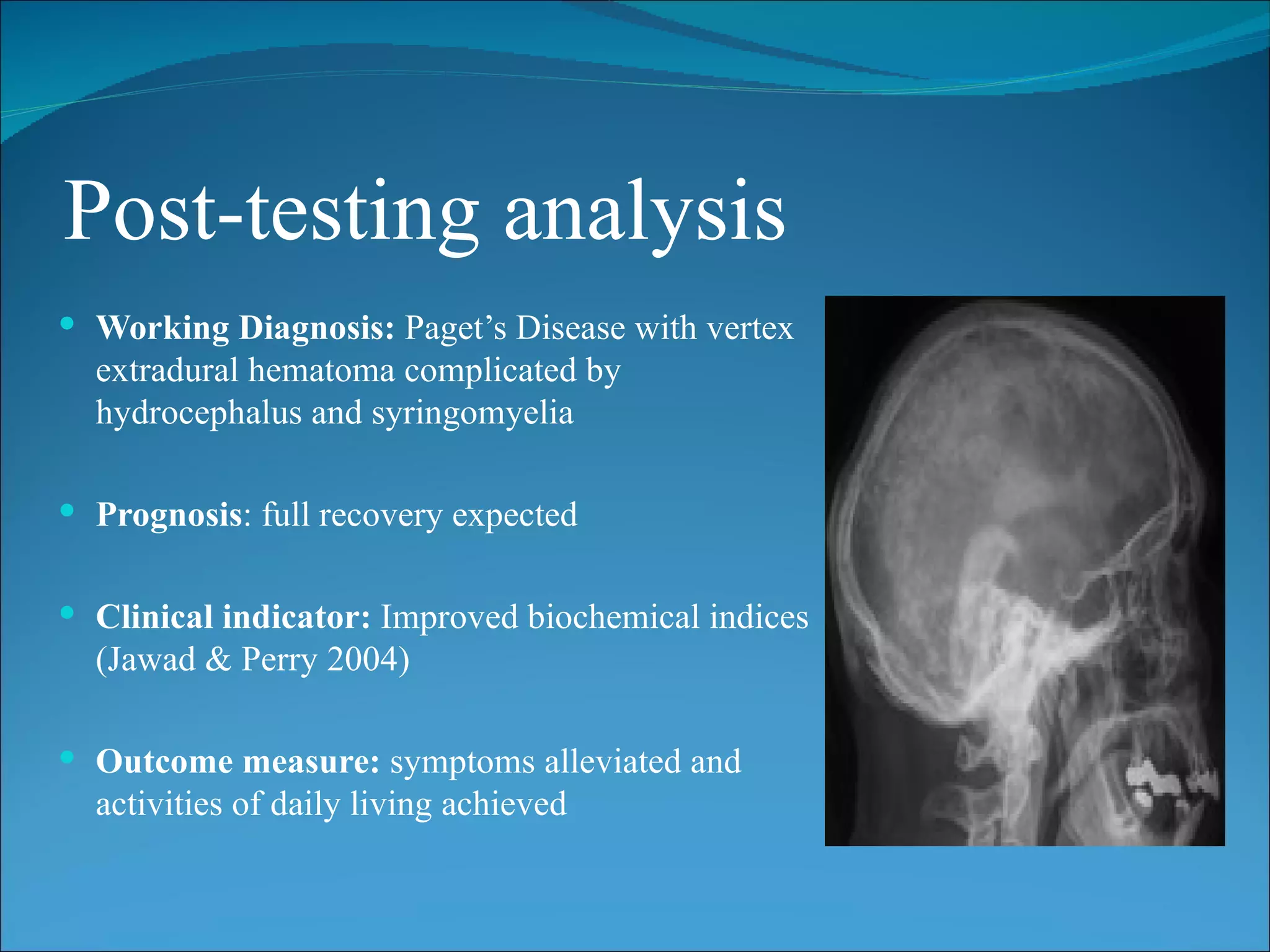
Case Study:
Paget’s Disease
Subjective Interview
                SH
                 Patient is a 48-year-old woman
                  with active lifestyle who lives
                  with family
                HPC
                 Fall from scooter occurred
                 Severe headache noted
                 Right upper limb weakness
                  noted
                 Transient loss of consciousness
                  during accident
                 Conscious and alert upon
                  admission
Subjective Interview
               PC
                With previous history of
               headaches
                With slow progressive hearing
               loss (Shukla 2010)

               Status: Stable after initial
               improvement

               Patient returned three years later
               with complaint of slow progressive
               numbness, weakness of left arm and
               difficulty walking
Planning

  Must: AROM of the shoulders/elbows and lower extremities
  Should: perform ADL’s with minimal support
  Could: slow progression of the disease and its complications
Objective Testing
Observation:
                                        Ct Scan:
 Mild bilateral sensorineural           Revealed moderately sized
    hearing loss                          extradural hematoma at the
   Aside from the noted                  vertex, towards left side
    progressive weakness of the          Skull vault showed widened
    left upper extremity and              diploic space suggestive of
    difficulties in walking, no other     Paget’s disease of the skull
    neurologic deficit noted
   Right upper limb – Grade 3
    strength
   Normal vital parameters
   No external scalp injury seen
   Normal optic fundus
Objective Testing
Skull Radiograph:
 Evidence of platybasia and
  basilar impression
 Widespread alteration of bone
  structure
 Elevated markers of bone
  turnover:
    alkaline phosphatase >1000
      units/l
    24h urinary hydroxyproline
      >2000μmol/day
Post-testing analysis
 Working Diagnosis: Paget’s Disease with vertex
  extradural hematoma complicated by
  hydrocephalus and syringomyelia

 Prognosis: full recovery expected


 Clinical indicator: Improved biochemical indices
  (Jawad & Perry 2004)

 Outcome measure: symptoms alleviated and
  activities of daily living achieved
Aims / Objectives of Treatment
Short Term
 Improve muscle weakness            Relieve present symptoms
    Encourage stretching               Educate on surgical and
      exercises/ROM                      pharmacologic options
    Encourage patient compliance       Encourage to verbalize fears or
    Promote normal movement             doubts on intervention to further
                                         promote communication
 Improve neurological conditions
    Encourage prophylactic            (Raubenheimer, Taylor & Soule
     treatment                           2002)
    Encourage patient compliance
Aims / Objectives of Treatment
Long Term
 Avoid increased or further complications
 Achieve remission
    Continued treatment if needed
    Encourage patient compliance


   (Roux & Dougados 1999)
Initial Management & Rationale
 Frontoparietal vertex                  Pharmacologic treatment
  craniotomy                                Clodronate (Ostac) IV and
    Evacuate extradural hematoma            Pamidronate ( Aredia) IV
    Excise diffusely oozing bone           Resolve bone pain
     flap that caused recollection of       Significantly improve
     hematoma and diffuse brain              biochemical indices
     swelling
    Improved right upper limb
                                           (Raubenheimer, Taylor & Soule
     weakness                                2002)

   (Ramesh, Deiveegan &
     Soundappan 2005)
Retrospective Analysis
 Outcome: full recovery
 Reflection
    Interventions done were appropriate. All expectations after the
     interventions were met. Complications that occurred were not expected
     but was dealt with properly. Continued observation and patient
     encouragement should be done to enhance continuity of care.
References
Jawad, A & Perry, J 2004, Paget’s Disease, Rheumatology, viewed 29 March
  2011, http://www.grandrounds-e-med.com/articles/gr049007.htm
Shukla, A 2010, Paget’s Disease – Complications of Paget’s Disease,
  Orthopedics, viewed 28 March 2011, http://orthopedics.ygoy.com/paget
  %E2%80%99s-disease-%E2%80%93-complications-of-paget
  %E2%80%99s-disease/
Paget Foundation 2010, Paget’s Disease of the Bone, viewed 29 March 2011,
  http://www.paget.org/index.php/healthcare-professionals/pagets-disease-of-
  bone.html
Ramesh, V, Deiveegan, K & Soundappen V 2005, vertex extradural hematoma
  in association with Paget’s disease of the skull, Neurology India, Vol, 53,
  No. 1, pp.115-116
Roux, C & Dougados, M 1999, Treatment of Patients with Paget’s Disease of
  the Bone, Drugs, Vol. 58, No. 5, pp.823-830
Sheil, W 2011, Paget’s Disease of Bone, Medicinenet.com, viewed 28 March
  2011, http://www.medicinenet.com/pagets_disease/article.htm

1

  • 1.
  • 2.
    Subjective Interview SH  Patient is a 48-year-old woman with active lifestyle who lives with family HPC  Fall from scooter occurred  Severe headache noted  Right upper limb weakness noted  Transient loss of consciousness during accident  Conscious and alert upon admission
  • 3.
    Subjective Interview PC  With previous history of headaches  With slow progressive hearing loss (Shukla 2010) Status: Stable after initial improvement Patient returned three years later with complaint of slow progressive numbness, weakness of left arm and difficulty walking
  • 4.
    Planning Must:AROM of the shoulders/elbows and lower extremities Should: perform ADL’s with minimal support Could: slow progression of the disease and its complications
  • 5.
    Objective Testing Observation: Ct Scan:  Mild bilateral sensorineural  Revealed moderately sized hearing loss extradural hematoma at the  Aside from the noted vertex, towards left side progressive weakness of the  Skull vault showed widened left upper extremity and diploic space suggestive of difficulties in walking, no other Paget’s disease of the skull neurologic deficit noted  Right upper limb – Grade 3 strength  Normal vital parameters  No external scalp injury seen  Normal optic fundus
  • 6.
    Objective Testing Skull Radiograph: Evidence of platybasia and basilar impression  Widespread alteration of bone structure  Elevated markers of bone turnover:  alkaline phosphatase >1000 units/l  24h urinary hydroxyproline >2000μmol/day
  • 7.
    Post-testing analysis  WorkingDiagnosis: Paget’s Disease with vertex extradural hematoma complicated by hydrocephalus and syringomyelia  Prognosis: full recovery expected  Clinical indicator: Improved biochemical indices (Jawad & Perry 2004)  Outcome measure: symptoms alleviated and activities of daily living achieved
  • 8.
    Aims / Objectivesof Treatment Short Term  Improve muscle weakness  Relieve present symptoms  Encourage stretching  Educate on surgical and exercises/ROM pharmacologic options  Encourage patient compliance  Encourage to verbalize fears or  Promote normal movement doubts on intervention to further promote communication  Improve neurological conditions  Encourage prophylactic (Raubenheimer, Taylor & Soule treatment 2002)  Encourage patient compliance
  • 9.
    Aims / Objectivesof Treatment Long Term  Avoid increased or further complications  Achieve remission  Continued treatment if needed  Encourage patient compliance (Roux & Dougados 1999)
  • 10.
    Initial Management &Rationale  Frontoparietal vertex  Pharmacologic treatment craniotomy  Clodronate (Ostac) IV and  Evacuate extradural hematoma Pamidronate ( Aredia) IV  Excise diffusely oozing bone  Resolve bone pain flap that caused recollection of  Significantly improve hematoma and diffuse brain biochemical indices swelling  Improved right upper limb (Raubenheimer, Taylor & Soule weakness 2002) (Ramesh, Deiveegan & Soundappan 2005)
  • 11.
    Retrospective Analysis  Outcome:full recovery  Reflection  Interventions done were appropriate. All expectations after the interventions were met. Complications that occurred were not expected but was dealt with properly. Continued observation and patient encouragement should be done to enhance continuity of care.
  • 12.
    References Jawad, A &Perry, J 2004, Paget’s Disease, Rheumatology, viewed 29 March 2011, http://www.grandrounds-e-med.com/articles/gr049007.htm Shukla, A 2010, Paget’s Disease – Complications of Paget’s Disease, Orthopedics, viewed 28 March 2011, http://orthopedics.ygoy.com/paget %E2%80%99s-disease-%E2%80%93-complications-of-paget %E2%80%99s-disease/ Paget Foundation 2010, Paget’s Disease of the Bone, viewed 29 March 2011, http://www.paget.org/index.php/healthcare-professionals/pagets-disease-of- bone.html Ramesh, V, Deiveegan, K & Soundappen V 2005, vertex extradural hematoma in association with Paget’s disease of the skull, Neurology India, Vol, 53, No. 1, pp.115-116 Roux, C & Dougados, M 1999, Treatment of Patients with Paget’s Disease of the Bone, Drugs, Vol. 58, No. 5, pp.823-830 Sheil, W 2011, Paget’s Disease of Bone, Medicinenet.com, viewed 28 March 2011, http://www.medicinenet.com/pagets_disease/article.htm

Editor's Notes

  • #3 The client is a 48-year old woman who has a very active lifestyle. She lives with her family, along with her husband and their two teenage daughters. The client’s history includes falling from her scooter on her way to the grocery store. After her fall, she verbalized her complain of severe headache. Also, upon assessment, it was observed that there was right upper lime weakness. Furthermore, there was a transient loss of consciousness occurring to the patient after the accident. However, upon admission to the health care facility, the client was deemed to be conscious, alert and coherent.
  • #4 During the interview, it was found that the client had a previous history of headache complaints. Alongside this complaint is the progressive hearing loss that the client also experienced. After the client’s initial improvement since the progression of the disease, the client’s condition was already stable. However, after three years, the client sought health care complaining of slow progressive numbness, weakness of the left arm and difficulty of walking.
  • #5 After the interview, it was deemed that the client must perform Active Range of Motion of the shoulders and the elbows and the lower extremities. Included in the plan for the patient is that the client perform Activities of Daily Living with little dependence to her significant others. Finally, the health care team could slow the progression of the disease and its complications through meticulous treatment interventions.
  • #6 Observations from the client included bilateral sensorineural hearing loss. Included in the observations is the progressive weakness of the upper extremities as evidenced by the Grade 3 out of 5 muscle strength of the right arm and perceived weakness of the left arm before the accident. In addition, abnormality was also experienced by the lower extremities, which was evidenced by the client’s difficulty in walking. There is normal optic fundus, as well as vital parameters. Aside from the complaint of severe headache, no external scalp injury was seen. The Computed Tomography Scan of the client shows evident moderately sized extradural hematoma at the vertex of the brain, toward the left hemisphere. The skull vault showed widened diploic space suggestive of Paget’s disease of the skull.
  • #7 The skull radiograph shows evident platybasia and basilar impression. There is also widespread alteration of bone structure which shows disease progression. Laboratory tests also show increased alkaline phosphatase and elevated levels of the 24-hour urinary hydroxyproline. Elevated serum alkaline phosphatase (Paget Foundation 2010) rules out the disease to being arthritis or other disorders. Thus, with elevated SAP, the disease can be considered to be Paget’s disease. Since there is disorganized bone remodeling, SAP is highly excreted. To further validate the SAP lab result, the urinary hydroxyproline test is done. As expected, the client’s result is also elevated which leads to the diagnosis of Paget’s Disease.
  • #8 Basing from the evident signs and symptoms, and the diagnostic work-ups done, the working diagnosis of the client is Paget’s disease. Defining symptoms include vertex epidural hematomas as complicated by hydrocephalus and syringomyelia. Paget’s disease (Roux & Dougados 1999) is a progressive disease of the musculoskeletal system which is characterized by abnormal enlargement of the affected bones and fast-paced disorganized bone alterations. The disease can complicate and lead to bone pain, deformities, and fractures; which in turn lead to muscular difficulties and irregularities. The prognosis for the disease is good if appropriate treatment is done at an early time (Shukla 2010). As mentioned earlier, the disease was diagnosed based from the evident symptoms and the validating changes in the biochemical levels of the patient. The main outcome hoped for the client is alleviated symptoms and tolerated activities of daily living.
  • #9 Since the client is currently experiencing muscle weakness, neurosurgical conditions, and skeletal complaints, the short term goals for the client is the relief of the current complaints. The client is encouraged to actively participate in range of motion exercises and stretching exercises to promote circulation and improve muscle and bone movement. In addition, a peaceful and low-stimulus environment is also encouraged to relieve the client of difficulties since she is experiencing progressive hearing loss. To improve neurosurgical conditions, the client and her significant others is offered the suggestion of undergoing prophylactic treatment to prevent Paget’s disease progression. In addition, the client is also encouraged to verbalize fears and anxieties regarding her condition. This would promote improved nurse-client interaction, which in turn, could further improve the client’s care.
  • #10 Aside from the short term needs of the client, long term goals are also considered and planned. This promotes the client to look forward to being cured in the future. Included in the long term goals for the client are avoidance of increased complications and avoidance of remissions. This goal can be achieved if complete client and health care team collaboration is done. Planning and implementing treatment interventions to prevent further complications are suggested.
  • #11 The client’s immediate management includes frontoparietal vertex craniotomy and introduction of pharmacologic treatment. The craniotomy is don to evacuate extradural hematoma and relieve the client of increased intracranial pressure resulting from hydrocephalus. Since the client has increase intracranial pressure (ICP) as evidenced by her severe headache, the initial intervention is to relieve the client of increased ICP. Relief of the hematoma provides improved left brain circulation, which in turn, can improve the right upper limb weakness of the client. Aside from the surgical intervention, pharmacologic interventions are encouraged to supplement the client’s care. Clondronate and Pamidroante is given intravenously to relieve the client of severe bone pain.
  • #12 After the intensive medical and nursing interventions, the client is hoped to have a full recovery from the accident.