The seminar discussed ankle injuries, focusing on anatomy, classification systems, and common injuries. The ankle is supported by strong ligaments and tendons and permits dorsiflexion and plantar flexion. Common injuries include ligament sprains and fractures of the medial and lateral malleoli. Injury patterns are classified using systems like Lauge-Hansen which consider the mechanism of force and resulting bone and soft tissue injuries. Proper treatment aims to restore normal ankle alignment and joint surfaces.
Tendoachilles rupture and its managementRohan Vakta
Achilles tendon is the strongest tendon of body. There are many causes of its rupture. It can be acute or chronic rupture. Management of chronic rupture by semitendinosus tendon is mentioned here.
Arthroscopic ACL Reconstruction By Dr Shekhar ShrivastavDelhiArthroscopy
Arthroscopic Acl Reconstruction By Dr Shekhar Shrivastav.
HOW NORMAL KNEE WORKS ?
The knee is the largest joint in the body, and one of the most easily injured. It is made up of the lower end of the thigh bone(femur), the upper end of the shin bone (tibia), and the knee cap (patella), which slides in a groove on the end of the femur. Four bands of tissue, the anterior and posterior cruciate ligaments, and the medial and lateral collateral ligaments connect the femur and the tibia and provide joint stability. The surfaces where the femur, tibia and patella touch are covered with articular cartilage, a smooth substance that cushions the bones and enables them to glide freely. Semicircular rings of tough fibrous-cartilage tissue called the lateral and medial menisci act as shock absorbers and stabilizers.
WHAT IS THE ROLE OF ACL ?
ACL along with other ligaments of the knee joint and meniscus provides stability to the knee joint.
WHAT IS LIGAMENT RECONSTRUCTION ( ACL ) ?
Ligament reconstruction involves replacing the torn ligament with a tendon (graft) from your knee and fixing the graft in place with screws. This procedure is performed with the use of the arthroscope. The anterior cruciate ligament (ACL) is the most common ligament requiring reconstruction procedures. The torn ligament is excised arthroscopically and new ligament is prepared by ligament grafts taken from your own body. Bony tunnels are prepared in femur and tibia using specialized instruments through which the new ligament is passed and fixed with special screws. This procedure requires relative rest or leave from your work or studies for about 2-3 weeks after which you will be allowed normal day to day activities.
WHEN CAN THE PATIENT BE AMBULATED AFTER SURGERY ?
The patient can walk from the same evening of the surgery. Initially the patient is advised to walk with a brace and a walking cane. Strengthening and range of motion exercises for the knee are started from the next day. The patient is discharged from the hospital 2nd or 3rd day after surgery. The patient can walk without support by 10-14 days depending on muscle strengthening. Slow Jogging and other strenuous activities are permitted after 3 months and the patient can return to active sports only 8-9 months after surgery.
Torn ACL Reconstructed ACL
For Further Queries contact your Orthopedic Surgeon at
+ 91 9971192233
The Ilizarov apparatus is a type of external fixation used in orthopedic surgery to lengthen or reshape limb bones; as a limb-sparing technique to treat complex and/or open bone fractures; and in cases of infected nonunions of bones that are not amenable with other techniques. It is named after the orthopedic surgeon Gavriil Abramovich Ilizarov from the Soviet Union, who pioneered the technique.
The Ankle Joint.pptx Dr Haki Selaj Residency in Orthopedic and Traumatology i...HakiSelaj1
it is one of the joints most often attacked by injury, in this case it is distorted. for this reason, accurate evaluation and diagnosis is required. for this reason, this presentation will help young doctors for access, exam tests and radiology around the TC joint
knee joint
Functionally, the knee joint is a condylar & modified hinge joint.
Transverse axis of movement is not fixed, & moves forward during extension & translates backward in flexion;
Along with extension & flexion, there is a conjunct rotation of femur on tibia(or vice versa) around a more or less vertical axis.
1. Capsular ligament
2. Synovial membrane
3. Ligamentum patellae
4. Tibial collateral ligament
5. Fibular collateral ligament
6. Oblique popliteal ligament
Arcuate popliteal ligament
Medial & lateral menisci
TIBIAL COLLATERAL LIGAMENT
The ligament consist of superficial & deep part . Both part are attached above to the medial epicondyle of femur. The superficial part extends downward & forward as a flattened band & is attached to the medial condyle & upper part of medial border of shaft of tibia along a rough strip of bone.
Ligaments of ankle joint (Ankle complex)Ajith lolita
this will be more informative for you.The collateral ligaments are fully explained in this PPT and it gives clear & prospect information about ankle complex.
Total hip replacement,ARTHROPLASTY OF THE HIP: APPLIED BIOMECHANICS, DESIGN AND SELECTION OF TOTAL HIP COMPONENTS, ALTERNATE BARRINGS INDICATIONS, CONTRAINDICATIONS OF THR & TEMPLETING AND PRE-OP EVALUATION.
paediatric injuries around the elbow
supracondylar elbow injuries
pulled elbow in paediatric age r
radiological signs around elbow in supracondylar fracture humerus
TEST BANK for Operations Management, 14th Edition by William J. Stevenson, Ve...kevinkariuki227
TEST BANK for Operations Management, 14th Edition by William J. Stevenson, Verified Chapters 1 - 19, Complete Newest Version.pdf
TEST BANK for Operations Management, 14th Edition by William J. Stevenson, Verified Chapters 1 - 19, Complete Newest Version.pdf
New Drug Discovery and Development .....NEHA GUPTA
The "New Drug Discovery and Development" process involves the identification, design, testing, and manufacturing of novel pharmaceutical compounds with the aim of introducing new and improved treatments for various medical conditions. This comprehensive endeavor encompasses various stages, including target identification, preclinical studies, clinical trials, regulatory approval, and post-market surveillance. It involves multidisciplinary collaboration among scientists, researchers, clinicians, regulatory experts, and pharmaceutical companies to bring innovative therapies to market and address unmet medical needs.
Flu Vaccine Alert in Bangalore Karnatakaaddon Scans
As flu season approaches, health officials in Bangalore, Karnataka, are urging residents to get their flu vaccinations. The seasonal flu, while common, can lead to severe health complications, particularly for vulnerable populations such as young children, the elderly, and those with underlying health conditions.
Dr. Vidisha Kumari, a leading epidemiologist in Bangalore, emphasizes the importance of getting vaccinated. "The flu vaccine is our best defense against the influenza virus. It not only protects individuals but also helps prevent the spread of the virus in our communities," he says.
This year, the flu season is expected to coincide with a potential increase in other respiratory illnesses. The Karnataka Health Department has launched an awareness campaign highlighting the significance of flu vaccinations. They have set up multiple vaccination centers across Bangalore, making it convenient for residents to receive their shots.
To encourage widespread vaccination, the government is also collaborating with local schools, workplaces, and community centers to facilitate vaccination drives. Special attention is being given to ensuring that the vaccine is accessible to all, including marginalized communities who may have limited access to healthcare.
Residents are reminded that the flu vaccine is safe and effective. Common side effects are mild and may include soreness at the injection site, mild fever, or muscle aches. These side effects are generally short-lived and far less severe than the flu itself.
Healthcare providers are also stressing the importance of continuing COVID-19 precautions. Wearing masks, practicing good hand hygiene, and maintaining social distancing are still crucial, especially in crowded places.
Protect yourself and your loved ones by getting vaccinated. Together, we can help keep Bangalore healthy and safe this flu season. For more information on vaccination centers and schedules, residents can visit the Karnataka Health Department’s official website or follow their social media pages.
Stay informed, stay safe, and get your flu shot today!
Ozempic: Preoperative Management of Patients on GLP-1 Receptor Agonists Saeid Safari
Preoperative Management of Patients on GLP-1 Receptor Agonists like Ozempic and Semiglutide
ASA GUIDELINE
NYSORA Guideline
2 Case Reports of Gastric Ultrasound
These simplified slides by Dr. Sidra Arshad present an overview of the non-respiratory functions of the respiratory tract.
Learning objectives:
1. Enlist the non-respiratory functions of the respiratory tract
2. Briefly explain how these functions are carried out
3. Discuss the significance of dead space
4. Differentiate between minute ventilation and alveolar ventilation
5. Describe the cough and sneeze reflexes
Study Resources:
1. Chapter 39, Guyton and Hall Textbook of Medical Physiology, 14th edition
2. Chapter 34, Ganong’s Review of Medical Physiology, 26th edition
3. Chapter 17, Human Physiology by Lauralee Sherwood, 9th edition
4. Non-respiratory functions of the lungs https://academic.oup.com/bjaed/article/13/3/98/278874
Title: Sense of Smell
Presenter: Dr. Faiza, Assistant Professor of Physiology
Qualifications:
MBBS (Best Graduate, AIMC Lahore)
FCPS Physiology
ICMT, CHPE, DHPE (STMU)
MPH (GC University, Faisalabad)
MBA (Virtual University of Pakistan)
Learning Objectives:
Describe the primary categories of smells and the concept of odor blindness.
Explain the structure and location of the olfactory membrane and mucosa, including the types and roles of cells involved in olfaction.
Describe the pathway and mechanisms of olfactory signal transmission from the olfactory receptors to the brain.
Illustrate the biochemical cascade triggered by odorant binding to olfactory receptors, including the role of G-proteins and second messengers in generating an action potential.
Identify different types of olfactory disorders such as anosmia, hyposmia, hyperosmia, and dysosmia, including their potential causes.
Key Topics:
Olfactory Genes:
3% of the human genome accounts for olfactory genes.
400 genes for odorant receptors.
Olfactory Membrane:
Located in the superior part of the nasal cavity.
Medially: Folds downward along the superior septum.
Laterally: Folds over the superior turbinate and upper surface of the middle turbinate.
Total surface area: 5-10 square centimeters.
Olfactory Mucosa:
Olfactory Cells: Bipolar nerve cells derived from the CNS (100 million), with 4-25 olfactory cilia per cell.
Sustentacular Cells: Produce mucus and maintain ionic and molecular environment.
Basal Cells: Replace worn-out olfactory cells with an average lifespan of 1-2 months.
Bowman’s Gland: Secretes mucus.
Stimulation of Olfactory Cells:
Odorant dissolves in mucus and attaches to receptors on olfactory cilia.
Involves a cascade effect through G-proteins and second messengers, leading to depolarization and action potential generation in the olfactory nerve.
Quality of a Good Odorant:
Small (3-20 Carbon atoms), volatile, water-soluble, and lipid-soluble.
Facilitated by odorant-binding proteins in mucus.
Membrane Potential and Action Potential:
Resting membrane potential: -55mV.
Action potential frequency in the olfactory nerve increases with odorant strength.
Adaptation Towards the Sense of Smell:
Rapid adaptation within the first second, with further slow adaptation.
Psychological adaptation greater than receptor adaptation, involving feedback inhibition from the central nervous system.
Primary Sensations of Smell:
Camphoraceous, Musky, Floral, Pepperminty, Ethereal, Pungent, Putrid.
Odor Detection Threshold:
Examples: Hydrogen sulfide (0.0005 ppm), Methyl-mercaptan (0.002 ppm).
Some toxic substances are odorless at lethal concentrations.
Characteristics of Smell:
Odor blindness for single substances due to lack of appropriate receptor protein.
Behavioral and emotional influences of smell.
Transmission of Olfactory Signals:
From olfactory cells to glomeruli in the olfactory bulb, involving lateral inhibition.
Primitive, less old, and new olfactory systems with different path
New Directions in Targeted Therapeutic Approaches for Older Adults With Mantl...i3 Health
i3 Health is pleased to make the speaker slides from this activity available for use as a non-accredited self-study or teaching resource.
This slide deck presented by Dr. Kami Maddocks, Professor-Clinical in the Division of Hematology and
Associate Division Director for Ambulatory Operations
The Ohio State University Comprehensive Cancer Center, will provide insight into new directions in targeted therapeutic approaches for older adults with mantle cell lymphoma.
STATEMENT OF NEED
Mantle cell lymphoma (MCL) is a rare, aggressive B-cell non-Hodgkin lymphoma (NHL) accounting for 5% to 7% of all lymphomas. Its prognosis ranges from indolent disease that does not require treatment for years to very aggressive disease, which is associated with poor survival (Silkenstedt et al, 2021). Typically, MCL is diagnosed at advanced stage and in older patients who cannot tolerate intensive therapy (NCCN, 2022). Although recent advances have slightly increased remission rates, recurrence and relapse remain very common, leading to a median overall survival between 3 and 6 years (LLS, 2021). Though there are several effective options, progress is still needed towards establishing an accepted frontline approach for MCL (Castellino et al, 2022). Treatment selection and management of MCL are complicated by the heterogeneity of prognosis, advanced age and comorbidities of patients, and lack of an established standard approach for treatment, making it vital that clinicians be familiar with the latest research and advances in this area. In this activity chaired by Michael Wang, MD, Professor in the Department of Lymphoma & Myeloma at MD Anderson Cancer Center, expert faculty will discuss prognostic factors informing treatment, the promising results of recent trials in new therapeutic approaches, and the implications of treatment resistance in therapeutic selection for MCL.
Target Audience
Hematology/oncology fellows, attending faculty, and other health care professionals involved in the treatment of patients with mantle cell lymphoma (MCL).
Learning Objectives
1.) Identify clinical and biological prognostic factors that can guide treatment decision making for older adults with MCL
2.) Evaluate emerging data on targeted therapeutic approaches for treatment-naive and relapsed/refractory MCL and their applicability to older adults
3.) Assess mechanisms of resistance to targeted therapies for MCL and their implications for treatment selection
Recomendações da OMS sobre cuidados maternos e neonatais para uma experiência pós-natal positiva.
Em consonância com os ODS – Objetivos do Desenvolvimento Sustentável e a Estratégia Global para a Saúde das Mulheres, Crianças e Adolescentes, e aplicando uma abordagem baseada nos direitos humanos, os esforços de cuidados pós-natais devem expandir-se para além da cobertura e da simples sobrevivência, de modo a incluir cuidados de qualidade.
Estas diretrizes visam melhorar a qualidade dos cuidados pós-natais essenciais e de rotina prestados às mulheres e aos recém-nascidos, com o objetivo final de melhorar a saúde e o bem-estar materno e neonatal.
Uma “experiência pós-natal positiva” é um resultado importante para todas as mulheres que dão à luz e para os seus recém-nascidos, estabelecendo as bases para a melhoria da saúde e do bem-estar a curto e longo prazo. Uma experiência pós-natal positiva é definida como aquela em que as mulheres, pessoas que gestam, os recém-nascidos, os casais, os pais, os cuidadores e as famílias recebem informação consistente, garantia e apoio de profissionais de saúde motivados; e onde um sistema de saúde flexível e com recursos reconheça as necessidades das mulheres e dos bebês e respeite o seu contexto cultural.
Estas diretrizes consolidadas apresentam algumas recomendações novas e já bem fundamentadas sobre cuidados pós-natais de rotina para mulheres e neonatos que recebem cuidados no pós-parto em unidades de saúde ou na comunidade, independentemente dos recursos disponíveis.
É fornecido um conjunto abrangente de recomendações para cuidados durante o período puerperal, com ênfase nos cuidados essenciais que todas as mulheres e recém-nascidos devem receber, e com a devida atenção à qualidade dos cuidados; isto é, a entrega e a experiência do cuidado recebido. Estas diretrizes atualizam e ampliam as recomendações da OMS de 2014 sobre cuidados pós-natais da mãe e do recém-nascido e complementam as atuais diretrizes da OMS sobre a gestão de complicações pós-natais.
O estabelecimento da amamentação e o manejo das principais intercorrências é contemplada.
Recomendamos muito.
Vamos discutir essas recomendações no nosso curso de pós-graduação em Aleitamento no Instituto Ciclos.
Esta publicação só está disponível em inglês até o momento.
Prof. Marcus Renato de Carvalho
www.agostodourado.com
These lecture slides, by Dr Sidra Arshad, offer a quick overview of physiological basis of a normal electrocardiogram.
Learning objectives:
1. Define an electrocardiogram (ECG) and electrocardiography
2. Describe how dipoles generated by the heart produce the waveforms of the ECG
3. Describe the components of a normal electrocardiogram of a typical bipolar leads (limb II)
4. Differentiate between intervals and segments
5. Enlist some common indications for obtaining an ECG
Study Resources:
1. Chapter 11, Guyton and Hall Textbook of Medical Physiology, 14th edition
2. Chapter 9, Human Physiology - From Cells to Systems, Lauralee Sherwood, 9th edition
3. Chapter 29, Ganong’s Review of Medical Physiology, 26th edition
4. Electrocardiogram, StatPearls - https://www.ncbi.nlm.nih.gov/books/NBK549803/
5. ECG in Medical Practice by ABM Abdullah, 4th edition
6. ECG Basics, http://www.nataliescasebook.com/tag/e-c-g-basics
Lung Cancer: Artificial Intelligence, Synergetics, Complex System Analysis, S...Oleg Kshivets
RESULTS: Overall life span (LS) was 2252.1±1742.5 days and cumulative 5-year survival (5YS) reached 73.2%, 10 years – 64.8%, 20 years – 42.5%. 513 LCP lived more than 5 years (LS=3124.6±1525.6 days), 148 LCP – more than 10 years (LS=5054.4±1504.1 days).199 LCP died because of LC (LS=562.7±374.5 days). 5YS of LCP after bi/lobectomies was significantly superior in comparison with LCP after pneumonectomies (78.1% vs.63.7%, P=0.00001 by log-rank test). AT significantly improved 5YS (66.3% vs. 34.8%) (P=0.00000 by log-rank test) only for LCP with N1-2. Cox modeling displayed that 5YS of LCP significantly depended on: phase transition (PT) early-invasive LC in terms of synergetics, PT N0—N12, cell ratio factors (ratio between cancer cells- CC and blood cells subpopulations), G1-3, histology, glucose, AT, blood cell circuit, prothrombin index, heparin tolerance, recalcification time (P=0.000-0.038). Neural networks, genetic algorithm selection and bootstrap simulation revealed relationships between 5YS and PT early-invasive LC (rank=1), PT N0—N12 (rank=2), thrombocytes/CC (3), erythrocytes/CC (4), eosinophils/CC (5), healthy cells/CC (6), lymphocytes/CC (7), segmented neutrophils/CC (8), stick neutrophils/CC (9), monocytes/CC (10); leucocytes/CC (11). Correct prediction of 5YS was 100% by neural networks computing (area under ROC curve=1.0; error=0.0).
CONCLUSIONS: 5YS of LCP after radical procedures significantly depended on: 1) PT early-invasive cancer; 2) PT N0--N12; 3) cell ratio factors; 4) blood cell circuit; 5) biochemical factors; 6) hemostasis system; 7) AT; 8) LC characteristics; 9) LC cell dynamics; 10) surgery type: lobectomy/pneumonectomy; 11) anthropometric data. Optimal diagnosis and treatment strategies for LC are: 1) screening and early detection of LC; 2) availability of experienced thoracic surgeons because of complexity of radical procedures; 3) aggressive en block surgery and adequate lymph node dissection for completeness; 4) precise prediction; 5) adjuvant chemoimmunoradiotherapy for LCP with unfavorable prognosis.
HOT NEW PRODUCT! BIG SALES FAST SHIPPING NOW FROM CHINA!! EU KU DB BK substit...GL Anaacs
Contact us if you are interested:
Email / Skype : kefaya1771@gmail.com
Threema: PXHY5PDH
New BATCH Ku !!! MUCH IN DEMAND FAST SALE EVERY BATCH HAPPY GOOD EFFECT BIG BATCH !
Contact me on Threema or skype to start big business!!
Hot-sale products:
NEW HOT EUTYLONE WHITE CRYSTAL!!
5cl-adba precursor (semi finished )
5cl-adba raw materials
ADBB precursor (semi finished )
ADBB raw materials
APVP powder
5fadb/4f-adb
Jwh018 / Jwh210
Eutylone crystal
Protonitazene (hydrochloride) CAS: 119276-01-6
Flubrotizolam CAS: 57801-95-3
Metonitazene CAS: 14680-51-4
Payment terms: Western Union,MoneyGram,Bitcoin or USDT.
Deliver Time: Usually 7-15days
Shipping method: FedEx, TNT, DHL,UPS etc.Our deliveries are 100% safe, fast, reliable and discreet.
Samples will be sent for your evaluation!If you are interested in, please contact me, let's talk details.
We specializes in exporting high quality Research chemical, medical intermediate, Pharmaceutical chemicals and so on. Products are exported to USA, Canada, France, Korea, Japan,Russia, Southeast Asia and other countries.
HOT NEW PRODUCT! BIG SALES FAST SHIPPING NOW FROM CHINA!! EU KU DB BK substit...
04 ankle fractures ppt
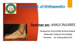
1. .
Department of Orthopaedics
Seminar on: ANKLE INJURIES.
Chairperson: Prof. & HOD: Dr.Kiran Kalaiah
Moderator: Professor Dr.J K Reddy
Presenter : Dr. Yashavardhan.T.M
2. Introduction:
Ankle injuries apart from road traffic accidents can also result from a slip while
walking or getting down from stairs or a twisting injury in sports and fall from a
height.
All these tend to produce ankle injuries when one attempts to turn violently over
a fixed foot or the foot being used as lever to produce twist at the ankle .
Great majority of ankle injuries are caused by indirect violence.
If not treated properly the ankle injuries are a source of disability in the form of
pain, instability and early degenerative arthritis of the ankle.
Injuries around the ankle joint cause destruction of not only the bony
architecture but also often the ligamentous and soft tissue components.
3. Aims:
(1) The normal relationships of the ankle mortise must be restored
(2) The weight-bearing alignment of the ankle must be at a right angle
to the longitudinal axis of the leg.
(3) The contours of the articular surface must be satisfactorily reduced.
The best results are obtained by anatomic joint restoration, and the
method used to accomplish this may be either closed manipulation or
open reduction and internal fixation (ORIF). For most fractures, the
latter method most often ensures anatomic joint restoration and union.
4. Anatomy:
Articular surfaces ?
1. Structurally, the joint is very strong. The stability of the joint is ensured by:
2. (i) Close interlocking of the articular surfaces;
3. (ii) strong collateral ligaments on the sides; and
4. (iii) the tendons that cross the joint, four in front,
and five behind
5. Ligaments
The joint is supported by:
(i) Fibrous capsule,
(ii) the deltoid or medial ligament, and
(iii) a lateral ligament.
6. Fibrous Capsule
It surrounds the joint and is attached all around the articular margins with
exceptions.
(1) Poster-superiorly, it is attached to the inferior transverse tibiofibular
ligament; and
2) anteroinferiorly, it is attached to the dorsum of the neck of the talus at
some distance from the trochlear surface.
7. Deltoid or Medial Ligament
This is a very strong triangular ligament present on the medial side of the ankle.
superficial layer
tibiocalcaneal ligament
tibionavicular ligament
posterior superficial tibiotalar ligament
tibiospring ligament
deep layer: this layer is intra-articular and is covered by synovium
anterior tibiotalar ligament (ATTL)
posterior deep tibiotalar ligament (PDTL)
8. Deltoid or Medial Ligament
This is a very strong triangular ligament present on the medial side of the ankle.
superficial layer
tibiocalcaneal ligament
tibionavicular ligament
posterior superficial tibiotalar ligament
tibiospring ligament
deep layer: this layer is intra-articular and is covered by synovium
anterior tibiotalar ligament (ATTL)
posterior deep tibiotalar ligament (PDTL)
9. Deltoid or Medial Ligament
This is a very strong triangular ligament present on the medial side of the ankle.
superficial layer
tibiocalcaneal ligament
tibionavicular ligament
posterior superficial tibiotalar ligament
tibiospring ligament
deep layer: this layer is intra-articular and is covered by synovium
anterior tibiotalar ligament (ATTL)
posterior deep tibiotalar ligament (PDTL)
10. Lateral Ligament
This ligament consists of three bands as follows.
1. The anterior talofibular ligament is a flat band which passes from the anterior margin of
the lateral malleolus to the neck of the talus, just in front of the fibular facet.
2. The posterior talofibular ligament passes from the lower part of the malleolar fossa of
the fibula to the lateral tubercle of the talus.
3. The calcaneofibular ligament is a long rounded cord which passes from the notch on
lower border of the lateral malleolus to the tubercle on the lateral surface of the
calcaneum.
11. Lateral Ligament
This ligament consists of three bands as follows.
1. The anterior talofibular ligament is a flat band which passes from the anterior margin of the
malleolus to the neck of the talus, just in front of the fibular facet.
2. The posterior talofibular ligament passes from the lower part of the malleolar fossa of the fibula
the lateral tubercle of the talus.
3. The calcaneofibular ligament is a long rounded cord which passes from the notch on the lower
border of the lateral malleolus to the tubercle on the lateral surface of the calcaneum.
12. Movements
movements are dorsiflexion and plantar-flexion
(a) dorsiflexion
(b) plantar flexion
Muscles producing movements
Movement Principal muscles Accessory muscles
A. Dorsiflexion Tibialis anterior 1. Extensor digitorum anterior longus
2. Extensor hallucis longus
3. Peroneus tertius
B. Plantar 1. Gastrocnemius 1. Plantaris flexion
2. Soleus 2. Tibialis posterior
3. Flexor hallucis longus
4. Flexor digitorum longus
13. BLOOD SUPPLY
The arteries crossing the ankle sends branches which form arterial anastomosis
around the ankle, tarsus and metatarsus
14. BLOOD SUPPLY
The arteries crossing the ankle sends branches which form arterial anastomosis
around the ankle, tarsus and metatarsus
15. BLOOD SUPPLY
The arteries crossing the ankle sends branches which form arterial anastomosis
around the ankle, tarsus and metatarsus
16. NERVE SUPPLY
The talocrural joint is innervated by branches from the deep peroneal,
sural and tibial nerves (or medial and lateral plantar nerves, depending on the
level of division of the tibial nerve). Occasionally, the superficial peroneal nerve
also supplies the ankle joint.
20. Ligament Injuries
The common injuries are the lateral ligament injury, the medial or the deltoid ligament injury and
the syndesmotic ligament injury (the ligament binding the inferior tibio-fibular syndesmosis).
• Grade 1—Stretching of the ligaments.
• Grade 2—Partial tear of the ligaments.
• Grade 3—Complete tear of the ligaments.
Management
Complete and severe tears require surgical repair. Less severe strains and sprains heal well with
adequate immobilization. Physiotherapy and gradual mobilization is always essential for a good
recovery.
25. Bowsworth Fracture
The distal end of the proximal fragment of the fibula may be displaced posterior to the
tibia and locked by the tibia's posterolateral ridge; the bone cannot be released
by manipulation because of the pull of the intact interosseous membrane
26. Classification.
1. Lauge-Hansen Classification*
1. Supination-Adduction (SA)
Transverse avulsion-type fracture of the fibula below the level of the joint or tear of the lateral collateral
ligaments Vertical fracture of the medial malleolus
2. Supination-Eversion (External) Rotation (SER)
Disruption of the anterior tibiofibular ligament Spiral oblique fracture of the distal fibula. Disruption of the
posterior tibiofibular ligament or fracture of the posterior malleolus. Fracture of the medial malleolus or
rupture of the deltoid ligament
27. Classification.
1. Lauge-Hansen Classification*
1. Supination-Adduction (SA)
Transverse avulsion-type fracture of the fibula below the level of the joint or tear of the lateral collateral
ligaments Vertical fracture of the medial malleolus
2. Supination-Eversion (External) Rotation (SER)
Disruption of the anterior tibiofibular ligament Spiral oblique fracture of the distal fibula. Disruption of the
posterior tibiofibular ligament or fracture of the posterior malleolus. Fracture of the medial malleolus or
rupture of the deltoid ligament
28. 3. Pronation-Abduction (PA)
Transverse fracture of the medial malleolus or rupture of the deltoid ligament. Rupture of the
syndesmotic ligaments or avulsion fracture of their insertions Short, horizontal, oblique fracture of
the fibula above the level of the joint
4. Pronation-Eversion (External) Rotation (PER)
Transverse fracture of the medial malleolus or disruption of the deltoid ligament. Disruption of the
anterior tibiofibular ligament Short oblique fracture of the fibula above the level of the joint.
Rupture of posterior tibiofibular ligament or avulsion fracture of the posterolateral tibia
29. 3. Pronation-Abduction (PA)
Transverse fracture of the medial malleolus or rupture of the deltoid ligament. Rupture of the
syndesmotic ligaments or avulsion fracture of their insertions Short, horizontal, oblique fracture of
the fibula above the level of the joint
4. Pronation-Eversion (External) Rotation (PER)
Transverse fracture of the medial malleolus or disruption of the deltoid ligament. Disruption of the
anterior tibiofibular ligament Short oblique fracture of the fibula above the level of the joint.
Rupture of posterior tibiofibular ligament or avulsion fracture of the posterolateral tibia
30. 5. Pronation-Dorsiflexion (PD)
Fracture of the medial malleolus. Fracture of the anterior margin of the tibia.
Supramalleolar fracture of the fibula. Transverse fracture of the posterior tibial
surface.
31. 5. Pronation-Dorsiflexion (PD)
Fracture of the medial malleolus. Fracture of the anterior margin of the tibia.
Supramalleolar fracture of the fibula. Transverse fracture of the posterior tibial
surface.
34. 3. AO CLASSIFICATION:
AO Classification of fractures has further divided each of these three types into
three groups - to quantify the spectrum of injury within each type.
35. Ruedi-Allgower Classification
Type 1: No significant articular incongruity;
cleavage fractures without displacement of
bony fragments.
Type 2: Significant articular incongruity with
minimal impaction or comminution.
Type 3: Significant articular comminution
with metaphysical Impaction
44. Ottawa Rules: When to Image
Ottawa Ankle Rules: 98% sensitivity for fracture, decrease
radiographs
Validated in ED and PCP Office
Do not apply rules if:
Age < 18 yo
Pregnancy
Multiple painful
injuries
Compromised
sensation
53. MANAGEMENT
Definitive
Aim- restoration of complete normal anatomical alignment of ankle.
Patients if needs operation should be operated within 24hrs of injury or after one
week once the swelling subsides.
Undisplaced fracture medial malleolus :
Below knee POP cast for 6 weeks.
Reduction fails (may be due to soft tissue (periosteal) inter position)
54. Displaced:
Open reduction and internal fixation by
Cancellous screws group
Tension band wiring
Fracture lateral malleolus:
Lateral Malleolus helps in length maintenance & maintenance of ankle mortice.
Hence, lateral malleolus has to be fixed internally
57. GATELLIER AND CHASTANG for LATERAL MALLEOLUS
■ Begin the incision about 12 cm proximal to the tip of the lateral malleolus
and extend it distally along the posterior margin of the fibula to the tip of
the malleolus. Curve the incision anteriorly for 2.5 to 4 cm in the line of the
peroneal tendons ■ Expose the fibula, including the lateral malleolus
subperiosteally, and incise the sheaths of the peroneal retinacula and
tendons, permitting the tendons to be displaced anteriorly.
■ if the fibula is not fractured, divide it 10 cm proximal to the tip of the
lateral malleolus and free the distal fragment by dividing the interosseous
membrane and the anterior and posterior tibiofibular ligaments.
58. GATELLIER AND CHASTANG for LATERAL MALLEOLUS
■ Begin the incision about 12 cm proximal to the tip of the lateral malleolus
and extend it distally along the posterior margin of the fibula to the tip of
the malleolus. Curve the incision anteriorly for 2.5 to 4 cm in the line of the
peroneal tendons ■ Expose the fibula, including the lateral malleolus
subperiosteally, and incise the sheaths of the peroneal retinacula and
tendons, permitting the tendons to be displaced anteriorly.
■ if the fibula is not fractured, divide it 10 cm proximal to the tip of the
lateral malleolus and free the distal fragment by dividing the interosseous
membrane and the anterior and posterior tibiofibular ligaments.
59. ■ carefully preserve the calcaneofibular and talofibular ligaments to serve
as a hinge and to maintain the integrity of the ankle after operation. Turn
the fibula laterally on this hinge and expose the lateral and posterior
aspects of the distal tibia and the lateral aspect of the ankle joint. Great
care should be used in children to avoid creating a fracture through the
distal fibular physis when reflecting the fibula.
■ When closing the incision, replace the fibula and secure it with a screw
extending transversely from the proximal part of the lateral malleolus
through the tibiofibular syndesmosis into the tibia just proximal and
parallel to the ankle joint.
60. ■ carefully preserve the calcaneofibular and talofibular ligaments to serve
as a hinge and to maintain the integrity of the ankle after operation. Turn
the fibula laterally on this hinge and expose the lateral and posterior
aspects of the distal tibia and the lateral aspect of the ankle joint. Great
care should be used in children to avoid creating a fracture through the
distal fibular physis when reflecting the fibula.
■ When closing the incision, replace the fibula and secure it with a screw
extending transversely from the proximal part of the lateral malleolus
through the tibiofibular syndesmosis into the tibia just proximal and
parallel to the ankle joint.
61. ■ Overdrill the hole made in the fibula to allow for compression across the
syndesmosis. Dorsiflex the ankle joint as the screw is tightened because the
talar dome is wider at its anterior half than its posterior half. Failure to overdrill
the fibula can result in widening of the syndesmosis and ankle mortise, with
resulting arthritic degeneration of the
tibiotalar joint. Add additional fixation with a small plate and screws if desired.
■ Replace the tendons, repair the tendon sheaths and retinacula, and close the
incision.
After the osteotomy or fracture has healed, remove the screw to prevent its
becoming loose or breaking
63. KOENIG AND SCHAEFER
■ Curve the incision just proximal to the medial malleolus and divide the malleolus with
an osteotome or small power saw; preserve the attachment of the deltoid ligament.
■ Subluxate the talus and malleolus laterally to reach the joint surfaces.
■ Later replace the malleolus and fix it with one or two cancellous screws. To make
replacement easier, drill the holes for the screws before the osteotomy, insert the screw,
and then remove it. At the end of the operation, reinsert the screws and close the
wound.
■ The surfaces of the osteotomized bone are smooth, and the malleolus can rotate on a
single screw. Two screws are used to prevent rotation of the osteotomized medial
malleolus Interfragmentary technique should be used for screw fixation of the medial
malleolus to provide compression across the osteotomy site
64. COLONNA AND RALSTON
■ Begin the incision at a point about 10 cm proximal and 2.5 cm posterior to the
medial malleolus and curve it anteriorly and inferiorly across the center of the
medial malleolus and inferiorly and posteriorly 4 cm toward the heel (Fig. 1-38C).
■ Expose the medial malleolus by reflecting the periosteum, but preserve the
deltoid ligament.
■ Divide the flexor retinaculum and retract the flexor halluces longus tendon and
the neurovascular bundle posteriorly and laterally.
■ Retract the tibial posterior and flexor digitorum longus tendons medially and
anteriorly to expose the posterior tibial fracture.
65. COLONNA AND RALSTON
■ Begin the incision at a point about 10 cm proximal and 2.5 cm posterior to the
medial malleolus and curve it anteriorly and inferiorly across the center of the
medial malleolus and inferiorly and posteriorly 4 cm toward the heel (Fig. 1-38C).
■ Expose the medial malleolus by reflecting the periosteum, but preserve the
deltoid ligament.
■ Divide the flexor retinaculum and retract the flexor halluces longus tendon and
the neurovascular bundle posteriorly and laterally.
■ Retract the tibial posterior and flexor digitorum longus tendons medially and
anteriorly to expose the posterior tibial fracture.
Military Disability Rating
Code 5270: If the ankle joint is frozen in place and cannot be moved, then it is rated depending on where it is frozen. If it is frozen in plantar flexion more than 40°, in dorsiflexion more than 10°, or in abduction, adduction, inversion or eversion, then it is rated 40%.
it is frozen in plantar flexion between 30° and 40° or in dorsiflexion between 0° and 10°, then it is rated 30%. A 20% rating is given if the ankle is frozen in plantar flexion less than 30°.
Code 5271: If the ankle is not frozen, but limited in motion, then it is rated under this code. Normal range of motion for the ankle is 0° to 20° dorsiflexion and 0° to 45° plantar flexion. A 20% rating is given for a markedly limited range of motion and a 10% is given for a moderately limited range of motion.
The synovial membrane lines the capsule
OSTEOCHONDRAL INJURY
Most commonly located in the posteromedial or anterolateral talar dome. A crecentric
low T1/T2 fracture line is identified on MR imaging. Unstable lesions have fluid (high T2
weighted signal) between the fracture fragment and underlying bone. Surrounding edema
is noted in acute injuries. Surgery is reserved for unstable lesions or stable lesions that
fail conservative management