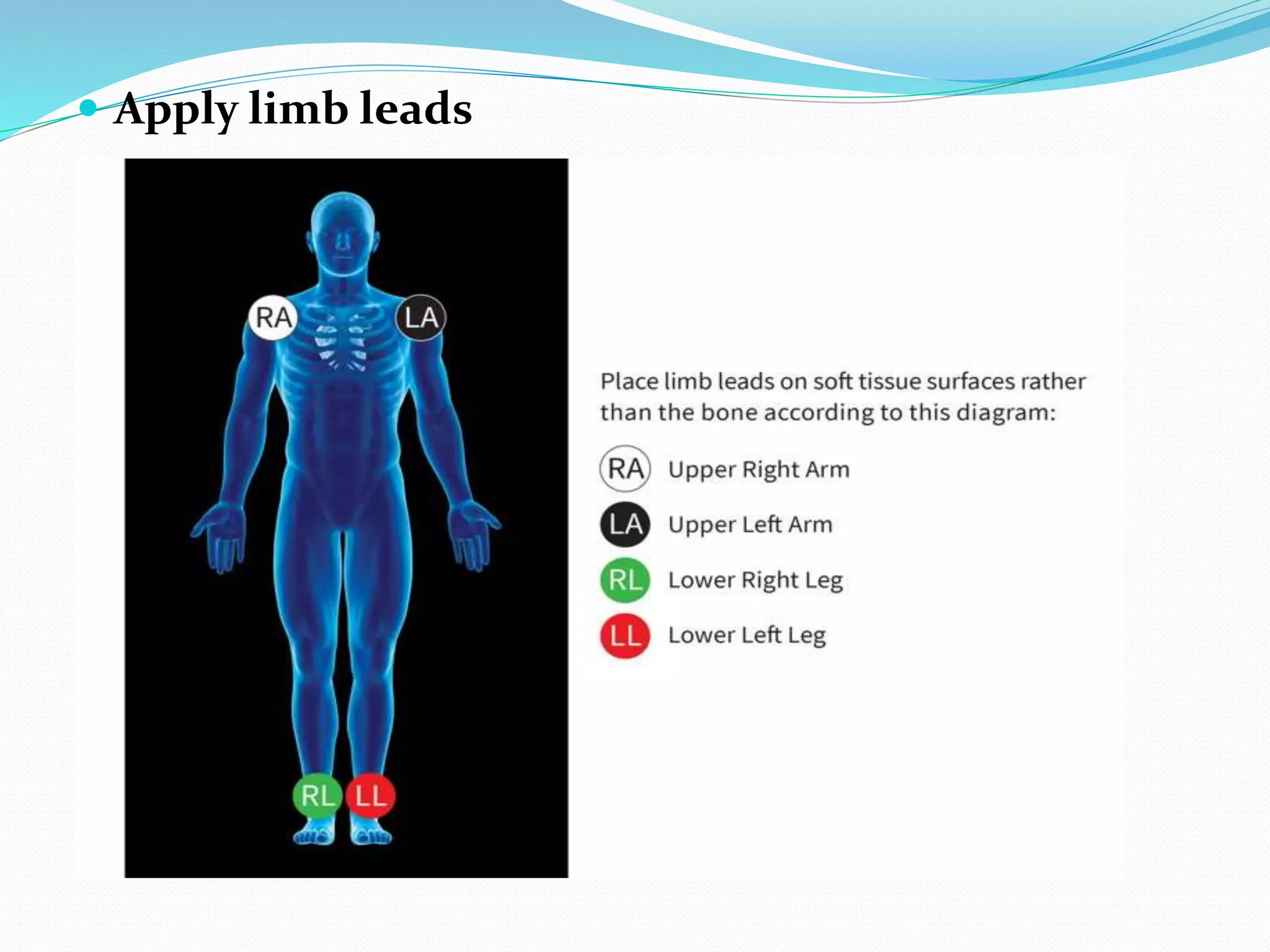
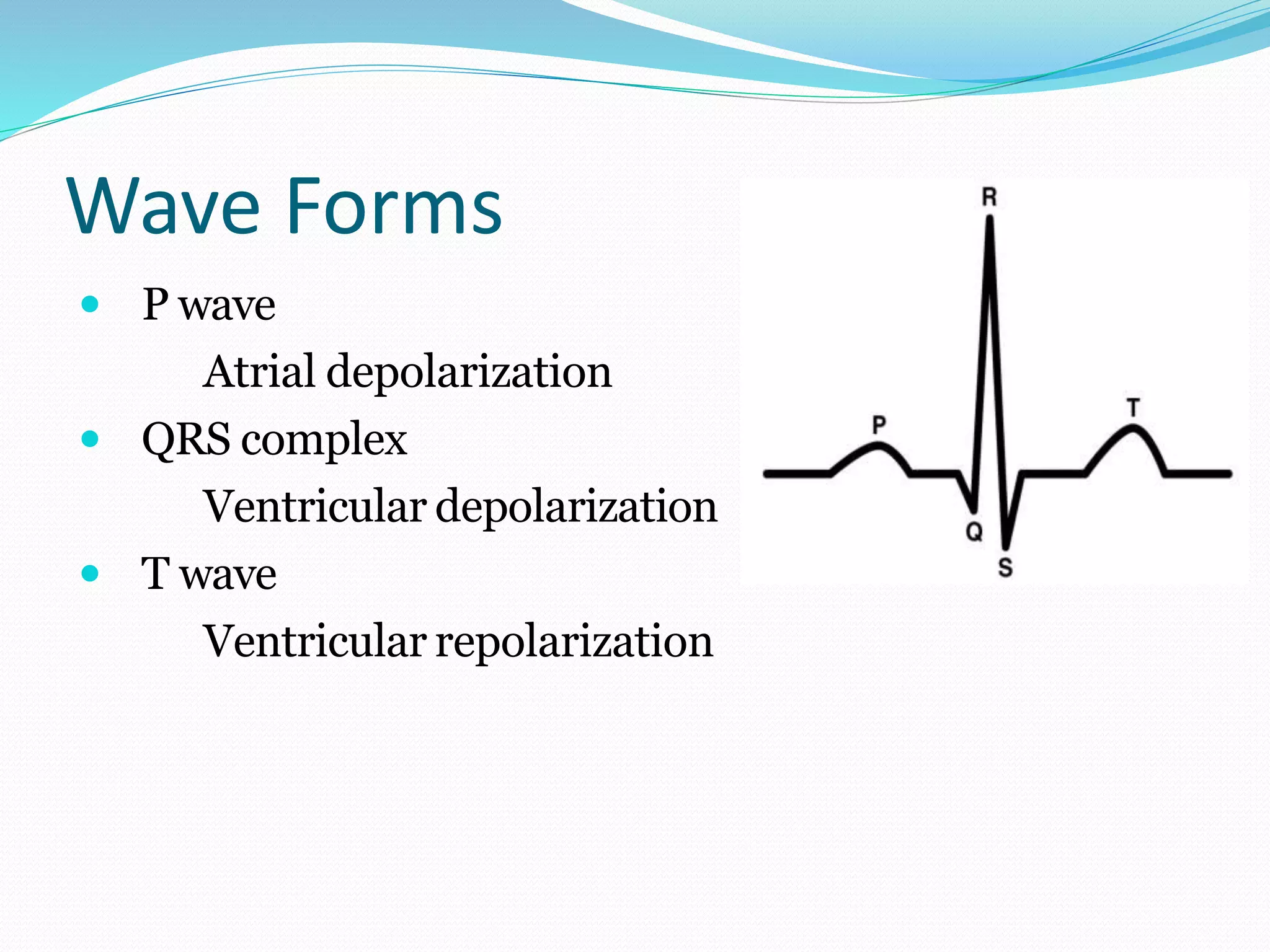
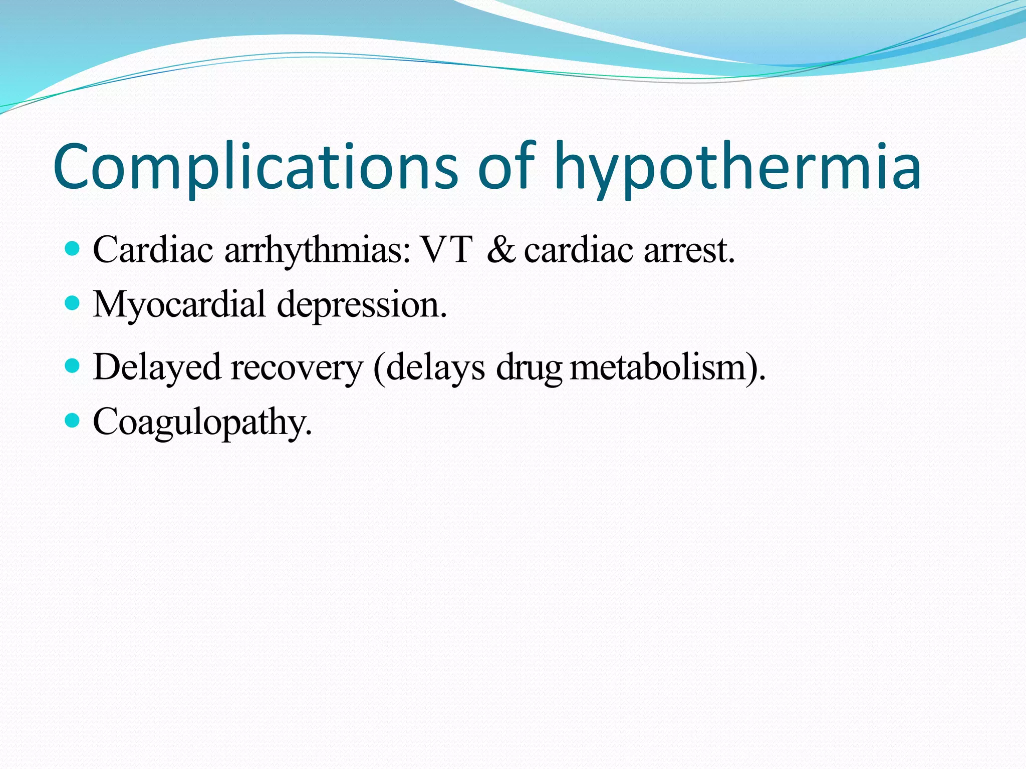
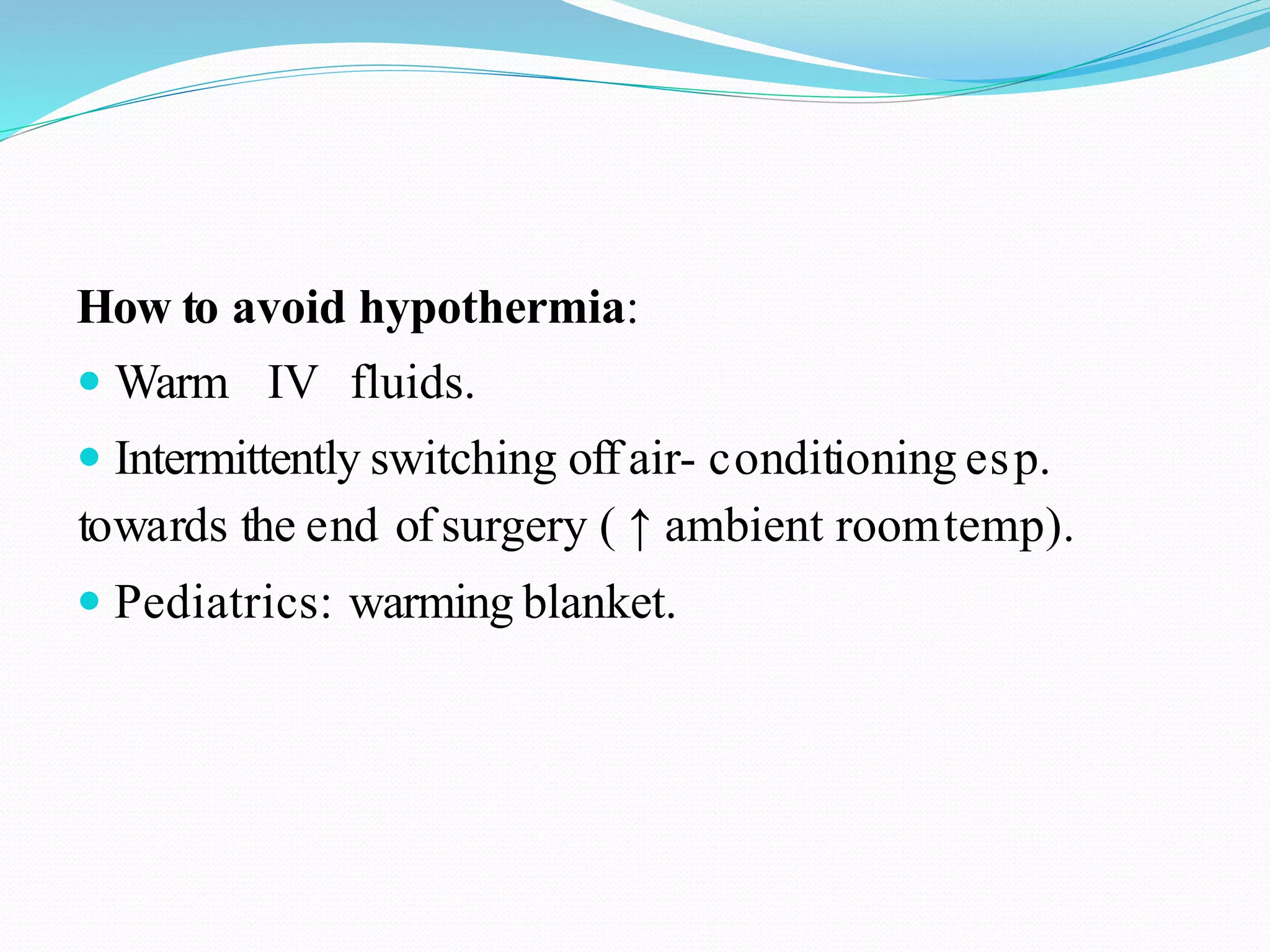
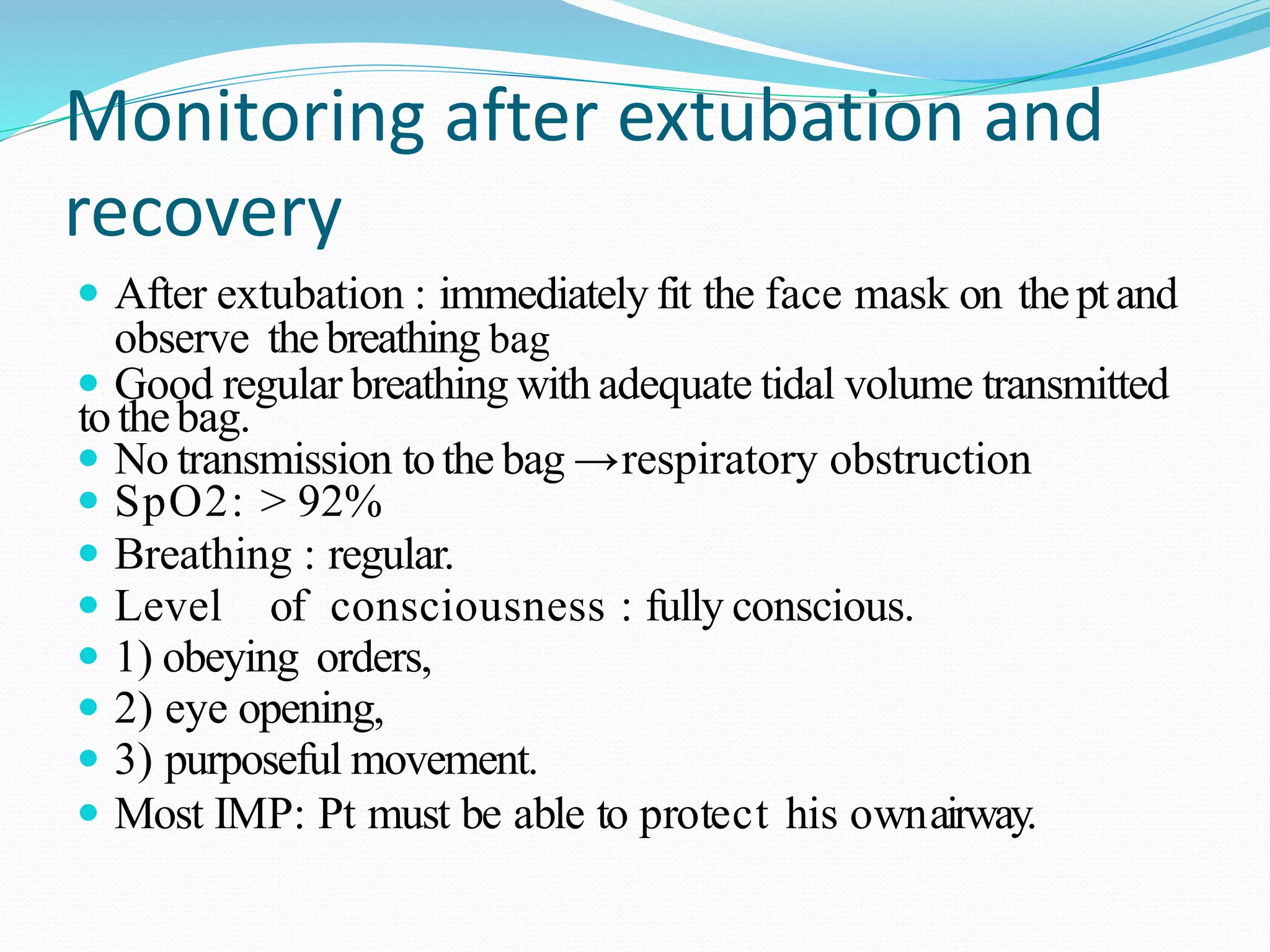
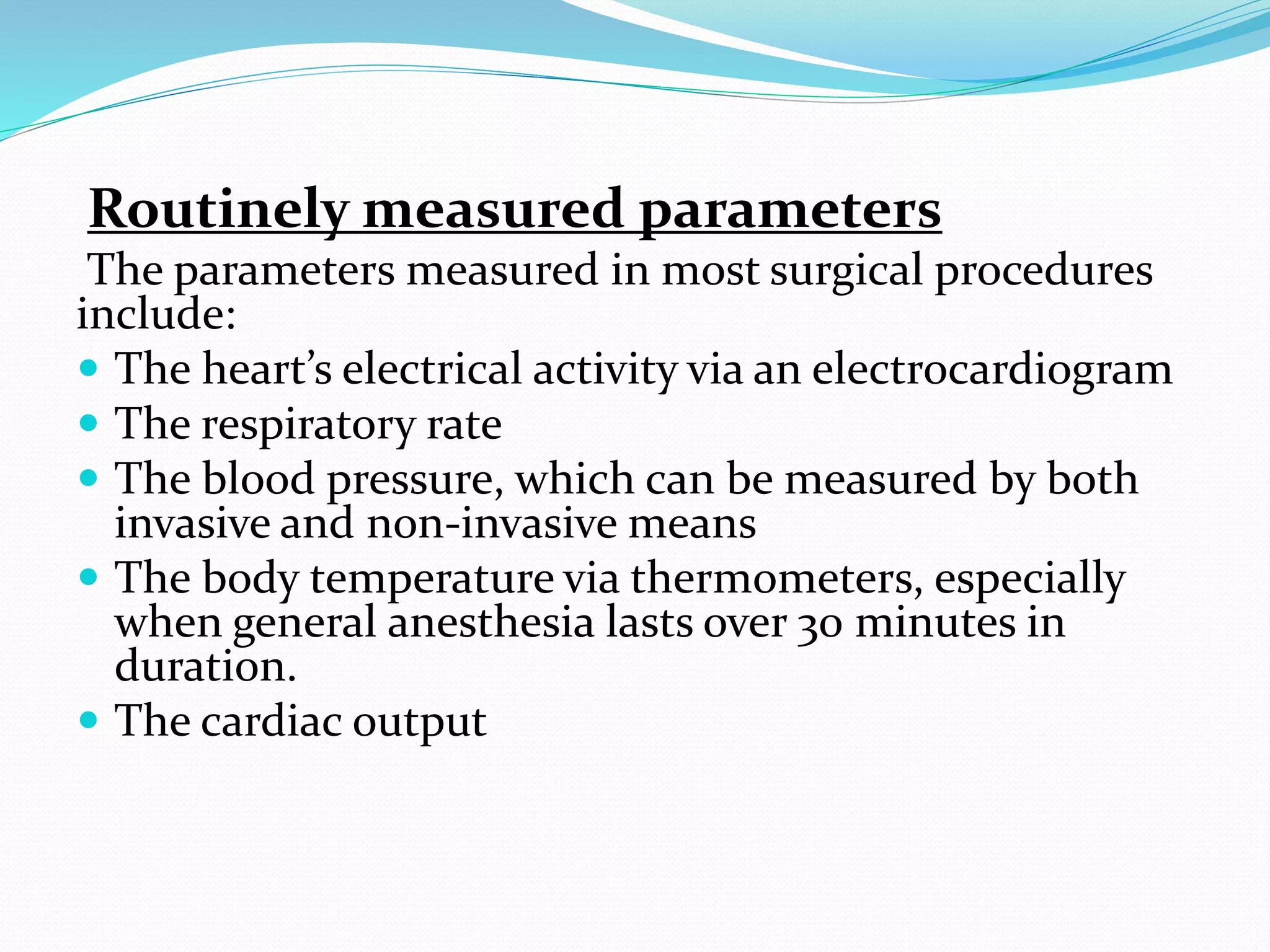
This document discusses parameters that are routinely monitored during surgical procedures, including electrocardiography (ECG), blood oxygen saturation levels (SpO2), blood pressure, end-tidal carbon dioxide (EtCO2), and temperature. Key parameters like ECG, SpO2, and blood pressure must be monitored throughout surgery. Precise measurement requires properly attaching sensors and being aware of potential errors from issues like loose or misplaced sensors. Monitoring continues in recovery to track patient status after the procedure.
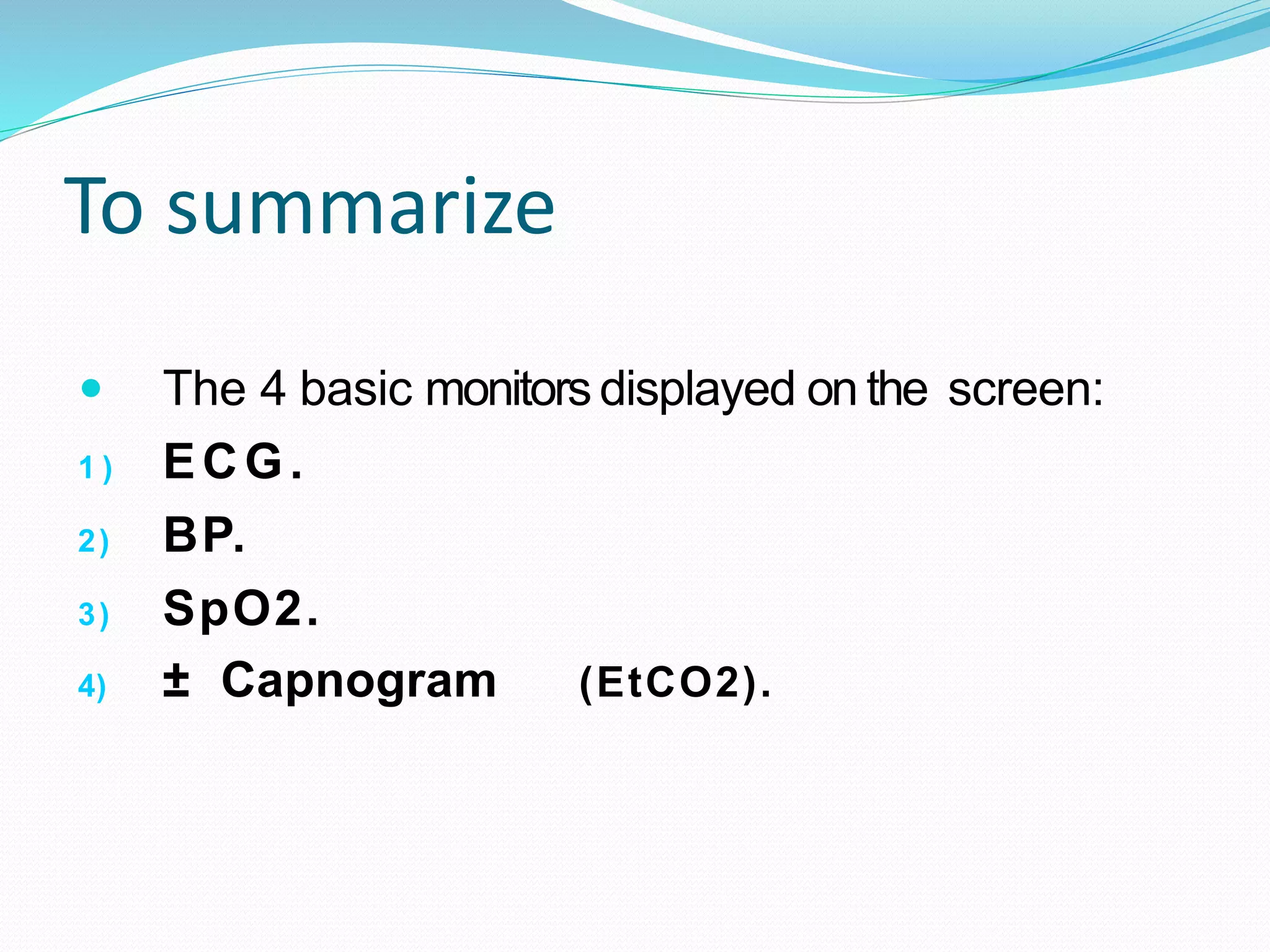
This document provides information on applying anti-embolic stockings, including their purpose to prevent deep vein thrombosis, contraindications for use, assessment steps, equipment needed, and application procedure and rationale. Key points are that stockings promote blood flow from the legs and prevent clots, should be properly fitted to avoid constriction or looseness, and removed twice daily to assess skin while encouraging leg exercises in between.
Intraoperative monitoring involves monitoring key patient vital signs throughout surgery to ensure patient safety and well-being. The four basic monitors are ECG to monitor heart rate and rhythm, pulse oximetry (SpO2) to monitor oxygen saturation and perfusion, and blood pressure (either non-invasive or invasive). Modern monitors make monitoring easier but clinical judgement is still most important. Any monitor readings require correlation with the patient's clinical condition.
The document discusses operation tables, which are specialized surgical tables used to support patients during procedures. Operation tables have several key features - they are height adjustable, can tilt in various positions, and some are compatible with imaging equipment. There are different types of operation tables designed for specific specialties like general surgery, orthopedics, neurosurgery, and bariatrics. Overall, operation tables provide an essential tool for safely positioning patients and allowing surgeons access to surgical sites.
1. The operating room table is fully adjustable to position patients in various surgical positions and orientations. It has movable sections, articulating joints, and tilt capabilities to manipulate the patient as needed.
2. Accessories like armboards, leg holders, and overhead tables provide support and access around the patient. The table also has features like smooth contours, radiolucency, and electrical or hydraulic controls for adjustments.
3. Proper positioning is important for patient safety, surgical access, and physiological needs. It prevents pressure points, maintains circulation and breathing, and positions anatomical structures for procedures. Common positions include supine, prone, lateral, lithotomy and their variations.
This document provides an overview of intraoperative patient monitoring. It defines monitoring as warning or recognizing issues. Key aspects of monitoring discussed include the cardiovascular, respiratory and central venous pressure systems. Specific monitoring modalities covered are ECG, blood pressure, pulse oximetry, capnography and blood gas analysis. The roles of monitoring in assessing oxygenation, ventilation and perfusion are emphasized.
This document discusses medicated baths, which involve dispersing medication in water for therapeutic purposes to treat dermatological conditions. It describes different types of medicated baths like Epsom salt baths, oat meal baths, and sulfur baths. The document provides indications for various medicated baths to treat issues like psoriasis, eczema, itching, and arthritis pain. It outlines the preparatory, performance, and follow-up phases of administering a medicated bath and notes supplies needed. Potential complications are mentioned along with nursing responsibilities to monitor patients during and after medicated baths.
This document discusses cardiovascular drugs, including inotropes, vasodilators, and vasopressors. It lists various drug types for each category and describes their modes of action, uses, side effects, and dosages. The objectives are to describe cardiovascular drugs, list the types, explain their uses and side effects, and discuss dosage. Examples of inotropes discussed are catecholamines, phosphodiesterase inhibitors, and cardiac glycosides. Nitrates, ACE inhibitors, and calcium channel blockers are some vasodilators covered. The vasopressors section outlines alpha-adrenergic drugs and vasopressin analogs.
Paracentesis is a procedure to remove fluid from the peritoneal cavity for diagnostic and therapeutic purposes. Large amounts of ascites fluid can cause respiratory compromise by exerting pressure on the diaphragm and abdominal organs. Paracentesis relieves this pressure and improves breathing. Key steps include ultrasound assessment, local anesthesia, inserting a needle into the abdomen, draining fluid, and analyzing laboratory samples of the fluid. Complications may include infection, bleeding, or organ damage.
A suction machine, also known as an aspirator, is a medical device that uses suction to remove obstructions like mucus, blood, or secretions from a person's airway. It maintains a clear airway for individuals unable to clear their own secretions due to lack of consciousness or an ongoing medical procedure. Precautions must be taken when using suction machines to avoid potential complications like hypoxia, airway trauma, infection, or bradycardia.
The document discusses surgical draping, including its definition, aims, importance, types of drapes, draping procedures, positions, rules, and precautions. Specifically, surgical draping involves using sterile fabric to isolate the surgical site and maintain a sterile field. It aims to prevent contamination and maintain sterility. Various draping materials, techniques, and standards are covered to outline best practices for ensuring a sterile environment.
Intravenous
Cannulation
A intravenous cannula is a flexible tube which when inserted
into the body is used either to withdraw fluid or insert
medication.
• IV Cannula normally comes with a trocar ( a sharp pointed
needle ) attached which allows puncture of the body to get
into the intended space.
This document discusses methods for preventing venous stasis when mobility is limited. Venous stasis can lead to blood clots and leg swelling. To prevent this, proper positioning, leg exercises, anti-embolic stockings, and sequential compression devices are recommended. Anti-embolic stockings help circulation by compressing the legs to push blood back to the heart. They are useful after surgery or for those who are immobile or have vascular issues. Sequential compression devices work similarly by alternately filling and deflating sleeves on the legs to mimic muscle pumping of the calves.
The document provides information on basic life support (BLS) procedures. It outlines the steps of BLS which include ensuring safety, checking for response, activating emergency services, performing chest compressions, checking airway and ventilating, and defibrillating if needed. It describes how to check for a response using the COWS method (Can you hear me, Open your eyes, What is your name, Squeeze my hand). Instructions are given on performing chest compressions, opening the airway, performing rescue breathing with different devices, and using an automated external defibrillator (AED).
This document discusses various oxygen therapy delivery systems including nasal cannulas, nasal catheters, face masks, and oxygen tents. It describes the parts of each system and how they work to deliver different concentrations of oxygen at various flow rates. Face masks discussed include simple masks, partial rebreather masks, and non-rebreather masks. The venturi mask is described as delivering concentrations from 24-40% oxygen at flow rates from 4-10 L/min depending on its color coding. Proper use and any special considerations for each system are provided.
This document discusses monitoring techniques for surgical patients, including conventional methods like blood pressure, heart rate, and urine output as well as more advanced techniques. It describes the ideal monitoring technique as non-invasive, accurate, and safe. Selection of a monitoring method depends on patient characteristics and severity of injury. Advanced techniques discussed include pulmonary artery catheters, which can measure cardiac output and pressures, but carry risks like pneumothorax. Lactate levels and tonometry can also help assess tissue oxygenation. The goal of monitoring is to assess the patient's response to treatment and guide therapy to avoid organ failure.
Patient positioning in operating theatre -gihsgangahealth
This document discusses proper patient positioning during surgery. It outlines common surgical positions like supine, prone, Trendelenburg, and lithotomy. For each position, it describes how to position the patient, nursing precautions to take, and potential complications to avoid. The goal of positioning is to provide optimal surgical access while maintaining patient safety, comfort, and dignity. Careful positioning can prevent injuries, but risks increase for patients with certain medical factors.
This document provides instructions for caring for various types of drainage tubes and recording output. It specifies that drains should be emptied regularly, the fluid measured and disposed of properly, and the output recorded. A special Stryker reinfusion drain is also described which collects and filters blood post-operatively to allow for reinfusion by a nurse, as long as certain volume and timing requirements are met.
This document provides information and guidelines for emergency medical responders (EMRs) on maintaining peripheral intravenous lines for stable patients during transport. It discusses the EMR's role in safely handling and transporting patients with existing IVs. It outlines personal safety concerns, required skills like adjusting drip rates and changing IV bags, and goals around keeping the IV patent and monitoring for complications. The document specifies considerations for IV transport, authorized and unauthorized IV solutions, complications, stabilization techniques, flow rate factors, drip rate calculations, troubleshooting problems, and required documentation.
Cleaning and disinfection of p atient care equipmentMEEQAT HOSPITAL
This document provides definitions and guidelines for cleaning, disinfecting, and sterilizing patient care equipment. It outlines that cleaning removes foreign material, disinfection eliminates most pathogens, and sterilization destroys all microbes. It distinguishes between critical equipment that enters sterile tissues and non-critical equipment that touches intact skin. The goals are to eliminate transmission between patients and staff. It provides policies on sterilizing critical items and disinfecting high-touch non-critical surfaces between each patient use. Responsibilities and proper procedures are defined to ensure effective cleaning and protection of staff.
I hope that the content of my ppt will be very good for all of you in which ppt subject is "sterile & non- sterile trolley setup" in which we have describe how to use sterile & non-sterile trolley up in surgical procedures
This document discusses nasogastric tube feeding, including:
- The purpose is to remove fluid/gas from the GI tract, prevent/relieve nausea/vomiting after surgery, and administer medications/feedings directly into the GI tract.
- The procedure involves measuring and lubricating the tube, inserting it through the nose and down the esophagus into the stomach, and checking placement by aspirating contents.
- Potential complications include pulmonary aspiration, diarrhea, tube occlusion, constipation, abdominal issues, and tube displacement. Nursing care focuses on monitoring outputs, tube care, and skin assessment.
This document discusses central venous catheters. It describes central lines as flexible tubes inserted into large veins near the heart to deliver fluids, medications, blood products, and monitor central venous pressure. It outlines different types of central lines including non-tunneled, tunneled, and implanted ports. The document discusses indications, contraindications, complications, and proper insertion and maintenance techniques to prevent infections like chlorhexidine skin antisepsis and dressing changes. The goal is to promote infection prevention best practices for central lines.
Cardiac catheterization is an invasive procedure used to visualize the heart chambers, valves, and vessels to diagnose and treat abnormalities. It can be done for both diagnostic and interventional purposes. The nurse's role is important in pre, intra, and post-procedure care. During the procedure, a catheter is inserted into the appropriate vessel and advanced under fluoroscopy while contrast dye is injected to image the heart and vessels. The patient is monitored closely for any complications like arrhythmias, bleeding, or reaction to contrast dye. After the procedure, the patient requires bed rest, monitoring of the insertion site, and observation for complications.
Nurse /doctor will insert two lubricated, gloved fingers into your vagina with one hand, while the other hand presses gently on the outside of your lower abdomen. During this part of the exam, your doctor will check the size and shape of your uterus and ovaries, noting any tender areas or unusual growths.
Cardiac monitoring(presentation ) for medical studentsNehaNupur8
presentation on cardiac monitoring , different tools and mechanism used for monitoring one of the vital organ of our body that is heart. specially for medical students, made by basic bsc students of nursing
Thoracentesis is a procedure to remove fluid or air from the pleural cavity through insertion of a needle into the chest wall. It is indicated for conditions such as pleural effusion, pneumonia, or trauma. The nurse prepares equipment like syringes, needles, and specimen containers and assists the physician by administering local anesthetic, observing for complications, and providing aftercare like monitoring for bleeding or infection. Precise technique and positioning are important to safely drain fluid and avoid injuries to lungs during the procedure.
cannulation and introduction, sizes and site of cannulasonia dagar
Intravenous cannulation is a technique where a cannula is inserted into a vein to provide venous access for administering fluids, medications, blood products, and collecting blood samples. Different sized cannulas from 16 gauge to 24 gauge are used depending on the procedure and patient factors. Common sites for cannulation include the cephalic, basilic, and median veins in the arm. The procedure involves identifying a vein, inserting the cannula at a 30 degree angle until blood is seen, securing the cannula in place, and checking patency by flushing with saline. Potential complications include hematoma, infiltration, embolism, and phlebitis.
The document summarizes various monitoring devices used during anesthesia, including essential monitors like ECG, non-invasive blood pressure, pulse oximetry, capnography, and vapor concentration analyzers. It also discusses immediately available monitors like peripheral nerve stimulators and temperature monitors. Additional monitors that may be required in some cases include invasive blood pressure, urine output, central venous pressure, pulmonary artery pressure, and cardiac output, which can be measured using a pulmonary artery catheter.
This document discusses the importance of monitoring patients during anesthesia. It outlines the basic monitoring requirements which include having an anesthesiologist present, checking anesthesia equipment, and monitoring the patient. Both clinical and instrumental monitoring are described in detail, with arterial blood pressure, ECG, CVP, pulse oximetry, capnography, and temperature being some of the key instrumental monitors discussed. Normal ranges and indications for each monitor are provided.
Monitoring in anaesthesia is important to assess the patient's physiological status and response to interventions. Basic monitoring includes clinical assessments while advanced monitoring uses instruments. Instrumental monitoring can assess the cardiovascular, respiratory, temperature, central nervous, and neuromuscular systems. Electrocardiography, blood pressure monitoring, capnography, pulse oximetry, and central nervous system monitors like the bispectral index and entropy are commonly used advanced monitoring methods. Each method has advantages and limitations that should be considered during anaesthesia.
1. Pulse oximetry and capnography are important monitoring tools during procedures involving sedation. Pulse oximetry monitors oxygenation but not ventilation, while capnography directly monitors ventilation.
2. Various factors can limit the accuracy of pulse oximetry, including abnormal hemoglobins, low perfusion, and certain dyes. Capnography provides early warning of respiratory issues through waveform analysis.
3. In addition to oxygenation and ventilation, sedation monitoring should include assessment of heart rate, blood pressure and level of consciousness to rapidly identify potential complications.
Capnography measures carbon dioxide in exhaled breath through capnography and can be used by paramedics to objectively evaluate a patient's ventilation and indirectly their circulation and metabolism. It provides important information about respiratory status and can help confirm endotracheal tube placement, guide CPR efforts, and predict resuscitation outcomes. Capnography is a useful vital sign monitoring tool for paramedics.
Measurement of blood pressure is one of the oldest physiological measurements. It originates from the heart and depends on three factors: cardiac output, artery diameter, and blood quantity. Normal values are below 120/80 mmHg. Indirect non-invasive methods like auscultation and oscillometry use an occlusive cuff on the brachial artery. Direct invasive methods involve catheter insertion but are needed for continuous accurate readings in dynamic situations. Both methods rely on measuring pressures as a cuff is inflated and deflated over the artery.
cardiac output measurment and monitoring ppt-1.pptxshekinah41
Cardiac output monitoring provides essential information about heart function and tissue perfusion. There are various methods of monitoring cardiac output, ranging from non-invasive to invasive. The pulmonary artery catheter, which involves thermodilution, has long been the gold standard but is invasive. Less invasive methods like lithium dilution and pulse contour analysis are also used. The ideal method would be non-invasive, accurate, continuous and reproducible across patients, but no single technique currently meets all these criteria.
This document discusses various methods for measuring lung volumes and oxygen levels in the body. It begins by explaining how spirometry is used to measure lung volumes like tidal volume, inspiratory reserve volume, expiratory reserve volume, vital capacity, and residual volume. It then covers different methods for measuring oxygen levels, including pulse oximetry, co-oximetry, photoplethysmography, mixed venous oxygen saturation, and regional oxygen saturation. The document also discusses capnography and its use in monitoring end-tidal carbon dioxide levels and waveforms both for intubated and non-intubated patients. It covers how capnography can be used to confirm endotracheal tube placement and detect displacement.
Pulse Oxymetry , Inspired & Expired Gas MonitoringAleenaGigiYU
Monitoring devices such as pulse oximetry and inspired and expired gas monitoring are used to monitor a patient's oxygen saturation, respiratory rate, and end-tidal carbon dioxide levels. Pulse oximetry uses light absorption to noninvasively measure oxygen saturation and heart rate, while capnography monitors end-tidal carbon dioxide levels via mainstream or sidestream methods. Blood gas analysis directly measures pH, PCO2, PO2, and HCO3 levels in arterial blood to evaluate respiratory and metabolic function.
The 4 basic monitors displayed in the operating room are:
1) ECG
2) Blood pressure
3) Pulse oximetry
4) Capnogram (end-tidal CO2)
It is important to never start induction without these monitors and to never remove any monitors before extubation and recovery. The best monitor is always the anesthesiologist using their clinical judgement.
An electrocardiogram (ECG) measures the electrical activity of the heart. Electrodes are placed on the skin to record the heart's electrical signals. An ECG can detect abnormalities in heart rate and rhythm. It involves preparing the patient, attaching electrodes to record 12 leads of the heart, and interpreting the tracing to assess rhythm, rate, intervals, and segments. An ECG is a safe, noninvasive test used to evaluate chest pain, heart function, and the effects of medications or surgery.
Cardiac output is the product of heart rate and stroke volume measured in liters per minute. Measurement of cardiac output is important for managing hemodynamically unstable patients by assessing cardiac preload and response to interventions. Methods for measuring cardiac output include thermodilution, Doppler ultrasound, echocardiography, Fick principle, impedance cardiography, and arterial pressure waveform analysis. Each method uses different physiological indicators like temperature change, blood flow, ultrasound imaging, oxygen consumption, electrical impedance, or arterial pressure to calculate cardiac output.
Cardiac output is the product of heart rate and stroke volume measured in liters per minute. Measurement of cardiac output is important for managing hemodynamically unstable patients by assessing cardiac preload and response to interventions. Methods for measuring cardiac output include thermodilution, Doppler ultrasound, echocardiography, Fick principle, impedance cardiography, and arterial pressure waveform analysis. Thermodilution involves injecting cold fluid and measuring downstream temperature change, while Doppler ultrasound and echocardiography use sound waves to evaluate blood flow noninvasively.
The document provides information about electrocardiography (ECG), including:
1. It describes the history of the ECG machine and how it was invented in 1903 by Willem Einthoven, who received a Nobel Prize for his work.
2. Modern ECG machines have evolved to be compact electronic systems that often include computerized interpretation of readings.
3. Performing an ECG involves placing 10 electrodes on the patient's limbs and chest to record the electrical activity of the heart over time.
4. The leads from the electrodes are used to analyze different views and angles of the heart's electrical activity and determine the rate, rhythm, and other diagnostic information about cardiac function.
The document provides information about electrocardiography (ECG) including:
- An ECG records the electrical activity of the heart over time through electrodes placed on the skin. Willem Einthoven invented the first practical ECG machine in 1903.
- A standard ECG uses 10 electrodes placed in specific locations to produce 12 leads that view the heart from different angles. The leads help determine the heart's electrical axis.
- An ECG trace is analyzed by examining characteristics like rate, rhythm, P wave, QRS complex, and ST segment to identify normal sinus rhythm or potential abnormalities.
- Common rhythms include sinus bradycardia, sinus tachycardia, atrial
The electrocardiogram (ECG) is a graphical presentation of the electrical activity of the heart during each heartbeat. The ECG indicates the rate and rhythm of heartbeats as well as blood flow in heart muscles. The ECG produces distinct waves - P, QRS, and T - which represent atrial and ventricular depolarization and repolarization during each heartbeat. The ECG is recorded using either a pen recorder system or oscilloscope recorder system, which detect the tiny electrical changes on the skin that are produced with each heartbeat. Abnormalities in the size of the ECG waves can help detect various heart conditions.
This document discusses various methods of monitoring patients under anaesthesia. It covers basic monitoring including vital signs and advanced instrumental monitoring of cardiovascular, respiratory, temperature, neuromuscular and central nervous systems. For each system, both non-invasive and invasive monitoring techniques are described along with their clinical indications, principles of operation, normal values and potential complications. Maintaining vigilance through multimodal monitoring is important to prevent anaesthesia complications.
Trauma Outpatient Center is a comprehensive facility dedicated to addressing mental health challenges and providing medication-assisted treatment. We offer a diverse range of services aimed at assisting individuals in overcoming addiction, mental health disorders, and related obstacles. Our team consists of seasoned professionals who are both experienced and compassionate, committed to delivering the highest standard of care to our clients. By utilizing evidence-based treatment methods, we strive to help our clients achieve their goals and lead healthier, more fulfilling lives.
Our mission is to provide a safe and supportive environment where our clients can receive the highest quality of care. We are dedicated to assisting our clients in reaching their objectives and improving their overall well-being. We prioritize our clients' needs and individualize treatment plans to ensure they receive tailored care. Our approach is rooted in evidence-based practices proven effective in treating addiction and mental health disorders.
LGBTQ+ Adults: Unique Opportunities and Inclusive Approaches to CareVITASAuthor
This webinar helps clinicians understand the unique healthcare needs of the LGBTQ+ community, primarily in relation to end-of-life care. Topics include social and cultural background and challenges, healthcare disparities, advanced care planning, and strategies for reaching the community and improving quality of care.
Under Pressure : Kenneth Kruk's StrategyKenneth Kruk
Kenneth Kruk's story of transforming challenges into opportunities by leading successful medical record transitions and bridging scientific knowledge gaps during COVID-19.
This particular slides consist of- what is Pneumothorax,what are it's causes and it's effect on body, risk factors, symptoms,complications, diagnosis and role of physiotherapy in it.
This slide is very helpful for physiotherapy students and also for other medical and healthcare students.
Here is a summary of Pneumothorax:
Pneumothorax, also known as a collapsed lung, is a condition that occurs when air leaks into the space between the lung and chest wall. This air buildup puts pressure on the lung, preventing it from expanding fully when you breathe. A pneumothorax can cause a complete or partial collapse of the lung.
INFECTION OF THE BRAIN -ENCEPHALITIS ( PPT)blessyjannu21
Neurological system includes brain and spinal cord. It plays an important role in functioning of our body. Encephalitis is the inflammation of the brain. Causes include viral infections, infections from insect bites or an autoimmune reaction that affects the brain. It can be life-threatening or cause long-term complications. Treatment varies, but most people require hospitalization so they can receive intensive treatment, including life support.
We are one of the top Massage Spa Ajman Our highly skilled, experienced, and certified massage therapists from different corners of the world are committed to serving you with a soothing and relaxing experience. Luxuriate yourself at our spas in Sharjah and Ajman, which are indeed enriched with an ambiance of relaxation and tranquility. We could confidently claim that we are one of the most affordable Spa Ajman and Sharjah as well, where you can book the massage session of your choice for just 99 AED at any time as we are open 24 hours a day, 7 days a week.
Visit : https://massagespaajman.com/
Call : 052 987 1315
Gemma Wean- Nutritional solution for Artemiasmuskaan0008
GEMMA Wean is a high end larval co-feeding and weaning diet aimed at Artemia optimisation and is fortified with a high level of proteins and phospholipids. GEMMA Wean provides the early weaned juveniles with dedicated fish nutrition and is an ideal follow on from GEMMA Micro or Artemia.
GEMMA Wean has an optimised nutritional balance and physical quality so that it flows more freely and spreads readily on the water surface. The balance of phospholipid classes to- gether with the production technology based on a low temperature extrusion process improve the physical aspect of the pellets while still retaining the high phospholipid content.
GEMMA Wean is available in 0.1mm, 0.2mm and 0.3mm. There is also a 0.5mm micro-pellet, GEMMA Wean Diamond, which covers the early nursery stage from post-weaning to pre-growing.
PET CT beginners Guide covers some of the underrepresented topics in PET CTMiadAlsulami
This lecture briefly covers some of the underrepresented topics in Molecular imaging with cases , such as:
- Primary pleural tumors and pleural metastases.
- Distinguishing between MPM and Talc Pleurodesis.
- Urological tumors.
- The role of FDG PET in NET.
Can coffee help me lose weight? Yes, 25,422 users in the USA use it for that ...nirahealhty
The South Beach Coffee Java Diet is a variation of the popular South Beach Diet, which was developed by cardiologist Dr. Arthur Agatston. The original South Beach Diet focuses on consuming lean proteins, healthy fats, and low-glycemic index carbohydrates. The South Beach Coffee Java Diet adds the element of coffee, specifically caffeine, to enhance weight loss and improve energy levels.
Empowering ACOs: Leveraging Quality Management Tools for MIPS and BeyondHealth Catalyst
Join us as we delve into the crucial realm of quality reporting for MSSP (Medicare Shared Savings Program) Accountable Care Organizations (ACOs).
In this session, we will explore how a robust quality management solution can empower your organization to meet regulatory requirements and improve processes for MIPS reporting and internal quality programs. Learn how our MeasureAble application enables compliance and fosters continuous improvement.
Rate Controlled Drug Delivery Systems, Activation Modulated Drug Delivery Systems, Mechanically activated, pH activated, Enzyme activated, Osmotic activated Drug Delivery Systems, Feedback regulated Drug Delivery Systems systems are discussed here.
The best massage spa Ajman is Chandrima Spa Ajman, which was founded in 2023 and is exclusively for men 24 hours a day. As of right now, our parent firm has been providing massage services to over 50,000+ clients in Ajman for the past 10 years. It has about 8+ branches. This demonstrates that Chandrima Spa Ajman is among the most reasonably priced spas in Ajman and the ideal place to unwind and rejuvenate. We provide a wide range of Spa massage treatments, including Indian, Pakistani, Kerala, Malayali, and body-to-body massages. Numerous massage techniques are available, including deep tissue, Swedish, Thai, Russian, and hot stone massages. Our massage therapists produce genuinely unique treatments that generate a revitalized sense of inner serenely by fusing modern techniques, the cleanest natural substances, and traditional holistic therapists.
This particular slides consist of- what is hypotension,what are it's causes and it's effect on body, risk factors, symptoms,complications, diagnosis and role of physiotherapy in it.
This slide is very helpful for physiotherapy students and also for other medical and healthcare students.
Here is the summary of hypotension:
Hypotension, or low blood pressure, is when the pressure of blood circulating in the body is lower than normal or expected. It's only a problem if it negatively impacts the body and causes symptoms. Normal blood pressure is usually between 90/60 mmHg and 120/80 mmHg, but pressures below 90/60 are generally considered hypotensive.
3. A patient undergoing surgery under anesthesia is
usually experiencing a number of disturbances
with regard to the normal functioning of several
body systems. This means that the patient’s
physiological status needs to be monitored at all
times
Various sensors and electrodes are attached to the
patient to gather information. These
measurements are displayed on monitors.
4. Routinely measured parameters
The parameters measured in most surgical procedures
include:
The heart’s electrical activity via an electrocardiogram
The respiratory rate
The blood pressure, which can be measured by both
invasive and non-invasive means
The body temperature via thermometers, especially
when general anesthesia lasts over 30 minutes in
duration.
The cardiac output
5. The arterial blood oxygen level measured by a
pulse oximeter, a photoelectric sensor clipped over
the finger or toe.
Pulmonary functions such as end-tidal carbon
dioxide (ETCO2).
Intracranial pressure monitor in patients suffering
from trauma to the head, or brain tumors, edema,
or intracranial hemorrhage. The sensor is inserted
through a burr hole made in the skull.
7. Purpose
To maintainthenormalpatientphysiology &
homeostasis throughoutanesthesia andsurgery.
Surgery is a very stressful conditiontachycardia,
arrhythmias.
Mostdrugsused for eneral ®ionalanesthesia
cause cardiogenicshock, myocardialdepression,
hypotension & arrhythmias.
Blood loss →anemia, hypotension. So itis necessary
to recognizewhenthepatientis inneed of blood
transfusion.
8. The FOUR basic monitors.
We are not authorizedto starta surgeryinthe absence ofany
ofthesemonitors:
ECG.
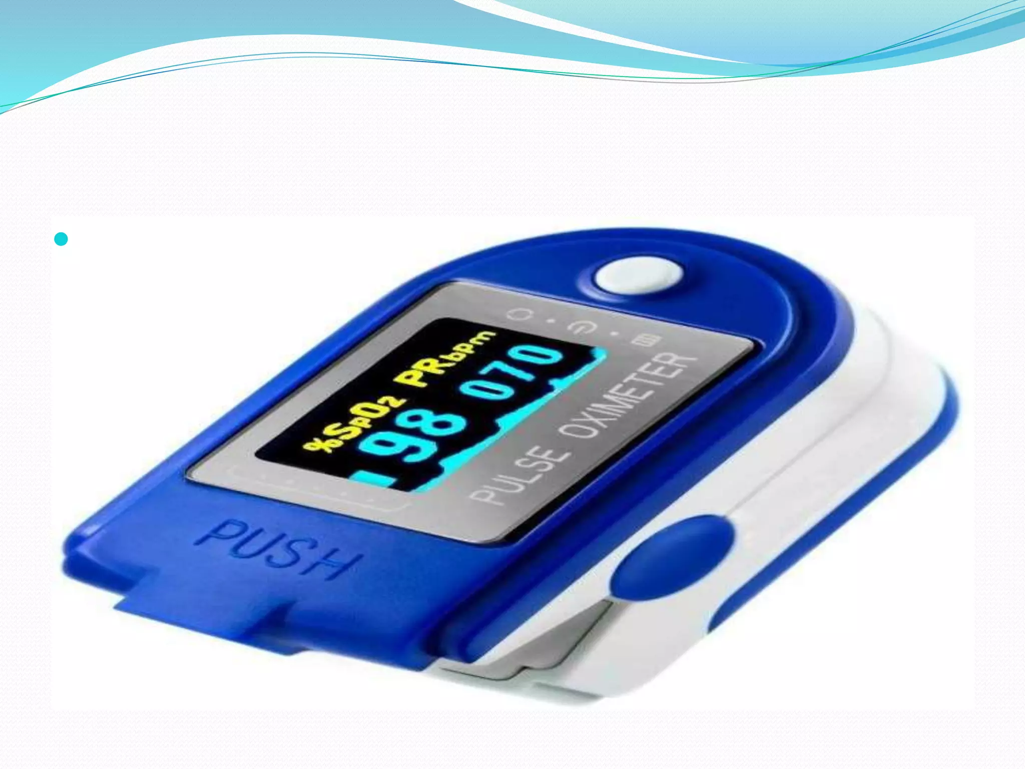
SpO2: arterial O2 saturation.
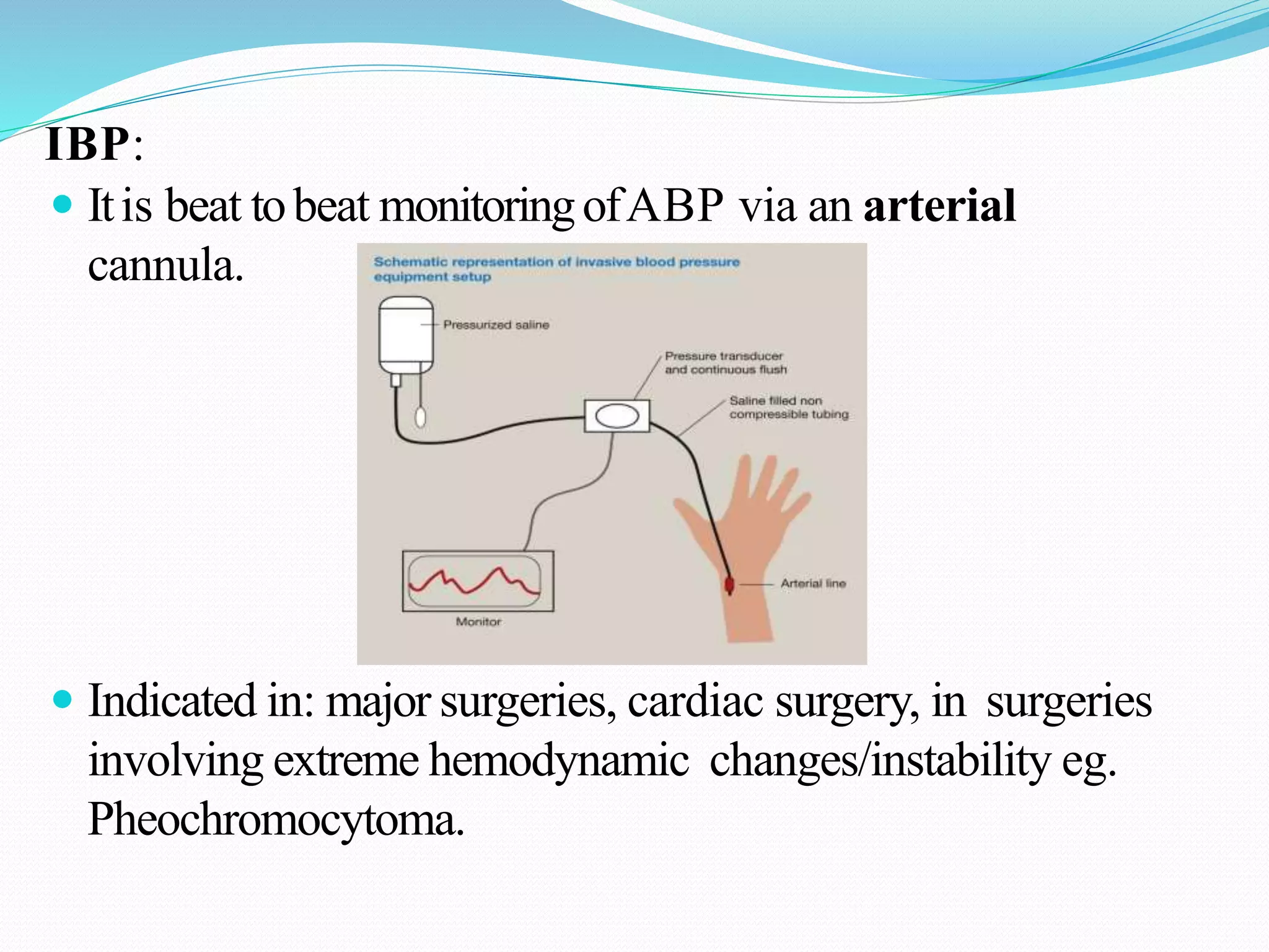
Blood Pressure: NIBP (non-invasive), IBP (invasive).
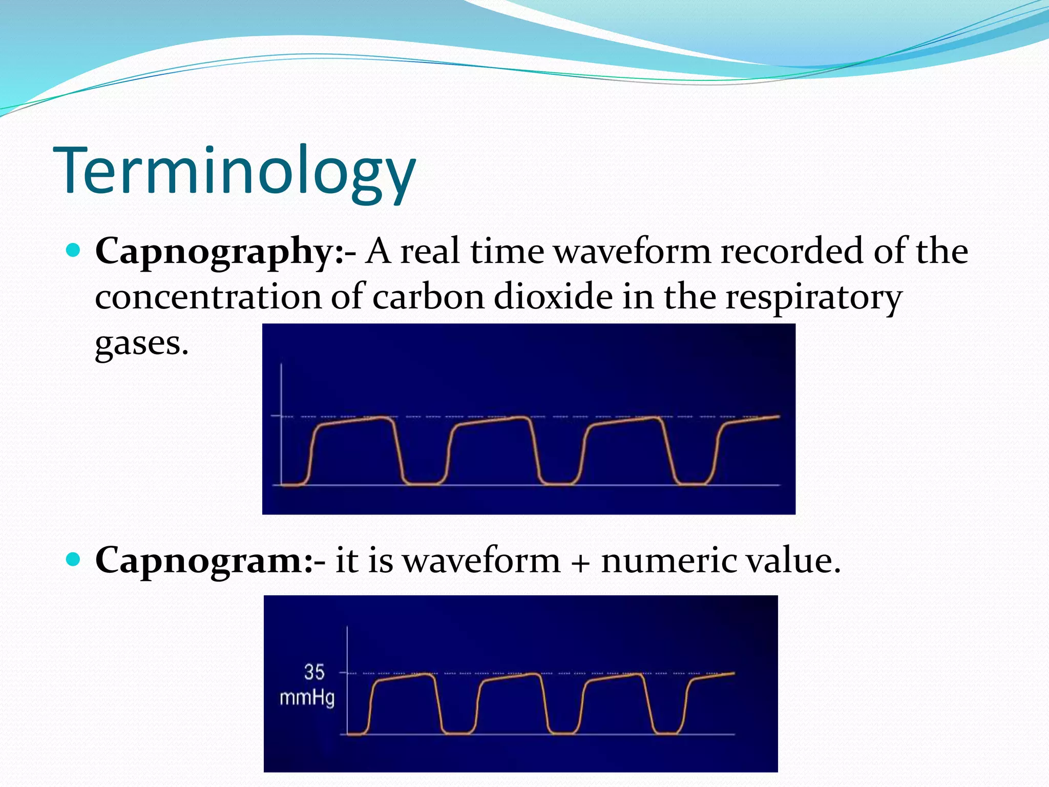
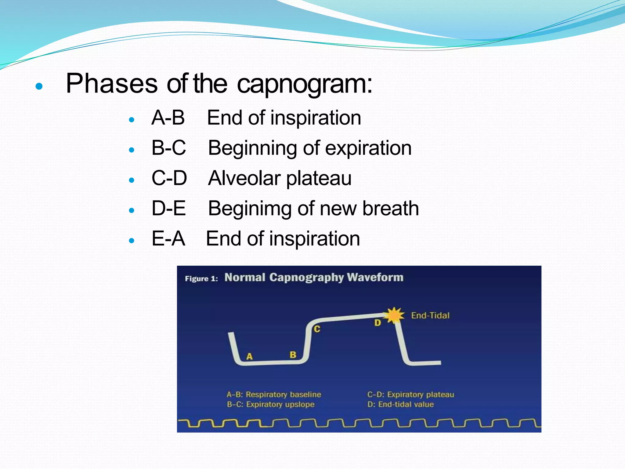
± [Capnography].
The mostcritical 2 timesduringanesthesiaare:
INDUCTION - RECOVERY.
Exactly like “flying aplane ” induction(= take off) &recovery
(= landing). The aimis to achieve a smooth induction&a
smooth recovery &a smooth intraoperative course.
9. (1) ECG
Graphical representation of electrical activity of heart.
The machine which is used to record the electrical
activity of heart is ELECTROCARDIOGRAPH.
The graph on which the electrical activity is recorded
is called ELECTROCARDIOGRAM.
10. Significance of ECG
ECG gives information about rate and rhythm of
the heart.
It is a diagnostic tool for various heart conditions like
hypertrophies , ischemia, infarction , arrhythmias and
pace maker activity.
Timing ofECG monitoring:Throughout the surgery:
before induction until recovery
11. ECG Paper
• This is long role of paper ,composed of small
squares.
• One square is 1mmwideand 1 mm high.
• On ECG paper there are thick lines, between two thick
lines there are 5 small squares.
• The speed of ECG machine is 25 mm per second.
12. • The time internal of each small square can be calculated
as
• 25 small sqrs are equal to 1 second.
• 1 small sqr is equal to 1/25 second.
• i.e 0.04 seconds.
14. • vertically the small square represent the amount of
electrical potential.
• One small sqr represent the potential of 0.1 mv.
• 10 small squares represent the potential of 1 mv.
15. ECG Leads
There are 10 electrodes in 12 lead ECG placed on
patients limbs and on the surface of chest.
16. How to attach ECG electrodes:
Prepare the skin
Before placing your electrodes, it is very important to
prepare the subject's skin by wiping the chest area
thoroughly with skin cleansing (alcohol) swabs. This
removes any oil that may be on the skin and which can
cause drift in your ECG/EKG signals.
Once the skin is clean, find and mark the placements for
the electrodes...
17. Find and mark the placements for the electrodes:
19. Wave Forms
P wave
Atrial depolarization
QRS complex
Ventricular depolarization
T wave
Ventricular repolarization
20. P Wave
• P Waveshows atrial
depolarization..
• Its duration is 0.1 sec (2 and half
small
sqr)
and height is 2.5 mv (2 and half
small sqr).
• Presence of p waves in ECG
strip shows the sinus rhythm.
21. QRS Complex
QRS complex represent the ventricular
depolarization.
its normal duration is about 0. 08 seconds
(less than 2 small sqr) and hight is about 5
to 20 small sqrs.
Q wave is first wave of this complex but
often absent.
22. Q wave present the interventricular
depolarization.
It is first wave in ECG with negative deflection.
Q wave greater than 1/3 the height of the R wave,
greater than 0.04 sec are abnormal and may represent
the old infarction.
23. T Wave
• it represent the ventricular repolarization.
• It is repolarizing wave but shows the upward
deflection.
24. • T wave should not be more than one third of R wave.
• T wave inversion represent ischemia of heart.
• Tall and peaked R wave is present in hyperkalemia.
25. (2) SpO2
SpO2, also known as blood oxygen saturation, is a measure
of the amount of oxygen-carrying hemoglobin in the blood.
The body needs there to be a certain level of oxygen in the
blood or it will not function as efficiently. In fact, very low
levels of SpO2 can result in very serious symptoms. This
condition is known as hypoxemia. There is a visible effect on
the skin, known as cyanosis due to the blue (cyan) tint it takes
on. Hypoxemia (low levels of oxygen in the blood) can turn
into hypoxia (low levels of oxygen in the tissue).
26. Timing ofSpO2 monitoring:
Throughoutthe surgery: It is the LAST monitortobe removed offthe
ptbeforetheptis transferredoutside the operatingroomtorecovery
room.SpO2 monitoringshouldbe continuedinrecovery room.
27. Measuring SpO2
• There are many ways that the blood can be tested to ensure it
contains normal oxygen levels. The most common way is to
use a pulse oximeter to measure the SpO2 levels in the blood.
Pulse oximeters are relatively easy to use, and are common in
health care facilities and at home.To use a pulse oximeter,
simply place it on your finger. Can also be applied to the
ear lobe.
• A percentage will be displayed on the screen. This percentage
should be between 94 percent and 100 percent, which
indicates a healthy level of hemoglobin carrying oxygen
through the blood. If it is less than 90 percent, you should see
a doctor.
30. Fallacies &Inaccuracies occur when:
Misplaced onthepts finger, slipped.
Pt movement, shivering.
Tissue perfusion (cold extremities) →warmthept, put
a glove filled withwarmwater in thepts hand (always
avoid hypothermia).
Cardiac arrest.
31. Rules
Keep thesound ofthepulse oximeterON at all times.
ALWAYS Remember thatyourclinical judgement is muchmore
superiortothe monitor.Check ptcolourforcyanosis: lips, nails.
Ifhypoxemiaoccurs immediately check theptscolour : nails &lips,
thenmanageaccordinglyandcallforhelp.
32. (3) Blood Pressure
Timing of BP monitoring :throughout the
surgery.
Frequency of measurement:
By default every 5 minutes.
Every 3 minutes: immediately after spinal
anesthesia.
Every 10 minutes: eg. Inawake pts under local
anesthesia.
33. Reading errors/Failures
Pressure line is disconnected.
Leakage from damaged cuff.
Line is compressed (under someone’s foot or under a
weal).
Line contains water from washing!
34. How to Attach
Correct cuff size : width of the cuff should be 1.5
times limb diameter and should occupy at least 2/3 of the
arm.
2 cuff sizes for adult: blue : for most adult individuals
(60-90 Kg), red : for morbid obese.
Selection of appropriate cuff size is importantbecause a
tight cuff leads to false high readings, while a L oose cuff
gives false Low readings.
35. IBP:
Itis beat tobeat monitoringofABP via an arterial
cannula.
Indicated in: major surgeries, cardiac surgery, in surgeries
involving extreme hemodynamic changes/instability eg.
Pheochromocytoma.
36. (4) Capnography
Definition
Continuous CO2 measurement displayed as a
waveform sampled fromthe patient’s airway during
ventilation.
Normal range: 35-40mmHg
What is EtCO2?
A point onthecapnogram. Itis thefinal measurement
at theendpointofthepts expiration before inspiration
begins again. Itis usually thehighest CO2
measurement during ventilation.
37. Terminology
Capnography:- A real time waveform recorded of the
concentration of carbon dioxide in the respiratory
gases.
Capnogram:- it is waveform + numeric value.
39. Phases of the capnogram:
A-B End of inspiration
B-C Beginning of expiration
C-D Alveolar plateau
D-E Beginimg of new breath
E-A End of inspiration
40. Factors that affect CO2 levels
Increase in ETCO2 Decrease in ETCO2
Increased muscular activity Decreased muscular activity
Increased cardiac output Decreased cardiac output
hypoventilation Hyperventilation
Partial airway obstruction Pulmonary embolism
48. How to avoid hypothermia:
Warm IV fluids.
Intermittently switching off air- conditioning esp.
towards the end ofsurgery ( ↑ ambient roomtemp).
Pediatrics: warming blanket.
49. Monitoring after extubation and
recovery
After extubation : immediately fit the face mask on theptand
observe thebreathing bag
Good regular breathing with adequate tidal volume transmitted
tothebag.
No transmission tothe bag →respiratory obstruction
SpO2: ˃ 92%
Breathing : regular.
Level of consciousness : fully conscious.
1) obeying orders,
2) eye opening,
3) purposeful movement.
Most IMP: Pt must be able to protect his ownairway.
50. To summarize
The 4 basic monitors displayed on the screen:
1 ) ECG.
2) BP.
3) SpO2.
4) ± Capnogram (EtCO2).