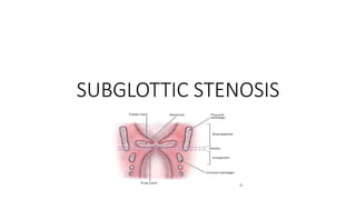
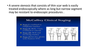
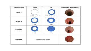
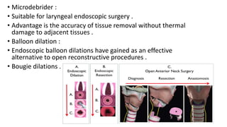
2. SUBGLOTTIC STENOSIS
• It is characterized by the narrowing of subglottis (portion of wind
pipe , just below vocal cords narrows ).
• It is usually characterized by inflammation and fibrosis such as scar
tissue in the area .
• Subglottic stenosis can affect newborn , babies and adults .
• People are either born with the condition or they develop it as
complication later on .
• Statistically , idiopathic subglottic stenosis affects females about 98
percent .
3. Types of subglottic stenosis :
• Congenital subglottic stenosis :
• Occurs when a baby is born with an airway smaller than
usual .
• Acquired subglottic stenosis :
• Not present at birth . It develops later because of trauma ,
infection or complications due to intubation etc.,
4. Symptoms of subglottic stenosis
• Shortness of birth
• Stridor
• Hoarseness of voice
• Increased mucous production
• Cough
• Symptoms common in infants :
• Cyanosis
• Poor weight gain
• Croup or lung infections
• Respiratory distress
5. Grading of subglottic stenosis :
• The classic grading of subglottic stenosis was proposed by
COTTON and is accepted world wide .
• Modified COTTON – MEYER grading scale was introduced in
1994 based on percentage of obstruction calculated by
passing an endotracheal tube through stenosis resulting in
approximation of stenotic diameter divided by age
appropriate endotracheal tube size .
• Grade 1 stenosis but with not bilateral vocal cord immobility,
treatment of stenosis will not result an adequate airway .
6. • A severe stenosis that consists of thin scar web is easily
treated endoscopically where as long but narrow segment
may be resistant to endoscopic procedures .
7.
8. Signs and symptoms
• The symptoms of subglottic stenosis in children
are closely related to degree of narrowing .
• Grade 1 is usually asymptomatic until an URTI
occurs
• Grade 2 and 3 causes biphasic stridor , air
hunger ,dyspnea ,and suprasternal ,intercoastal
and diaphragmatic retractions.
• It is important to recognize that compromised
airway in a child can lead to rapid deterioration
and requires rapid intervention to avoid
catastrophic outcome .
9. Evaluation :
• Relevant history includes birth weight , stridor , quality of cry and
voice ,feeding difficulties ,aspiration ,prior pulmonary and
cardiac status .
• Physical examination should include thorough head and neck
examination and assessment of craniofacial abnormalities
,micrognathia , macroglossia , laryngomalacia and choanal
atresia .
• Awake flexible fiberoptic laryngoscopy
• Direct laryngoscopy and rigid bronchoscopy with video
assistance under GA with spontaneous ventilation is gold
standard for diagnosis of subglottic stenosis .
10. Imaging
• CT neck or MRI usually not recommended in children in most
children .
• Magnetic resonance angiography – stenosis caused by
vascular anomaly or tumor.
• CT OR MRI is indicated when direct laryngoscopy shows
complete or near complete obstruction of subglottis in order
to measure the length of stenotic segment and asses
framework defects .
11. Congenital subglottic stenosis:
• It is the 3rd most common congenital disorder in larynx .
• Subglottic diameter if less than 4mm or in preterm it is less
than 3mm.
• In severe cases stridor present at birth , while in milder cases
symptoms present only after a few months .
• Congenital anterior glottic webs are frequently associated
with subglottic stenosis due to malformed cricoid ring .
• It is occasionally reported in children with Downs syndrome .
12. Acquired subglottic stenosis :
• It is usually due to endotracheal intubation
in children .
• The epithelium lining the subglottis is
delicate and is easily injured by
endotracheal tube .
• Secondly the cricoid cartilage is complete
circular ring , the edema caused by trauma
or pressure directly impinges on internal
diameter
• Third , subglottic region is the narrowest
area of airway in children .
13. • Next , significant edema can develop in subglottic region
quickly because of loose areolar tissue that comprises the
submucosa in the region .
• Minor injury causes a greater narrowing that compromises
the airway in children .
• Other causes are high tracheostomy causes damage to
cricoid ,emergent cricothyroidotomy , smoke inhalation ,
caustic ingestion , burns and trauma .
• Neoplasms can also causes subglottic stenosis commonly
subglottic hemangioma less commonly chondroma or
fibroma .
14. • Other causes include :
• Wegener’s granulomatosis, pemphigoid , relapsing
polychondritis , amyloidosis , aphthous ulcerations and
laryngo oculo cutaneous syndrome .
• Chronic infections such as tuberculosis and syphilis can also
lead to subglottic stenosis .
15. Management of subglottic stenosis :
• The treatment paradigm is based on presenting clinical
severity of breathing , vocal cord mobility ,comorbidities
including neurological and developmental aspiration and
coexisting airway lesions .
• Findings on direct laryngoscopy and tracheoscopy are
necessary in order to make appropriate treatment decisions .
16. Immediate interventions :
• Infant presenting to emergency in respiratory distress
requires immediate attention and intervention .
• The most important sign of impending disaster is increased
effort of breathing despite normal oxygen saturation levels .
• Child can decompensate suddenly .
• Therefore when there is stridor and increased effort of
bleeding , a quick decision of safest way to secure airway
must be made .
17. • The safest place for securing the airway in this situation is OT
room where direct laryngoscopy can be performed if
endotracheal intubation is difficult .
• Set up for emergent tracheostomy should be prepared as a
backup if attaining the airway is unsuccessful.
• As a stabilizing measure on the way to definitive treatment ,
inhalation of nebulized adrenaline with saline can help
reduce child’s work of breathing while preparing for airway
intervention .
• Cricothyroidotomy is unsafe in children due to size and
collapsibility of airway .
18. Observation :
• Children with grade I or a mild grade II may not require
surgical treatment and can be observed .
• Another reason for watchful waiting and avoiding surgical
intervention is a “ reactive larynx” due to inflammatory
process with edema and granulation tissue is identified .
• Children tend to heal poorly after surgery and hence open
airway reconstruction should be deferred until the larynx is
no longer reactive .
19. • Reconstructive airway surgery is relatively contraindicated in
children with low weight less than 10 kg , aspiration causing
recurrent pneumonia , in children been mainstay of airway
management .
• CO2 lasers , holmium lasers can be used to treat subglottic
stenosis .
• Laser tissue removal or incision and dilation can actually
increase scar formation and worsen the lesion .
• Alternatives such as microdebrider , balloon dilations have
emerged ., as they causes tendency to cause less amount of
scarring .
20. • Microdebrider :
• Suitable for laryngeal endoscopic surgery .
• Advantage is the accuracy of tissue removal without thermal
damage to adjacent tissues .
• Balloon dilation :
• Endoscopic balloon dilations have gained as an effective
alternative to open reconstructive procedures .
• Bougie dilations .
21. Adjuvant treatment :
• Mitomycin C has been used to prevent scarring and
granulation tissue formation so as to improve outcomes of
endoscopic procedures .
• It is applied directly to stenotic area towards the end of
procedure using cottonoids .
• Intralesional steroids
• Antireflux medications .
22. Open surgery :
• Tracheostomy , Laryngotracheoplasty ,laryngotracheal
reconstruction .
• Tracheostomy :
• Safest way to secure airway in children
• Serve as a bridge before reconstructive surgery becomes
feasible .
• Anterior cricoid split : alternative to tracheostomy in
premature neonates .
• Endoscopic anterior cricoid split combined with balloon
dilations are reported to have 80% success rate .
23. Laryngotracheal reconstruction and
laryngotracheoplasty :
• Includes splitting the cricoid cartilage the , the
lower third of thyroid cartilage and the first
tracheal ring and expanding the framework with
cartilage grafts , harvested from one of the
coastal ribs , preferably on the right side .
• Single or two step procedure .
• Single stage LTP is defined as not leaving a
tracheostomy tube at the end of surgery and
placing a nasotracheal tube as a stent for a
period of 5-7 days .
• Two stage LTP is defined as tracheostomy tube at
end of procedure that is removed several weeks
after the primary procedure .
24. • The decision to perform single or two
stage LTP is based on many variables
should be tailored individually .
• Also it can be decided based on direct
laryngoscopy that precedes the LTP/R.
• When single stage LTP is performed ,
nasotracheal tube is kept in place
throughout and after the surgery .
• In a two stage LTP various stent have
been used as adjuncts and kept in place
for 3-6 wks .
• To prevent scar contracture Montgomery
T tube is used as it has advantage of
serving both as a stent and as
tracheostomy tube .
25. Partial CTR :
• Seperation of trachea from esophagus , mobilization of upper
tracheal rings , excision of stenotic segment with preservation of
posterior cricoid plate .
• Next supralaryngeal release is performed followed by
thyrotracheal end to end anastomosis .
• It can be performed either single stage or two stage procedure
with or with out stenting .
• The procedure requires surgical expertise due to risk of damage
to recurrent laryngeal nerve and high precision that
thyrotracheal anastomosis requires .