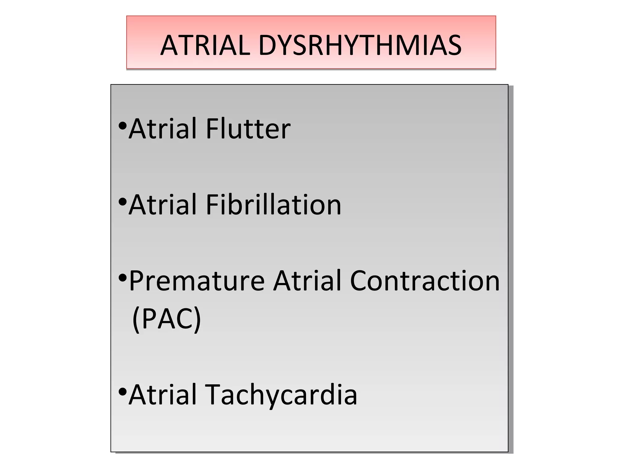
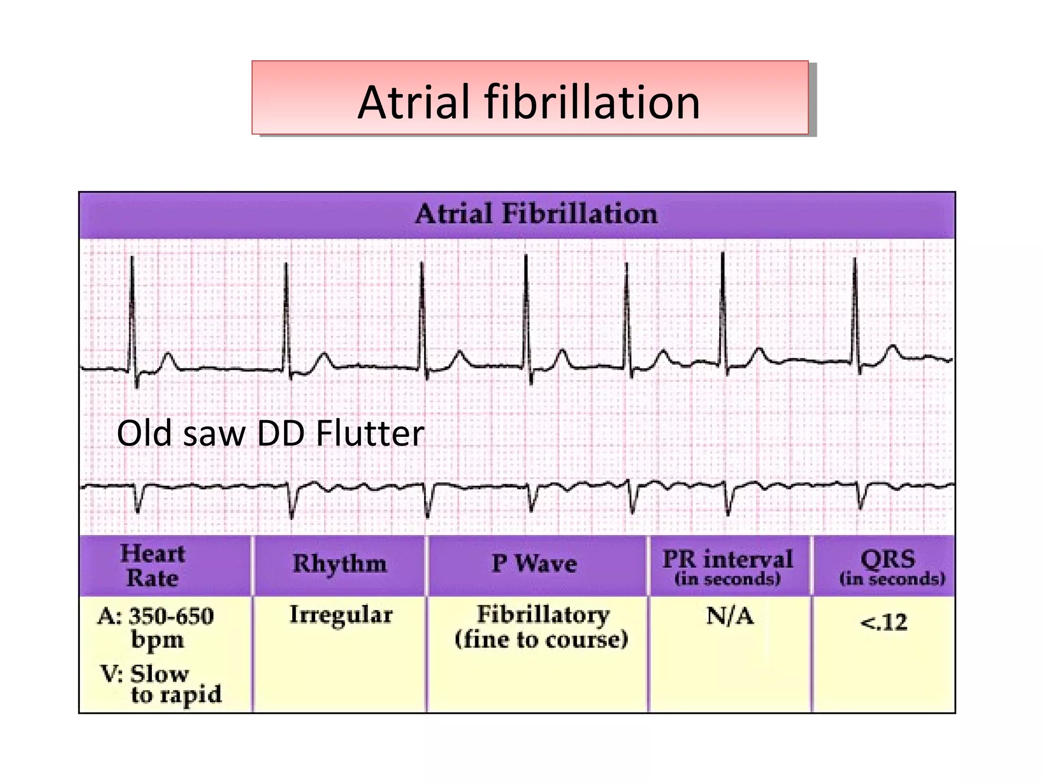
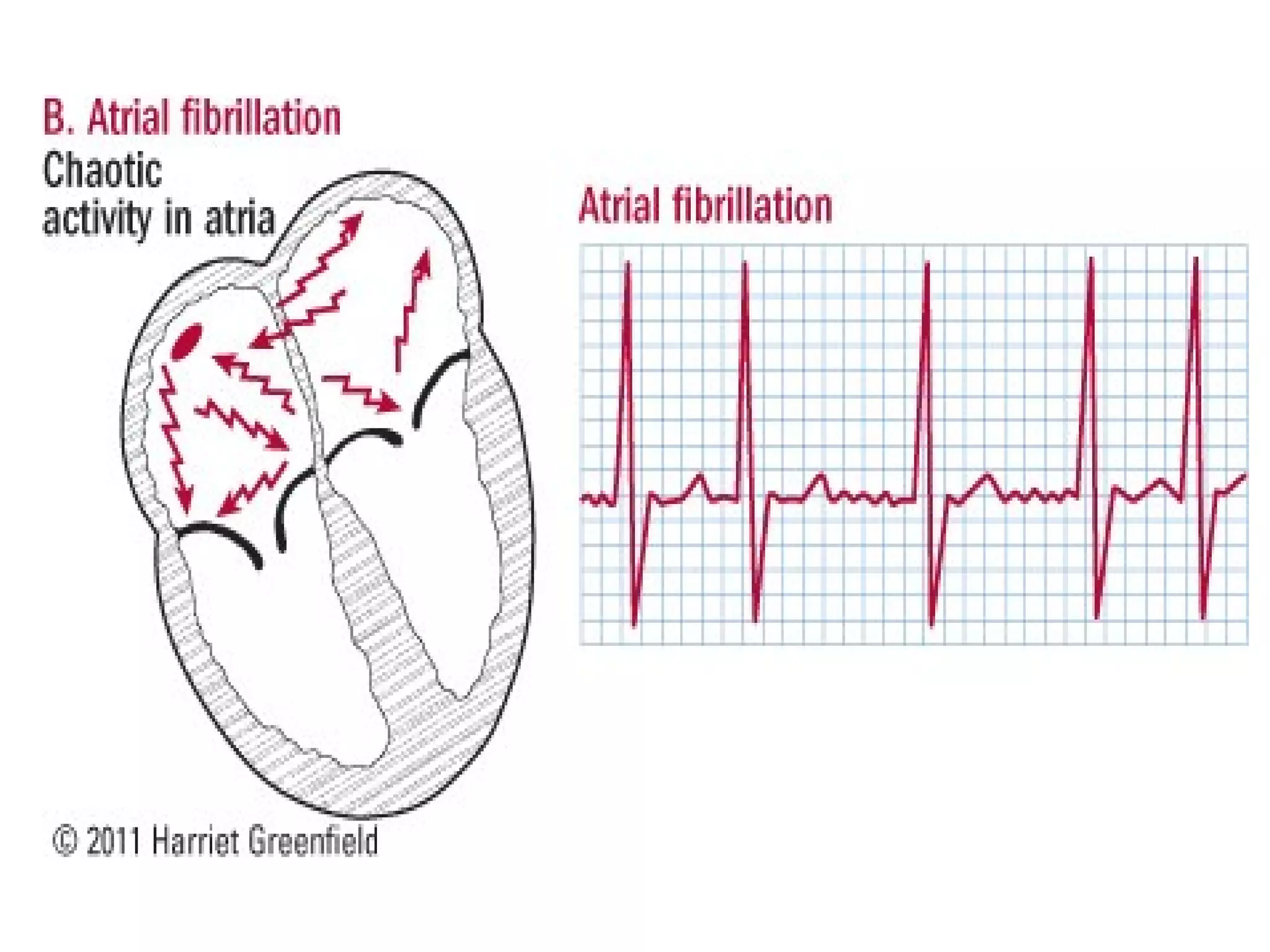
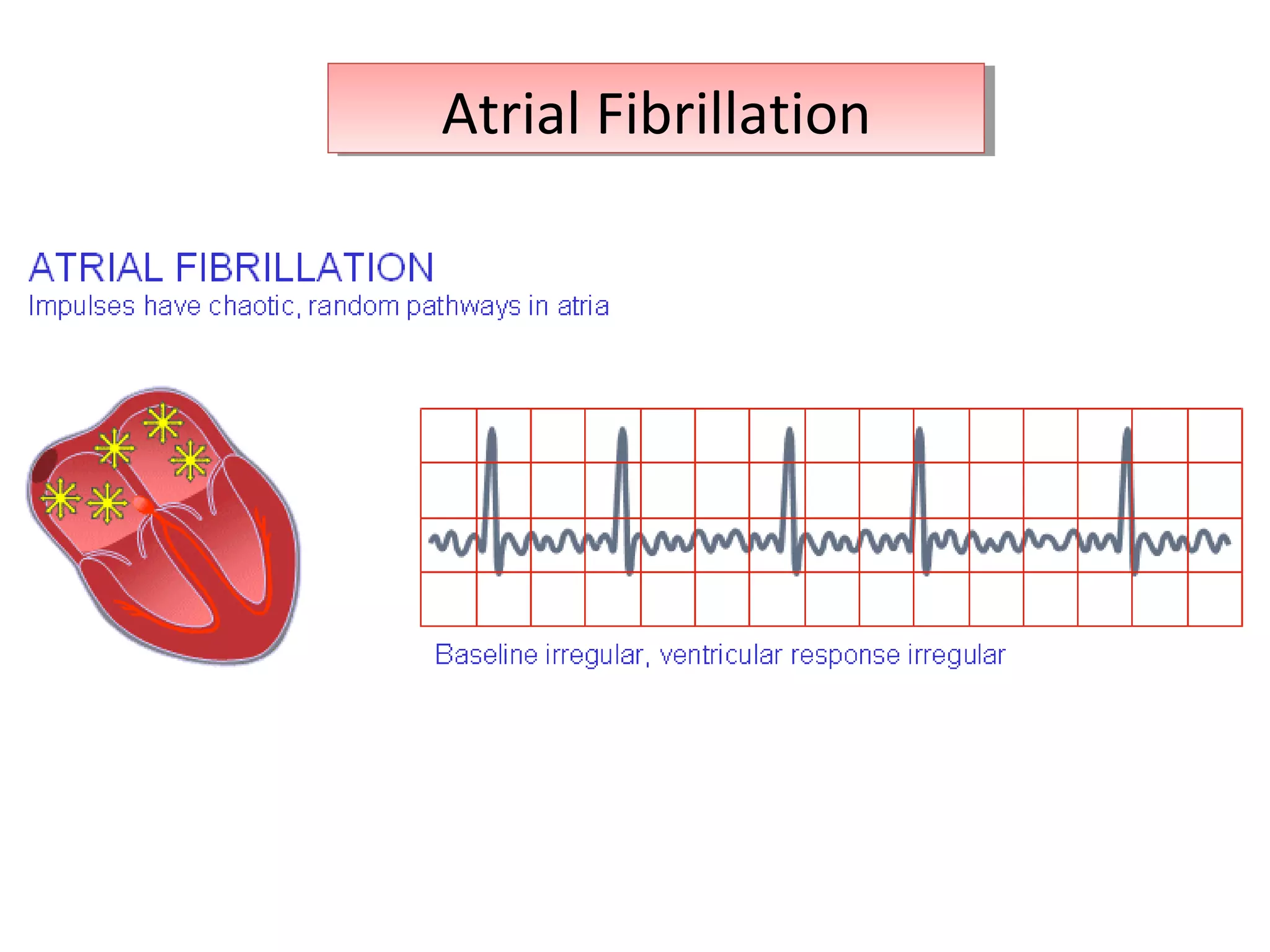
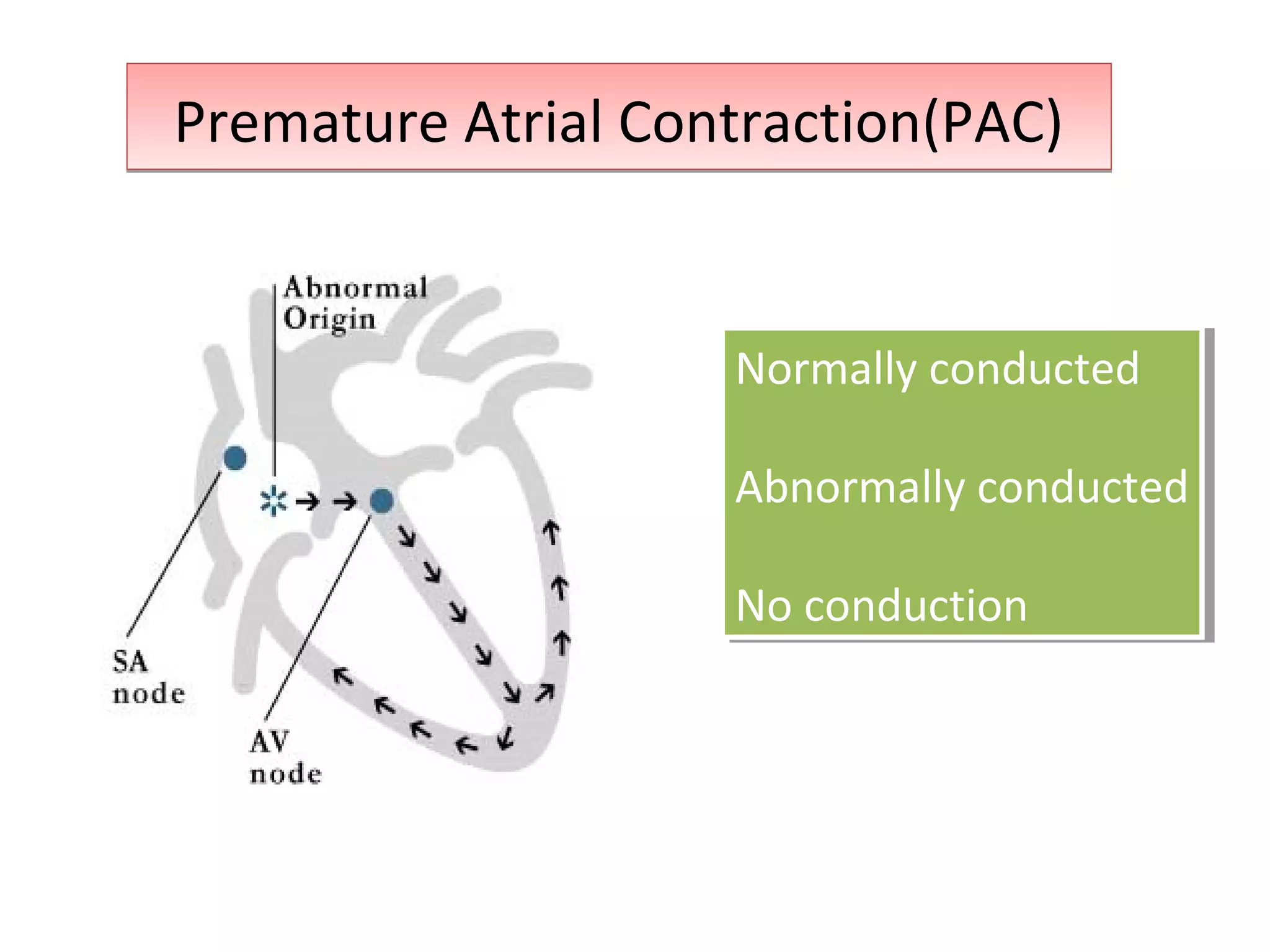
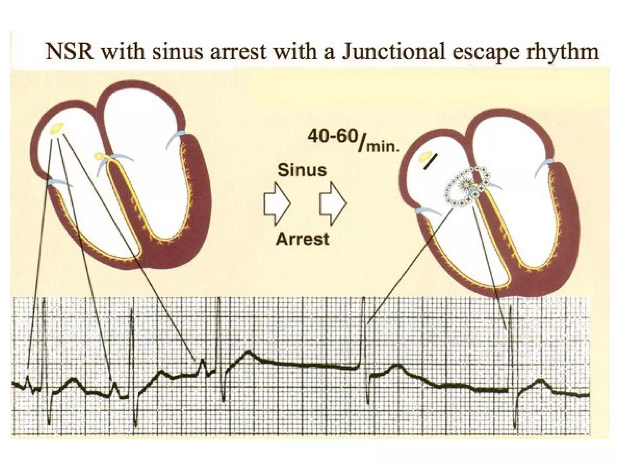
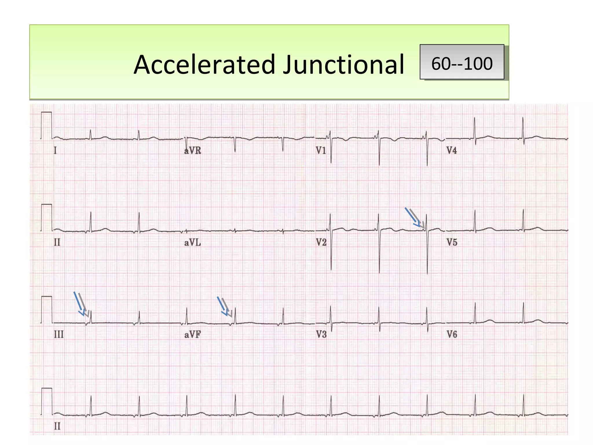
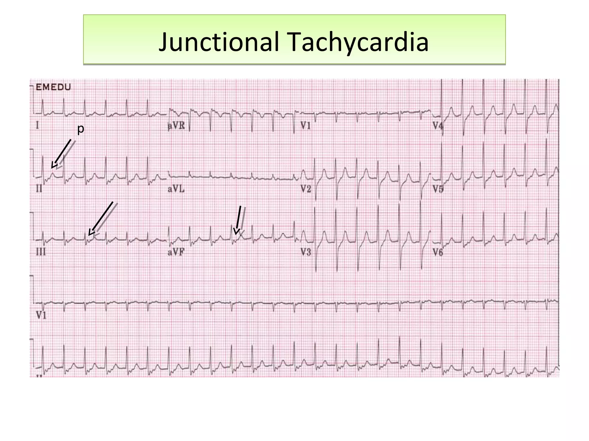
Call Girls Service In Shyam Nagar Whatsapp 8445551418 Independent Escort Service
ECG Atrial and Junctional rhythms Dr Sarfaraz
1. ATRIAL AND JUNCTIONAL
DYSRHYTHMIAS
ATRIAL AND JUNCTIONAL
DYSRHYTHMIAS
DR SARFARAZ AHMAD
ER PHYSICIAN
AFH--KANB
DR SARFARAZ AHMAD
ER PHYSICIAN
AFH--KANB
ECG
Interpretation
ECG
Interpretation
17. PAC
Conducted with aberration
PAC
Conducted with aberration
PAC WITH RBBB ABERRATION
Wide QRS called ASHMAN BEATWide QRS called ASHMAN BEAT
PAC makes it into the ventricles but finds a bundle branch or fascicle asleepPAC makes it into the ventricles but finds a bundle branch or fascicle asleep
22. Junctional DysrhythmiasJunctional Dysrhythmias
Junctional Rhythm(escape)
Accelerated Junctional
Junctional Tachycardia
Premature Junctional Contraction
(PJC)
Junctional Rhythm(escape)
Accelerated Junctional
Junctional Tachycardia
Premature Junctional Contraction
(PJC)
P wave inverted before or after QRS or hidden under QRS.P wave inverted before or after QRS or hidden under QRS.
24. Junctional RhythmJunctional Rhythm 40--6040--60
It is a normal response that may result from ----
•Excessive vagal tone on the SA node,
• A pathological slowing of the SA discharge,
• or a complete AV block.
28. Enhanced automaticity of the AV node
that supersedes the SA node rate.
Enhanced automaticity of the AV node
that supersedes the SA node rate.
Accelerated JunctionalAccelerated Junctional 60--10060--100
33. PSVTPSVT
Paroxysmal Supraventricular
(atrial) tachycardia is a regular,
fast (160 to 220 beats per minute)
heart rate that begins and ends
suddenly and originates in heart
tissue other than that in the
ventricles.
Paroxysmal Supraventricular
(atrial) tachycardia is a regular,
fast (160 to 220 beats per minute)
heart rate that begins and ends
suddenly and originates in heart
tissue other than that in the
ventricles.
34. P.S.V.T.P.S.V.T.
A narrow complex tachycardia/ atrial tachycardia
Originates in the 'atria‘
Not under direct control from the SA node.
SVT can occur in all age groups
• Types: depend on the location of reentry circuit---
1)atrioventricular nodal reentrant tachycardia
(AVNRT)
2)atrioventricular reentrant tachycardia (AVRT).
3) Sino-Atrial Reentrant Tachycardia.
49. CATHETER ABLATION OF ATRIAL
FLUTTER
A line is drAwn in the right Atrium
to block the flutter circuit.
Atrial flutter
circuit in the right atria
Atrial flutter
circuit in the right atria
Ablation lesionsAblation lesions
Ablation catheterAblation catheter
52. The achievement of the
electrocardiogram by
the physician and
nurses, in period of
crisis, is the
primordial element.
The achievement of the
electrocardiogram by
the physician and
nurses, in period of
crisis, is the
primordial element.
Editor's Notes
Discrete, multifocal P' waves occurring at rates of 100-250/min and with varying P'R intervals (should see at least 3 different P wave morphologies in a given lead). Ventricular response is irregularly irregular (i.e., often confused with A-fib). May be intermittent, alternating with periods of normal sinus rhythm. Seen most often in elderly patients with chronic or acute medical problems such as exacerbation of chronic obstructive pulmonary disease. If atrial rate is <100 bpm, call it multifocal atrial rhythm
Accelerated Junctional Rhythm: This is an active junctional pacemaker rhythm caused by events that perturb pacemaker cells (e.g., ischemia, drugs, and electrolyte abnormalities). The rate is 60-100 bpm).
Ischemia (usually from right coronary artery occlusion) and digitalis intoxication are the two most common causes.
It can appear similar to atrioventricular nodal reentrant tachycardia.
Rate: Underlying rhythmRhythm: Irregular with PJC'sPacemaker: Ectopic junctional pacemakerP wave: If present, negative in Lead 2PRI: .12 seconds or lessQRS: .08-.12 seconds, unless prolonged by aberrant conduction
Supraventricular tachycardia can be broadly defined as any tachycardia requiring the atrium or the atrioventricular (AV) node, either in whole or in part, for its perpetuation. Sino-Atrial Reentrant Tachycardia: This is a rare form of PSVT where the reentrant circuit is between the sinus node and the right atria. The ECG looks like sinus tachycardia, but the tachycardia is paroxysmal; i.e., it starts and ends abruptly.
Predictions about V:A time for DDX
Circuits require two pathways with different conduction vel and different refractory periods
Onset of AVNRT is triggered by a premature atrial impulse. The most frequent mechanism of SVT, usually a regular narrow QRS tachycardia . May occur in healthy, young individuals, and it most commonly occurs in women. Most patients do not have structural heart disease.
The AP conduction varies extensively from patient to patient.3 It can conduct antegradely during sinus rhythm (manifest Wolff-Parkinson-White (WPW) syndrome). Or it may conduct retrogradely from ventricle to atrium during tachycardia (concealed WPW syndrome). In manifest WPW syndrome the antegradely conducting AP excites the ventricle before the AV node, resulting in a delta wave on the surface electrocardiogram
Wolff-Parkinson-White syndrome (WPW) is a syndrome of pre-excitation of the ventricles of the heart due to an accessory pathway known as the bundle of Kent. This accessory pathway is an abnormal electrical communication from the atria to the ventricles. WPW is a type of atrioventricular reentrant tachycardia.
the abnormal electrical connection in the heart can be indentified and blocked either by burning or freezing the tissues.
Ablation can also be carried out for atrial flutter, where a line is drawn in the right atrium to block the flutter circuit. In recent years we have also learnt how to carry out ablation in atrial fibrillation.
Wolff-Parkinson-White syndrome is the name for episodes of fast heart rate caused by an extra electrical pathway between the atria and the ventricles. Electrical impulses can go down the normal pathway from the atria to the ventricles and then return to the atria through the extra pathway. Radiofrequency catheter ablation is the treatment of choice for Wolff-Parkinson-White syndrome. In this procedure, catheters are threaded through the blood vessels to the inside of the heart, and electrodes at the catheter tips are heated to destroy (ablate) a small spot of heart tissue, which blocks the extra electrical pathway.
Radiofrequency ablationIn most patients, atrial flutter is a macroreentrant circuit around the right atrium with a critical isthmus of tissue (right posteroseptal atria between inferior vena cava, tricuspid valve, and coronary sinus ostium) necessary for propagation of the flutter. This type of ablation is successful in over 90% of patients with very low complication rates. Therefore, ablation is the preferred method of treatment. This figure shows an atrial flutter circuit in the right atria (black arrow head) with the ablation lesions (red lines) delivered to terminate and prevent the atrial flutter.
All junctional tachycardia are Supraventricular tachycardia but all Supraventricular are not junctional.