Gonadal system

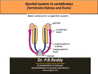
The male and female reproductive systems develop initially embryonically "indifferent", it is the product of the Y chromosome SRY gene that makes the "difference". ♂ - Male ♀ - Female The reproductive organs are developed from the intermediate mesoderm. The permanent organs of the adult are preceded by a set of structures which are purely embryonic, and which with the exception of the ducts disappear almost entirely before the end of fetal life. These embryonic structures are the mesonephric ducts (also known as Wolffian ducts) and the paramesonephric ducts, (also known as Müllerian ducts). The mesonephric duct remains as the duct in males which gives rise to seminal vesical, epididymes and vas deferens, and the paramesonephric duct as that of the female. Importantly its sex chromosome dependence, late embryonic/fetal differential development, complex morphogenic changes, long time-course, hormonal sensitivity and hormonal influences make it a system prone to many different abnormalities. Gonads: Gonads Produce eggs and sperm cells, transport and sustain egg and sperm cells, nurture developing offspring, and produce hormones. The gonads, ovary or testis, also develop in the intermediate mesoderm. They originally form as swellings that lie just ventral to the anterior mesonephric kidney. A mullarian duct also develops in the intermediate mesoderm near the mesonephric duct. Due to fusion or failure of 1st ridge to differentiate, some vertebrates (agnathans, some female lizards & crocodilians, & most female birds) have a single testis or ovary. Hormones cause differentiation of early gonads into either testes or ovaries. As males develop the mesonephric duct makes connection with the testis as the primary sperm conducting duct, and the mullerian duct is lost.

Recommended
More Related Content
What's hot
What's hot (20)
Similar to Gonadal system
Similar to Gonadal system (20)
More from Govt.college,Nagda, ujjain.M.P
More from Govt.college,Nagda, ujjain.M.P (20)
Recently uploaded
Recently uploaded (20)
Gonadal system
- 1. Genital system in vertebrates (Vertebrate Kidneys and Ducts) Dr. P.B.Reddy M.Sc,M.Phil,Ph.D, FIMRF,FICER,FSLSc,FISZS,FISQEM PG DEPARTMENT OF ZOOLOGY GOVERTNAMENT PG COLLEGE, RATLAM.M.P reddysirr@gmail.com
- 3. Introduction: The male and female reproductive systems develop initially embryonically "indifferent", it is the product of the Y chromosome SRY gene that makes the "difference". ♂ - Male ♀ - Female The reproductive organs are developed from the intermediate mesoderm. The permanent organs of the adult are preceded by a set of structures which are purely embryonic, and which with the exception of the ducts disappear almost entirely before the end of fetal life. These embryonic structures are the mesonephric ducts (also known as Wolffian ducts) and the paramesonephric ducts, (also known as Müllerian ducts). The mesonephric duct remains as the duct in males which gives rise to seminal vesical, epididymes and vas deferens, and the paramesonephric duct as that of the female. Importantly its sex chromosome dependence, late embryonic/fetal differential development, complex morphogenic changes, long time-course, hormonal sensitivity and hormonal influences make it a system prone to many different abnormalities.
- 4. Gonads: Gonads Produce eggs and sperm cells, transport and sustain egg and sperm cells, nurture developing offspring, and produce hormones. The gonads, ovary or testis, also develop in the intermediate mesoderm. They originally form as swellings that lie just ventral to the anterior mesonephric kidney. A mullarian duct also develops in the intermediate mesoderm near the mesonephric duct. Due to fusion or failure of 1st ridge to differentiate, some vertebrates (agnathans, some female lizards & crocodilians, & most female birds) have a single testis or ovary. Hormones cause differentiation of early gonads into either testes or ovaries. As males develop the mesonephric duct makes connection with the testis as the primary sperm conducting duct, and the mullerian duct is lost.
- 5. Ovaries: •In some teleosts, ovaries are hollow sacs, either because the ovary develops around coelom or the ovary becomes hollow at ovulation (eggs are discharged into cavity which is continuous with the oviduct). •In other teleosts plus agnathans, the ovaries are compact & eggs are discharged into coelom. •Amphibians - ovaries are hollow & eggs are discharged into the coelom. •Reptiles, birds, & monotremes - ovaries solid but develop irregular, fluid-filled lacunae (cavities); eggs discharged into coelom. •Mammals - ovaries compact; no large chambers or lacunae.
- 6. Testes: •Usually smaller than ovaries because sperm are much smaller than eggs (especially eggs with yolk). •In mammals, testes are larger than ovaries. Translocation of testes in mammals: •Testes descend permanently into scrotal sacs in many mammals. •Some mammals - testes lowered into scrotal sacs & retracted at will. •Inguinal canal - passage between abdominal cavity & scrotum. •Scrotal sacs - do not develop in some mammals; testes remain in abdomen.
- 7. Male genital ducts: •Some fishes (e.g., gar & sturgeon) & amphibians - mesonephric duct transmits sperm & urine. •Some amphibians - mesonephric duct transports only sperm; new accessory urinary duct drains the kidney. •Sharks - mesonephric duct is used primarily for sperm transport; accessory urinary duct develops. •Teleosts - mesonephric duct drains kidney; separate sperm duct develops. •Amniotes - embryonic mesonephric ducts transport sperm in adults.
- 8. Intromittent organs: Useful when fertilization is internal; introduce sperm into female reproductive tract. Found in some fish, some birds, reptiles, & mammals. Cartilaginous fish - appendages of pelvic fins called claspers direct sperm into female reproductive tract. Snakes & lizards - have pair of HEMIPENES (pocket like diverticula of wall of cloaca). Turtles, crocodilians, a few birds, & mammals - exhibit an unpaired erectile penis. Penis - usually a thickening of floor of cloaca consisting of spongy erectile tissue (corpus spongiosum) with grooves to direct sperm & ending in a glans penis (sensory endings that reflexly stimulate ejaculation). Mammals (except monotremes) - penis extends beyond body. The embryonic corpus spongiosum becomes a tube with urethra inside & 2 additional erectile masses develop (corpus cavernosa).
- 9. Female genital ducts: Consists of a pair of gonoducts (or oviducts) that extend from ostia to the cloaca. Different segments of ducts perform special functions. When internal, fertilization occurs near beginning of ducts, the mullerian duct becomes the passage for eggs. The females of fish and amphibians retain the mesonephric duct as a urinary duct. In reptiles, birds, and mammals (amniotes) the metanephric kidney replaces the mesonephric kidney.
- 10. Anatomy in various vertebrate groups: Cartilaginous fish - 2 ostia fuse to form single ostium (or osteum); shell gland secretes albumen & a shell; uterus holds eggs until laying. Teleosts - ducts are continuous with cavity of the ovary. Lungfish & amphibians - oviducts long & convoluted; lining secretes jelly-like material around each egg. Crocodilians, some lizards, & nearly all birds (diagram below) - 1 coiled oviduct lined with glands that add albumen, shells, &, sometimes, pigment Monotremes - tract is reptilian; caudal end secretes a shell before egg passes into the cloaca. Placental mammals - embryonic ducts give rise to oviducts, uteri, & vaginas. Adult tract is paired anteriorly & unpaired posteriorly (typically terminating as an unpaired vagina). oviducts (fallopian tubes) are relatively short, small in diameter, convoluted, & lined with cilia; begin at ostium bordered with fimbria. Uterus: Marsupials - no fusion of embryonic ducts so there are 2 tracts (DUPLEX UTERUS)
- 11. Other placental mammals - varying degrees of fusion: Bipartite uterus - 2 uterine horns, a uterine body (with 2 lumens), & a single vagina. Bicornuate uterus - 2 uterine horns, a uterine body (with a single lumen), & a single vagina. Simplex uterus - no uterine horns & oviducts open directly into body of uterus. Vagina - fused terminal portion of oviducts that opens either into urogenital sinus or to the exterior; receives male intromittent organ.
- 13. Both Müllerian and Wolffian ducts are present at the bipotential stage. In males, the Müllerian ducts degenerate under the influence of Anti-Müllerian hormone (AMH) secreted by the testicular Sertoli cells. Wolffian ducts differentiate into epididymides, vasa deferentia, and seminal vesicles under the control of androgens produced by Leydig cells. In females, the Wolffian duct regresses and the Müllerian duct differentiates into oviduct, uterus, and upper vagina.
- 14. Fish male FROG -MALE - URENOGENITAL SYSTEM L. Testes are very long and ribbon like. 1. Testes are small and rod-like. 2. Testes are attached to the kidneys anteriorly. 2. Testes are attached to the kidneys above the middle region with mesorchium. 3. Testes are connected with the rectal gland by epigonal organs. 3. The epigonal organs are absent. 4. Vasa efferentia leave the testis at its anterior end. 4. Vasa efferentia leave the testis alone its inner border. 5. These open into the wolffian duct. 5. open into .the Bidder's canal which drains into the wolffian duct. 6. Wolffian duct is differentiated into an anterior narrow closely convoluted epididymis and the posterior wide less convoluted vesiculus seminalis. 6.Wolffian duct is not differentiated into the parts except having a small seminal vesicle near its beginning. 7. Wolffian ducts act only as genital ducts. 7. Wolffian ducts act as both urinary and genital duct. Hence they are known as urinogenital ducts. 8. A pair of club-shaped sperm sac open into the urinogenital sinus 8. Sperm sacs are absent 9. The copulatory apparatus comprising siphons and claspers. 9. Copulatory apparatus is absent. 10. Sperms are released into the genita iuct of female, hence fertilization is internal. 10. Sperms are released over the eggs tithe fresh water, hence fertilization is external. Sperms are released as milt' 11. There are no fat bodies attachec to the testes. 11. There is a large branched fai-bodv attached to the anterior end of ead testis. 12. There is a single urino-genital 12. Urino-genital papillae are paired. SHARK -FEMALE - REPRODUCTIVE SYSTEM FROG -FEMALE - REPRODUCTIVE SYSTEM 1. Ovaries are small, tabulated bodies 1. Ovaries are large, hollow, lobed sacs 2. connected with rectal glands by long epigonal organs. 2. Epigonal organs are absent. 3. Oviducts (Mullerian ducts) are long but not convoluted. 3. Oviducts (Mullerian ducts) are very long and greatly convoluted. 4. Oviducts converge and unite in front of the ovaries leaving a slit 'ostium tubae! The oviducal funnels are on either side of ostium tubae. 4. Oviducts converge infront of the ovaries but do not unite. Each oviduct has its own ostium at the lip of oviducal funnel. 5. shell glands are present. 5. There is no shell gland 6. The oviducts are expanded to form large uteri in the region of renal part of kidneys 6. The oviducts are expanded to form small ovisacs behind the kidneys. 7. The oviducts join to form a median sac-'vagina' which opens into the cloaca. 7. The oviducts independently open into the cloaca. The vagina is absent. 8. There are no fat bodies. 8, There is a large fat body attached to the anterior end of each ovary. 9 Since the fertilization in internal, there is no question of releasing the egg out side the body 9. The mass of eggs is called "spawn* which is released outside the body Fertilization is external.
- 15. FEMALE-CALOTES FEMALE-PEGION FEMALE-RABBIT 1, Two ovaries are present. 1. Only left ovary is present. 1. A pair of ovaries are present on the left and right sides. They are small and compact bodies. 2. Ovaries are irregular bodies situated asymmetrically and hanging from the dorsal wall of the body cavity by mesovaria. 2. The single left ovary is attached to anterior lobe of the left kidney by mesovarium. 2. The ovaries lie behind the kidneys and attached to dorsal wall of the abdominal cavity by mesovaria. 3. Right ovaiy is a little anterior to the left one. 3. Right ovary is absent. 3. Right and left ovaries are at the same level. 4. Oviducts are paired. 4. Only left oviduct is developed. 4. Oviducts are paired. 5. Oviducts give striated appearance over greater part of their length. 5. Oviduct doesnot give striated appearance. 5: Striated appearance is absent. 6. Oviducts extend well ahead of ovaries and follow straight course. 6. Oviduct starts'just behind the ovary and follow a convoluted course. 6. Oviducts start just outside the ovaries and follow convoluted course. 7. Oviducal funnel are large and have externally directed Ostia with the entire margin. 7. Single Left oviducal funnel is very large and membranous. It has fimbricated margins it lies close to the ovary with it; ostium. 7. Oviducal funnel are small and have internally directed ostia with fimbricated margin. 8. Each oviduct dilates to form an oval shell gland along the ventral surface of the kidney uteri are not demarcated. Oviducts are enlarged into small ovisacs. These are two vaginae. Vaginae open into the urodaeum. 8. The oviduct is divided into anterior oviducal funnel, behind it magnum which is thick walled & secretes albumen. The posterior most part is the thick walled and muscular vagina. 8. The oviducal funnel leads distally in a narrow and convoluted tube - Fallopian tube. The posterior most parts of the two oviducts form uteri. The paired uteri open into median and highly muscular chamber-vagina. Associated with urethra, the vagina forms the urino-genital canal or vestibule. 9. Urodaeum opens outside by a cloaca 9. Urodaeum opens out by a cloaca. 9. Urino - genital canal opens outside by a longitudinal aperture - vulva in front of the anus. 10. There are no special glands associated with female reproductive system. 10. Same as in calotes. 10. Cowper's and perineal glands are associated with female reproductive system. 11. Milk glands are absent. 11. Milk Glands are absent. 11. On the ventral surface of trunk region 4 or 5 pairs of milk glands open through their tears. 12. Fertilization is internal oviparous animal. 12. Same as in calotes. Oviparous animal 12. Same as in calotes and columba. Viviparous animal
- 16. MALE-CALOTES (GARDENLIZARD) MALE- (PIGEON) MALE (RABBIT) 1. Testes are white ovoid bodies. 1. Testis white ovoid bodies. 1. Testes are pink, ovoid bodies. 2. Testes lie in the abdominal cavity much ahead of kidneys. Inguinal canal is absent. 2. Testes lie in the abdominal cavity under the anterior parts of kidneys. Inguinal canal is absent. 2. Testes are extra abdominal and lie in the scrotal sacs which are the folds of the skin. They are connected with perivisceral cavity by inguinal canals. 3 Right testis is a little ahead of the left one. 3. Left testis is a little bigger than the right one. 3. Right and left testes are symmetrical. 4. Spermatic cord is not formed. 4. Same as in calotes. 4. A spermatic cord extends from each testis to a little behind the kidney of its side. 5. Each testis is attached to the dorsal body wall by a double fold of peritoneum the mesorchium. 5. Each testis is attached to the kidney of its side by mesorchium. 5. Each testis is attached to the wall of scrotal sac by a short, thick, elastic cord 'gubernaculum'. 6. From the inner end of each testis arises a much convoluted tube-epididymis. 6. Epididymis is absent. 6. Epididymis is present. 7. Caput and cauda epididymis are not found 7. Same as in calotes. 7 Caput epididymis and cauda epididymes are the extensions of epididymis in front and behind the testis 8. Epididymis is continued behind as long, narrow, coiled and delicate vas deferens. It passes backwards along the ventral surface of the kidney of its side and joins with the ureter to form urino-genital sinus which opens into the cloaca. 8 The vas deferens arises directly from the inner border of the kidney in the form of a narrow convoluted tube. It runs backwards outside the ureter parallel to it and both open dorsally by separate aperture in urodaeum of the cloaca. 8. The vas deferens passes through the inguinal canal and runs forward and enters into the abdominal cavity. So that a loop around the ureter of its side to open into sac-uterus masculinus' which is present in the dorsal wall of the urinary bladder. 9. Seminal vesicles are absent. 9. Posterior end of each vas deferens enlarges to form seminal vesicle. 9. Seminal vesicles are absent. 10. There are no special glands associated with male genital system. 10. Same as in calotes. 10. There are prostate, couper's and perineal glands are associated with the male genital system. 11. In male a pair of eversible copulatory organs 'hemipenis' lie under the skin behind cloacal aperture 11. Copulatory organs are absent. 11. The copulatory organ in male is in the form of a thick muscular 'Penis'. It is covered by skin loose fold prepuce or foreskin penis is made up of a spongy tissue containing blood vessels and it is erectile.