SlideShare a Scribd company logo
1 of 46
Download to read offline
THE REPRODUCTIVE SYSTEMS
• Overall function is to produce offspring
• Testes produce sperm and male sex hormones
• Ovaries produce eggs and female sex hormones
• Mammary glands produce milk
COMPILED BY HOWIE BAUM
INTRODUCTION
The reproductive system is a collection of internal and external organs — in both males and
females — that work together for the purpose of procreating.
Due to its vital role in the survival of the species, many scientists feel that the
reproductive system is among the most important systems in the entire body.
Of the body’s major systems, the reproductive system is the one that differs most
between sexes, and the only system that does not function until puberty.
The male reproductive system is responsible for delivering sperm to the female reproductive
system
MALE REPRODUCTIVE SYSTEM
In males, the reproductive organs include the
penis, the testes, a number of storage and
transport ducts, and some supporting structures.
The two oval-shaped testes (also called
testicles) lie outside the body in a pouch of
skin called the scrotum, where they can
maintain the optimum temperature for
sperm production – approximately 5° F,
lower than body temperature.
Testes are oval-shaped glands responsible for
the manufacture of sperm and the sex hormone
testosterone.
From each testis, sperm pass into a coiled tube
– the epididymis – for the final stages of
maturation.
INSIDE THE SCROTUM
The scrotum contains two testes
(testicles) where sperm are
manufactured within tubes called
seminiferous tubules, and the
two epididymides where sperm
are stored.
Unlike female egg maturation,
which occurs in cycles and
ceases at menopause, sperm
production is continuous,
reducing gradually with age.
Each epididymis is a tube
about 20 feet long, which is
tightly coiled and bunched
into a length of just 2 inches.
MAKING SPERM
Each testis is a mass of more than 800 tightly
looped and folded vessels known as
seminiferous tubules.
Inside each tubule, sperm begin as blob-like
cells called spermatogonia lining the inner
wall.
These pass through a larger stage, as
primary spermatocytes, then become
smaller as secondary spermatocytes, and
begin to develop tails as spermatids.
As all of this happens, they move steadily
towards the middle of the tubule.
The spermatids finally develop into ripe sperm
with long tails.
Thousands of sperm are produced every
second, each taking about two months to
mature.
DIFFERENCE IN SEMEN AND SPERM
Semen, also known as seminal fluid, is
much more than just sperm.
Sperm is only about 5 to 10% of any
given male single ejaculation and the rest
is fructose (aka sugar), fatty acids, and
proteins to nourish the sperm during
their journey.
Sperm Count
A man will produce roughly 525
billion sperm during his whole
lifetime and close to 1 billion per
month.
There are around 200 to 500 million
sperm in an average in a single
human ejaculation !!
Sperm cell - A sperm is about 1/500 inch long,
but most of this is a tail.
The sperm head is only 1/5000 inch, about the
same size as a red blood cell.
Sperm develop in the
testes and consist of a
head, a midpiece, and a
tail.
The head contains the
nucleus with densely
coiled chromatin fibers
(chromosomes), with a
front section – the
Acrosome that contains
enzymes for penetrating
the female egg.
The midpiece has a central
filamentous core with many
Mitochondria spiraled
around it, to give it energy
to move the tail which
propels it forward.
The female reproductive system
includes external and internal
genitalia.
The vulva and its structures form
the external genitalia.
The internal genitalia include a
three-part system of ducts: the
uterine tubes, the uterus, and
the vagina.
This system of ducts connects to
the ovaries, the primary
reproductive organs.
The ovaries produce egg cells
and release them for
fertilization.
Fertilized eggs develop inside the
uterus.
Unlike the male, the female
reproductive organs are located
entirely inside the body.
From Puberty, their function is to ripen
and release an egg at regular intervals,
and, if the egg is fertilized, to protect
and nourish the embryo and fetus.
No eggs are manufactured after
birth – a female is born with a full
set.
Reproductive tract
The female reproductive glands (ovaries)
are located within the abdomen.
This release occurs roughly once a month
as part of the menstrual cycle.
The ripe egg travels along the fallopian
tube to the uterus, the muscular sac in
which it develops into an embryo and
then fetus.
EGG CELLS FROM THE OVARIES MOVE
THROUGH THE UTERINE TUBES
The uterine tubes (also called Fallopian
tubes or oviducts) connect the ovaries to
the uterus.
The walls of each tube have an external
serous layer, a middle muscular layer, and an
internal mucous layer that is continuous with
the inner lining of the uterus.
Each uterine tube can be divided into three
parts:
infundibulum
Isthmus connects with the uterus.
A dilated portion, the ampulla, curves over
the ovary.
Egg fertilization usually occurs in the
ampulla. The eggs then travel through
the isthmus into the uterus.
THE VAGINA: A TUNNEL WITH THREE
CORE FUNCTIONS
The vagina extends down from the cervix, the
lower part of the uterus, to the vestibule, which is
part of the vulva and the external genitalia.
It sits behind the bladder and in front of the
rectum.
An inner mucous membrane lines the smooth
muscle walls of the vagina.
This lining, like the inner layer of the uterine tubes,
is continuous with the mucous lining of the uterus.
The vagina has three core functions:
 It carries menstrual flow outside the body
 It receives the male penis during sexual
intercourse
 It serves as a birth canal during labor.
OVULATION
An ovary contains thousands of immature
egg cells.
During each menstrual cycle, follicle-
stimulating hormone (FSH) causes one
egg to begin development; this takes
place inside a primary follicle.
The follicle enlarges as its cells proliferate, and
begins to fill with fluid, becoming a secondary
follicle that moves to the ovary’s surface.
It also increases its production of the hormone
Estrogen.
A surge of luteinizing hormone (LH)
causes the follicle to rupture and release
the ripe egg–this is ovulation.
The lining of the empty follicle thickens into a
corpus luteum–a temporary source of
hormones.
https://www.youtube.com/watch?v=nLmg4
wSHdxQ
The ovary contains undeveloped eggs, eggs in follicles at
various stages of maturation, and empty follicles forming
corpora lutea. The bulk of the glandular tissue surrounding
these follicles is known as the stroma.
EGG AND SPERM HAVE DEVELOPED SOME PRETTY NIFTY TRICKS TO
MAKE CONCEPTION HAPPEN
Fortify the troops - The liquid portion of semen not only provides the sperm with nourishment for the
journey, it actually coagulates in a woman's vagina after ejaculation, forming a physical barrier that prevents
the sperm from wandering very far in the wrong direction. This protection disappears within half an hour,
when the semen becomes more fluid again.
Call in the transport unit - The cervical canal is a much more welcoming environment, and sperm that
make it there find themselves awash in a sea of cervical mucus. The mucus is specially designed to transport
sperm efficiently when you're most fertile. As you approach ovulation, your suddenly copious mucus becomes
stretchy, clear and thin and strings of molecules line up like train tracks so that sperm can hop on and ride to
their destination.
Picking up speed - A just-ejaculated sperm cell has to spend a couple of hours going through biochemical
changes, picking up tail-thrashing speed to help it make its way into the uterus and fallopian tubes to find its
target.
Timing is Important - Sperm must reach their destination within the right time frame for when the egg is
there.
They also need to pick their destination carefully - An egg is usually only present in one of the two
fallopian tubes in any given month. Pick the wrong tube, and the sperm end up hanging out partying together
with no guest of honor in sight.
May the best sperm win!
Note that the egg
has 2 layers that
the sperm needs to
get through – The
outer Corona
Radiata irregular
surface and then
the Zona Pellucida
which is a
transparent but
thicker layer.
The first sperm
to reach the
oocyte is never
the one to
fertilize it.
Rather, hundreds
of sperm cells
must undergo the
acrosomal
reaction, each
helping to degrade
the corona radiata
and zona pellucida
until a path is
created to allow
one sperm to
contact and fuse
with the plasma
membrane of the
oocyte (egg).
FERTILIZATION: A SPERM AND AN EGG
FORM A ZYGOTE
During sexual intercourse, some sperm ejaculated
from the male penis swim up through the female
vagina and uterus toward an oocyte (egg cell)
floating in one of the uterine tubes.
The sperm and the egg are called gametes.
They each contain half of the genetic
information necessary for reproduction.
When a sperm cell penetrates and fertilizes an
egg, that genetic information combines.
The 23 chromosomes from the sperm pair with 23
chromosomes in the egg, forming a 46-
chromosome cell called a zygote.
The zygote starts to divide and multiply.
As it travels toward the uterus it divides to become
a blastocyst, which will burrow into the uterine wall.
https://www.youtube.com/watch?v=_5OvgQW6FG4
THE ZYGOTE BECOMES AN EMBRYO: DEVELOPMENT
PRIOR TO AND DURING IMPLANTATION
A fertilized egg, or zygote, takes about five days to reach the uterus
from the uterine tube.
As it moves, the zygote divides and develops into a blastocyst, with
an inner mass of cells and a protective outer ring.
The blastocyst attaches to the wall of the uterus and gradually
implants itself into the uterine lining. During implantation, its cells
differentiate further.
At day 15 after conception, the cells that will form the embryo
become an embryonic disc. Other cells begin to form support
structures.
The yolk sac, on one side of the disc, will become part of the
digestive tract.
On the other side, the amnion fills with fluid and will surround the
embryo as it develops.
Other cell groups initiate the placenta and umbilical cord, which will
bring in nutrients and eliminate waste.
ZYGOTE
The fertilized egg
passes along the
fallopian tube.
Within 24–36 hours it
has divided into two
cells, then 12 hours
later into four cells,
and so on.
This process is known
as cleavage.
At each stage, the
resulting cells become
smaller, gradually
approaching normal
body cell size.
MORULA
The zygote divides
several times to form a
solid blackberry-like
cluster of 16–32 cells,
the morula (derived
from the Latin for
“mulberry”).
At around 3–4 days
after fertilization the
morula leaves the
fallopian tube and
enters the uterine
cavity.
BLASTOCYST
About six days after
fertilization, the cell
cluster forms a hollow
cavity and is known as a
blastocyst.
It floats within the uterus
for around 48 hours
before landing on the
thick uterus lining
(endometrium), which
softens to aid
implantation (burrowing
of the blastocyst into the
endometrium).
The inner group of cells
will become the embryo
itself.
EMBRYONIC DISC
Within the inner cell mass, an embryonic
disc forms.
This separates the cell cluster into the
amniotic cavity, which develops into a
sac that will fill with fluid and fold around
to cover the embryo, and the yolk sac,
which helps to transport nutrients to the
embryo during the second and third
weeks.
The disc develops three circular
sheets called the primary germ
layers –
 Ectoderm
 Mesoderm
 Endoderm
from which all body structures will
develop.
https://www.youtube.co
m/watch?v=dgPCDXmcQj
M 5 minutes
https://www.youtube.co
m/watch?v=UgT5rUQ9E
mQ 2.2 minutes
Early in development at the time of gastrulation a small group of cells are "put
aside" to later form oocytes (eggs) and sperm. These cells are described as
primordial germ cells (PGCs) and are a type of stem cell.
Primordial germ cells, the earliest recognizable precursors of gametes, arise outside the
gonads and migrate into the gonads during early embryonic development.
Human primordial germ cells first become readily recognizable at 24 days after
fertilization in a layer of the yolk sac.
Germ cells exit from the yolk sac into the hindgut tissue and then migrate until they
reach the location of the sex organs.
These cells differentiate at different times into male testis before puberty and female
ovary when the girl is young.
GROWING EMBRYO
As development proceeds, cells continue to
divide.
They move to form groups that will become
tissues and organs.
They also specialize to different cell types, as
genes in their chromosomes are switched off or
on.
In general, development is head-down, with the
brain and head taking shape early, then the body,
followed by the arms as small buds, and lastly
the legs.
By the end of the embryonic stage, eight
weeks after fertilization, all major organs
and body parts are formed.
From this time on, the baby is known as a fetus.
3 weeks - The neural tube forms. It will become
the spinal cord, enlarged at one end as the brain.
The simple tube-like heart pulsates. The embryo is
4/50– 5/50 inch long.
HUMANS MUST DEVELOP MALE OR FEMALE
GONADS AND GENITALIA TO BE CAPABLE OF
REPRODUCTION
Reproductive structures begin to form in the embryonic stage.
By week 6, gonads and genitalia are present but
undifferentiated. Whether they become male or female is
determined by one chromosome delivered by the sperm.
This pair contains an X sex chromosome from the female egg
and either an X or a Y sex chromosome from the male sperm.
If the chromosome pair is XY, the gonads develop into
testes starting in week 7.
If the chromosome pair is XX, the gonads become ovaries
starting in week 8.
Testes secrete testosterone, forming male genitalia
around week 10.
Without testosterone, female genitalia form.
All reproductive structures are in place at birth or shortly after.
At puberty, an increase in sex hormones will grow them to their
FOUR WEEKS
The four-chambered heart
beats, sending blood
through simple vessels.
Intestines, liver, pancreas,
lungs, and limb buds can be
seen.
The embryo is about 1/5
inch long.
EIGHT WEEKS
At this stage, the face and neck take
shape, the back straightens, and fingers
and toes can be differentiated clearly.
The embryo starts to move.
It is now around 1 to 1-1/5 inch in
length.
IN EIGHT WEEKS, THE EMBRYO DEVELOPS; BY
THE END OF WEEK 10, IT BECOMES A FETUS
Fifteen days after conception marks the beginning of the
embryonic period.
The embryo contains a flat embryonic disc that now
differentiates into three layers: the endoderm, the
mesoderm, and the ectoderm.
All organs of the human body derive from these
three tissues.
They begin to curve and fold and to form an oblong body.
By week 4, the embryo has a distinct head and tail and a
beating heart.
Over the next six weeks, limbs, eyes, brain regions, and
vertebrae form.
Primitive versions of all body systems appear. By the end
of week 10, the embryo is a fetus.
This is referring to the gestational age of the fetus.
The little groove or Philtrum that is in between our nose and upper lip is one of the
most important parts of the human face.
The Philtrum or also commonly called as ‘cupid’s bow’ is the hollow space that is in
between our nose and our upper lip.
http://www.youtube.com/watch?v=wFY_KPFS3LA
CHANGES IN THE FETUS
By 12 weeks the fetus has a large head compared
with the rest of its body, but its features are
distinctly human.
 All major internal organs are developed, and
even tiny nails exist as folds growing on the
fingers and toes.
 The external ears, eyelids, and 32 permanent
tooth buds have also formed.
 One month later, rapid development allows the
fetus to move its limbs vigorously, although this
is rarely felt by the mother at this stage.
 The external genitalia are visible, and a fine
downy hair (lanugo) grows over the body.
 As growth continues, the fetus becomes leaner
and wrinkly, but by the seventh to eighth month,
it starts to accumulate fat and assume the
“chubby” appearance of the newborn.
 The fetus is now somewhat restricted by the
uterus. The side of the placenta facing the fetus
is smooth and circular in outline, with the
umbilical cord attached at center.
CHANGES IN THE MOTHER
FIRST TRIMESTER
On average, pregnancy lasts for 40 weeks
from the first day of a woman’s last
menstrual period (usually 38 weeks from
fertilization).
By convention, the duration of
pregnancy is divided into thirds, or
trimesters, each lasting for about three
calendar months.
During this time, the mother’s body
undergoes many changes to support the
developing fetus, accommodate its increasing
size, and prepare itself for childbirth and
breast-feeding.
Breasts become tender and larger, with
darkened areolas; frequency of urination may
increase; nausea and vomiting are common.
SECOND TRIMESTER
The enlarging uterus shows
Heart rate increases
The mother’s forehead and cheek
skin may temporarily darken
(known as the “mask of
pregnancy”)
From week 10 of pregnancy,
the fetus grows inside the
uterus, fueled by nutrient-rich
blood supplied by the umbilical
cord.
The placenta provides oxygen
and nutrients to the fetus and
removes waste products from
the fetus’ blood.
THIRD TRIMESTER
Skin stretches over the abdomen
Slight contractions may be felt
Fatigue, back pain, heartburn,
and occasional breathlessness
may occur.
MULTIPLE PREGNANCY AND FETAL
POSITIONS
The presence of more than one fetus in the
uterus is called a multiple pregnancy.
Twins occur in about one in 80 pregnancies, and
triplets in about one in 8,000.
Both events are becoming more common, partly
due to improved antenatal care and also
increasing use of fertility methods such as IVF
(in vitro fertilization).
After about 30 weeks, the most common fetal
position is head down, facing the mother’s back,
with the neck flexed forward.
Such a position eases passage through the birth
canal.
However, about 1 in 30 full-term deliveries is
breech, in which the baby’s buttocks emerge
before the head.
Monozygotic twins - A single fertilized egg, or zygote,
forms an embryo that splits into two. Each develops into a
fetus. The two have the same genes and sex and share
one placenta. They look alike and are known as “identical”
twins.
DIZYGOTIC TWINS
Most of the time, a woman releases a
single egg during an ovulation cycle.
However, in approximately 1 percent of
ovulation cycles, two eggs are released
and both are fertilized.
Two zygotes form, implant, and develop,
resulting in the birth of dizygotic (or
fraternal) twins. Because dizygotic twins
develop from two eggs fertilized by two
sperm, they are no more identical than
siblings born at different times.
Two eggs are fertilized and develop
separately, each with its own placenta.
They may be different or the same sex.
Also called “fraternal twins”, they have the
same degree of resemblance as any
brothers and sisters.
FRANK BREECH
In frank breech, also called
incomplete breech, the baby
fails to turn head-down in
the uterus.
The hips are flexed and the
legs are straight, extending
alongside the body so that
the feet are positioned
beside the head.
COMPLETE BREECH
The baby’s legs are flexed at the
hips and knees, so the feet are next
to the buttocks.
This occurs less commonly than
frank breech.
The incidence of breech delivery is
much higher among premature
babies.
CHANGES IN THE CERVIX
The cervix is the firm band of muscle and
connective tissue that forms the neck-like
structure at the bottom of the uterus.
In late pregnancy, the cervix softens in
readiness for childbirth.
Sporadic uterine tightenings, known as
Braxton–Hicks contractions, help to thin the
cervix so that it merges with the uterus’s
lower segment.
Braxton–Hicks contractions are usually
painless, and occur through much of
pregnancy, becoming noticeable only after
mid-term.
Cervix softening
As labor nears, the cervix tissues lose their
firm consistency. They become softer and
more spongy, affected by natural
substances in the blood called prostaglandins.
CERVIX THINNING
The cervix becomes
wider and thinner, and
merges smoothly into
the uterus wall above.
The process of
softening and thinning is
known as effacement.
CONTRACTIONS
When pregnancy reaches full-term, the uterus
is the largest and strongest muscle in the
female body.
When its muscle fibers shorten, with the
eventual aim of expelling the fetus, it is known
as a uterine contraction or simply a
“contraction”.
True contractions, as opposed to Braxton–Hicks
contractions, are regular and become steadily
more frequent, more painful, and longer-
lasting.
The main area of contraction is in the uterine
fundus (upper uterus), which stretches, causing
the lower uterus and cervix to thin.
Judging when true labor has started can be
difficult due to “false alarms”.
EPIDURAL ANALGESIA
One of the most commonly used
methods of pain relief during labor
and delivery, epidural analgesia, is
delivered via a needle into the space
between the vertebrae and the
spinal column in the lower (lumbar)
region of the back.
It affects the nerve fibers that
detect contraction pains.
A new type of epidural, often
called a “walking epidural”,
reduces pain without removing
sensation, allowing women to
move around during labor and
participate actively in the
delivery.
BIRTH OF THE BABY
Around week 36 (usually), the process
of labor begins.
In the first stage, dilation, hormones
stimulate downward contractions of the
uterine walls.
The contractions push the head of the
fetus against the cervix at the lower
end of the uterus.
The cervix dilates.
In the second stage, expulsion,
powerful contractions push the head
and the rest of the body through the
dilated cervix, and out through the
vagina and the vulva.
The baby is born. Further contractions
expel the placenta to complete the
placental stage.
THE END

More Related Content

Similar to THE REPRODUCTION SYSTEMS.pdf

Reproduction in Man (Human Anatomy)
Reproduction in Man (Human Anatomy)Reproduction in Man (Human Anatomy)
Reproduction in Man (Human Anatomy)Marben Melencion
 
REPRODUCTION ANATOMYjjjjjjgjjjjjjhgui.pptx
REPRODUCTION ANATOMYjjjjjjgjjjjjjhgui.pptxREPRODUCTION ANATOMYjjjjjjgjjjjjjhgui.pptx
REPRODUCTION ANATOMYjjjjjjgjjjjjjhgui.pptxEmma910932
 
What is human reproduction
What is human reproductionWhat is human reproduction
What is human reproductionCherry Caralde
 
2. Human reproduction.pptx
2. Human reproduction.pptx2. Human reproduction.pptx
2. Human reproduction.pptxSrivatsanSP1
 
HUMAN REPRODUCTION.pptx
HUMAN  REPRODUCTION.pptxHUMAN  REPRODUCTION.pptx
HUMAN REPRODUCTION.pptxbarunmahto001
 
1 Basic medical facts about pregnancy.ppt
1 Basic medical facts about pregnancy.ppt1 Basic medical facts about pregnancy.ppt
1 Basic medical facts about pregnancy.pptShama
 
Basic medical facts about pregnancy.ppt
Basic medical facts about pregnancy.pptBasic medical facts about pregnancy.ppt
Basic medical facts about pregnancy.pptShama
 
15 Reproductive System.ppt
15 Reproductive System.ppt15 Reproductive System.ppt
15 Reproductive System.pptShama
 
178-Anatomy-Reproductive-System.ppt
178-Anatomy-Reproductive-System.ppt178-Anatomy-Reproductive-System.ppt
178-Anatomy-Reproductive-System.pptsadiaahmad30
 
Reproductionsystem 131109102239-phpapp01
Reproductionsystem 131109102239-phpapp01Reproductionsystem 131109102239-phpapp01
Reproductionsystem 131109102239-phpapp01Chethan Durgan
 
Reproductionsystem 131109102239-phpapp01
Reproductionsystem 131109102239-phpapp01Reproductionsystem 131109102239-phpapp01
Reproductionsystem 131109102239-phpapp01Chethan Durgan
 
Human Reproducton and biological change.pptx
Human Reproducton and biological change.pptxHuman Reproducton and biological change.pptx
Human Reproducton and biological change.pptxbholu803201
 
Reproductive system in Humans
Reproductive system in HumansReproductive system in Humans
Reproductive system in HumansBhavya Vashisht
 
Reproductive sys a&p
Reproductive sys a&pReproductive sys a&p
Reproductive sys a&pJay Patel
 
Organismal Reproduction and Development
Organismal Reproduction and DevelopmentOrganismal Reproduction and Development
Organismal Reproduction and DevelopmentFrezzy Vinson
 
Female reproductive anatomy
Female reproductive anatomyFemale reproductive anatomy
Female reproductive anatomyAhmed Ali
 
Chapter 4 Reproduction
Chapter 4 ReproductionChapter 4 Reproduction
Chapter 4 ReproductionBrandon Loo
 

Similar to THE REPRODUCTION SYSTEMS.pdf (20)

Reproduction in Man (Human Anatomy)
Reproduction in Man (Human Anatomy)Reproduction in Man (Human Anatomy)
Reproduction in Man (Human Anatomy)
 
REPRODUCTION ANATOMYjjjjjjgjjjjjjhgui.pptx
REPRODUCTION ANATOMYjjjjjjgjjjjjjhgui.pptxREPRODUCTION ANATOMYjjjjjjgjjjjjjhgui.pptx
REPRODUCTION ANATOMYjjjjjjgjjjjjjhgui.pptx
 
What is human reproduction
What is human reproductionWhat is human reproduction
What is human reproduction
 
2. Human reproduction.pptx
2. Human reproduction.pptx2. Human reproduction.pptx
2. Human reproduction.pptx
 
HUMAN REPRODUCTION.pptx
HUMAN  REPRODUCTION.pptxHUMAN  REPRODUCTION.pptx
HUMAN REPRODUCTION.pptx
 
1 Basic medical facts about pregnancy.ppt
1 Basic medical facts about pregnancy.ppt1 Basic medical facts about pregnancy.ppt
1 Basic medical facts about pregnancy.ppt
 
Basic medical facts about pregnancy.ppt
Basic medical facts about pregnancy.pptBasic medical facts about pregnancy.ppt
Basic medical facts about pregnancy.ppt
 
15 Reproductive System.ppt
15 Reproductive System.ppt15 Reproductive System.ppt
15 Reproductive System.ppt
 
Human Reproduction
Human ReproductionHuman Reproduction
Human Reproduction
 
178-Anatomy-Reproductive-System.ppt
178-Anatomy-Reproductive-System.ppt178-Anatomy-Reproductive-System.ppt
178-Anatomy-Reproductive-System.ppt
 
Reproductionsystem 131109102239-phpapp01
Reproductionsystem 131109102239-phpapp01Reproductionsystem 131109102239-phpapp01
Reproductionsystem 131109102239-phpapp01
 
Reproductionsystem 131109102239-phpapp01
Reproductionsystem 131109102239-phpapp01Reproductionsystem 131109102239-phpapp01
Reproductionsystem 131109102239-phpapp01
 
Human Reproducton and biological change.pptx
Human Reproducton and biological change.pptxHuman Reproducton and biological change.pptx
Human Reproducton and biological change.pptx
 
Reproductive system in Humans
Reproductive system in HumansReproductive system in Humans
Reproductive system in Humans
 
Reproductive sys a&p
Reproductive sys a&pReproductive sys a&p
Reproductive sys a&p
 
Organismal Reproduction and Development
Organismal Reproduction and DevelopmentOrganismal Reproduction and Development
Organismal Reproduction and Development
 
Human reproduction for grade 12
Human reproduction for grade 12Human reproduction for grade 12
Human reproduction for grade 12
 
REPRODUCTION IN ANIMALS.pptx
REPRODUCTION IN ANIMALS.pptxREPRODUCTION IN ANIMALS.pptx
REPRODUCTION IN ANIMALS.pptx
 
Female reproductive anatomy
Female reproductive anatomyFemale reproductive anatomy
Female reproductive anatomy
 
Chapter 4 Reproduction
Chapter 4 ReproductionChapter 4 Reproduction
Chapter 4 Reproduction
 

Recently uploaded

EPANDING THE CONTENT OF AN OUTLINE using notes.pptx
EPANDING THE CONTENT OF AN OUTLINE using notes.pptxEPANDING THE CONTENT OF AN OUTLINE using notes.pptx
EPANDING THE CONTENT OF AN OUTLINE using notes.pptxRaymartEstabillo3
 
AMERICAN LANGUAGE HUB_Level2_Student'sBook_Answerkey.pdf
AMERICAN LANGUAGE HUB_Level2_Student'sBook_Answerkey.pdfAMERICAN LANGUAGE HUB_Level2_Student'sBook_Answerkey.pdf
AMERICAN LANGUAGE HUB_Level2_Student'sBook_Answerkey.pdfphamnguyenenglishnb
 
Full Stack Web Development Course for Beginners
Full Stack Web Development Course  for BeginnersFull Stack Web Development Course  for Beginners
Full Stack Web Development Course for BeginnersSabitha Banu
 
ENGLISH6-Q4-W3.pptxqurter our high choom
ENGLISH6-Q4-W3.pptxqurter our high choomENGLISH6-Q4-W3.pptxqurter our high choom
ENGLISH6-Q4-W3.pptxqurter our high choomnelietumpap1
 
Difference Between Search & Browse Methods in Odoo 17
Difference Between Search & Browse Methods in Odoo 17Difference Between Search & Browse Methods in Odoo 17
Difference Between Search & Browse Methods in Odoo 17Celine George
 
Quarter 4 Peace-education.pptx Catch Up Friday
Quarter 4 Peace-education.pptx Catch Up FridayQuarter 4 Peace-education.pptx Catch Up Friday
Quarter 4 Peace-education.pptx Catch Up FridayMakMakNepo
 
ENGLISH 7_Q4_LESSON 2_ Employing a Variety of Strategies for Effective Interp...
ENGLISH 7_Q4_LESSON 2_ Employing a Variety of Strategies for Effective Interp...ENGLISH 7_Q4_LESSON 2_ Employing a Variety of Strategies for Effective Interp...
ENGLISH 7_Q4_LESSON 2_ Employing a Variety of Strategies for Effective Interp...JhezDiaz1
 
Roles & Responsibilities in Pharmacovigilance
Roles & Responsibilities in PharmacovigilanceRoles & Responsibilities in Pharmacovigilance
Roles & Responsibilities in PharmacovigilanceSamikshaHamane
 
Alper Gobel In Media Res Media Component
Alper Gobel In Media Res Media ComponentAlper Gobel In Media Res Media Component
Alper Gobel In Media Res Media ComponentInMediaRes1
 
Introduction to ArtificiaI Intelligence in Higher Education
Introduction to ArtificiaI Intelligence in Higher EducationIntroduction to ArtificiaI Intelligence in Higher Education
Introduction to ArtificiaI Intelligence in Higher Educationpboyjonauth
 
ECONOMIC CONTEXT - LONG FORM TV DRAMA - PPT
ECONOMIC CONTEXT - LONG FORM TV DRAMA - PPTECONOMIC CONTEXT - LONG FORM TV DRAMA - PPT
ECONOMIC CONTEXT - LONG FORM TV DRAMA - PPTiammrhaywood
 
Romantic Opera MUSIC FOR GRADE NINE pptx
Romantic Opera MUSIC FOR GRADE NINE pptxRomantic Opera MUSIC FOR GRADE NINE pptx
Romantic Opera MUSIC FOR GRADE NINE pptxsqpmdrvczh
 
Like-prefer-love -hate+verb+ing & silent letters & citizenship text.pdf
Like-prefer-love -hate+verb+ing & silent letters & citizenship text.pdfLike-prefer-love -hate+verb+ing & silent letters & citizenship text.pdf
Like-prefer-love -hate+verb+ing & silent letters & citizenship text.pdfMr Bounab Samir
 
MULTIDISCIPLINRY NATURE OF THE ENVIRONMENTAL STUDIES.pptx
MULTIDISCIPLINRY NATURE OF THE ENVIRONMENTAL STUDIES.pptxMULTIDISCIPLINRY NATURE OF THE ENVIRONMENTAL STUDIES.pptx
MULTIDISCIPLINRY NATURE OF THE ENVIRONMENTAL STUDIES.pptxAnupkumar Sharma
 
Hierarchy of management that covers different levels of management
Hierarchy of management that covers different levels of managementHierarchy of management that covers different levels of management
Hierarchy of management that covers different levels of managementmkooblal
 
Procuring digital preservation CAN be quick and painless with our new dynamic...
Procuring digital preservation CAN be quick and painless with our new dynamic...Procuring digital preservation CAN be quick and painless with our new dynamic...
Procuring digital preservation CAN be quick and painless with our new dynamic...Jisc
 
Proudly South Africa powerpoint Thorisha.pptx
Proudly South Africa powerpoint Thorisha.pptxProudly South Africa powerpoint Thorisha.pptx
Proudly South Africa powerpoint Thorisha.pptxthorishapillay1
 
Solving Puzzles Benefits Everyone (English).pptx
Solving Puzzles Benefits Everyone (English).pptxSolving Puzzles Benefits Everyone (English).pptx
Solving Puzzles Benefits Everyone (English).pptxOH TEIK BIN
 
HỌC TỐT TIẾNG ANH 11 THEO CHƯƠNG TRÌNH GLOBAL SUCCESS ĐÁP ÁN CHI TIẾT - CẢ NĂ...
HỌC TỐT TIẾNG ANH 11 THEO CHƯƠNG TRÌNH GLOBAL SUCCESS ĐÁP ÁN CHI TIẾT - CẢ NĂ...HỌC TỐT TIẾNG ANH 11 THEO CHƯƠNG TRÌNH GLOBAL SUCCESS ĐÁP ÁN CHI TIẾT - CẢ NĂ...
HỌC TỐT TIẾNG ANH 11 THEO CHƯƠNG TRÌNH GLOBAL SUCCESS ĐÁP ÁN CHI TIẾT - CẢ NĂ...Nguyen Thanh Tu Collection
 

Recently uploaded (20)

EPANDING THE CONTENT OF AN OUTLINE using notes.pptx
EPANDING THE CONTENT OF AN OUTLINE using notes.pptxEPANDING THE CONTENT OF AN OUTLINE using notes.pptx
EPANDING THE CONTENT OF AN OUTLINE using notes.pptx
 
AMERICAN LANGUAGE HUB_Level2_Student'sBook_Answerkey.pdf
AMERICAN LANGUAGE HUB_Level2_Student'sBook_Answerkey.pdfAMERICAN LANGUAGE HUB_Level2_Student'sBook_Answerkey.pdf
AMERICAN LANGUAGE HUB_Level2_Student'sBook_Answerkey.pdf
 
Full Stack Web Development Course for Beginners
Full Stack Web Development Course  for BeginnersFull Stack Web Development Course  for Beginners
Full Stack Web Development Course for Beginners
 
ENGLISH6-Q4-W3.pptxqurter our high choom
ENGLISH6-Q4-W3.pptxqurter our high choomENGLISH6-Q4-W3.pptxqurter our high choom
ENGLISH6-Q4-W3.pptxqurter our high choom
 
Difference Between Search & Browse Methods in Odoo 17
Difference Between Search & Browse Methods in Odoo 17Difference Between Search & Browse Methods in Odoo 17
Difference Between Search & Browse Methods in Odoo 17
 
Quarter 4 Peace-education.pptx Catch Up Friday
Quarter 4 Peace-education.pptx Catch Up FridayQuarter 4 Peace-education.pptx Catch Up Friday
Quarter 4 Peace-education.pptx Catch Up Friday
 
ENGLISH 7_Q4_LESSON 2_ Employing a Variety of Strategies for Effective Interp...
ENGLISH 7_Q4_LESSON 2_ Employing a Variety of Strategies for Effective Interp...ENGLISH 7_Q4_LESSON 2_ Employing a Variety of Strategies for Effective Interp...
ENGLISH 7_Q4_LESSON 2_ Employing a Variety of Strategies for Effective Interp...
 
Roles & Responsibilities in Pharmacovigilance
Roles & Responsibilities in PharmacovigilanceRoles & Responsibilities in Pharmacovigilance
Roles & Responsibilities in Pharmacovigilance
 
Alper Gobel In Media Res Media Component
Alper Gobel In Media Res Media ComponentAlper Gobel In Media Res Media Component
Alper Gobel In Media Res Media Component
 
Introduction to ArtificiaI Intelligence in Higher Education
Introduction to ArtificiaI Intelligence in Higher EducationIntroduction to ArtificiaI Intelligence in Higher Education
Introduction to ArtificiaI Intelligence in Higher Education
 
ECONOMIC CONTEXT - LONG FORM TV DRAMA - PPT
ECONOMIC CONTEXT - LONG FORM TV DRAMA - PPTECONOMIC CONTEXT - LONG FORM TV DRAMA - PPT
ECONOMIC CONTEXT - LONG FORM TV DRAMA - PPT
 
Romantic Opera MUSIC FOR GRADE NINE pptx
Romantic Opera MUSIC FOR GRADE NINE pptxRomantic Opera MUSIC FOR GRADE NINE pptx
Romantic Opera MUSIC FOR GRADE NINE pptx
 
Like-prefer-love -hate+verb+ing & silent letters & citizenship text.pdf
Like-prefer-love -hate+verb+ing & silent letters & citizenship text.pdfLike-prefer-love -hate+verb+ing & silent letters & citizenship text.pdf
Like-prefer-love -hate+verb+ing & silent letters & citizenship text.pdf
 
MULTIDISCIPLINRY NATURE OF THE ENVIRONMENTAL STUDIES.pptx
MULTIDISCIPLINRY NATURE OF THE ENVIRONMENTAL STUDIES.pptxMULTIDISCIPLINRY NATURE OF THE ENVIRONMENTAL STUDIES.pptx
MULTIDISCIPLINRY NATURE OF THE ENVIRONMENTAL STUDIES.pptx
 
Raw materials used in Herbal Cosmetics.pptx
Raw materials used in Herbal Cosmetics.pptxRaw materials used in Herbal Cosmetics.pptx
Raw materials used in Herbal Cosmetics.pptx
 
Hierarchy of management that covers different levels of management
Hierarchy of management that covers different levels of managementHierarchy of management that covers different levels of management
Hierarchy of management that covers different levels of management
 
Procuring digital preservation CAN be quick and painless with our new dynamic...
Procuring digital preservation CAN be quick and painless with our new dynamic...Procuring digital preservation CAN be quick and painless with our new dynamic...
Procuring digital preservation CAN be quick and painless with our new dynamic...
 
Proudly South Africa powerpoint Thorisha.pptx
Proudly South Africa powerpoint Thorisha.pptxProudly South Africa powerpoint Thorisha.pptx
Proudly South Africa powerpoint Thorisha.pptx
 
Solving Puzzles Benefits Everyone (English).pptx
Solving Puzzles Benefits Everyone (English).pptxSolving Puzzles Benefits Everyone (English).pptx
Solving Puzzles Benefits Everyone (English).pptx
 
HỌC TỐT TIẾNG ANH 11 THEO CHƯƠNG TRÌNH GLOBAL SUCCESS ĐÁP ÁN CHI TIẾT - CẢ NĂ...
HỌC TỐT TIẾNG ANH 11 THEO CHƯƠNG TRÌNH GLOBAL SUCCESS ĐÁP ÁN CHI TIẾT - CẢ NĂ...HỌC TỐT TIẾNG ANH 11 THEO CHƯƠNG TRÌNH GLOBAL SUCCESS ĐÁP ÁN CHI TIẾT - CẢ NĂ...
HỌC TỐT TIẾNG ANH 11 THEO CHƯƠNG TRÌNH GLOBAL SUCCESS ĐÁP ÁN CHI TIẾT - CẢ NĂ...
 

THE REPRODUCTION SYSTEMS.pdf

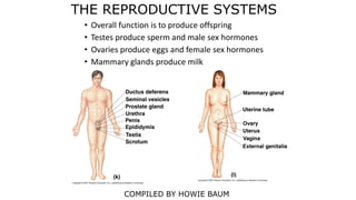
  • 1. THE REPRODUCTIVE SYSTEMS • Overall function is to produce offspring • Testes produce sperm and male sex hormones • Ovaries produce eggs and female sex hormones • Mammary glands produce milk COMPILED BY HOWIE BAUM
  • 2. INTRODUCTION The reproductive system is a collection of internal and external organs — in both males and females — that work together for the purpose of procreating. Due to its vital role in the survival of the species, many scientists feel that the reproductive system is among the most important systems in the entire body. Of the body’s major systems, the reproductive system is the one that differs most between sexes, and the only system that does not function until puberty. The male reproductive system is responsible for delivering sperm to the female reproductive system
  • 3. MALE REPRODUCTIVE SYSTEM In males, the reproductive organs include the penis, the testes, a number of storage and transport ducts, and some supporting structures. The two oval-shaped testes (also called testicles) lie outside the body in a pouch of skin called the scrotum, where they can maintain the optimum temperature for sperm production – approximately 5° F, lower than body temperature. Testes are oval-shaped glands responsible for the manufacture of sperm and the sex hormone testosterone. From each testis, sperm pass into a coiled tube – the epididymis – for the final stages of maturation.
  • 4.
  • 5. INSIDE THE SCROTUM The scrotum contains two testes (testicles) where sperm are manufactured within tubes called seminiferous tubules, and the two epididymides where sperm are stored. Unlike female egg maturation, which occurs in cycles and ceases at menopause, sperm production is continuous, reducing gradually with age. Each epididymis is a tube about 20 feet long, which is tightly coiled and bunched into a length of just 2 inches.
  • 6. MAKING SPERM Each testis is a mass of more than 800 tightly looped and folded vessels known as seminiferous tubules. Inside each tubule, sperm begin as blob-like cells called spermatogonia lining the inner wall. These pass through a larger stage, as primary spermatocytes, then become smaller as secondary spermatocytes, and begin to develop tails as spermatids. As all of this happens, they move steadily towards the middle of the tubule. The spermatids finally develop into ripe sperm with long tails. Thousands of sperm are produced every second, each taking about two months to mature.
  • 7. DIFFERENCE IN SEMEN AND SPERM Semen, also known as seminal fluid, is much more than just sperm. Sperm is only about 5 to 10% of any given male single ejaculation and the rest is fructose (aka sugar), fatty acids, and proteins to nourish the sperm during their journey. Sperm Count A man will produce roughly 525 billion sperm during his whole lifetime and close to 1 billion per month. There are around 200 to 500 million sperm in an average in a single human ejaculation !!
  • 8. Sperm cell - A sperm is about 1/500 inch long, but most of this is a tail. The sperm head is only 1/5000 inch, about the same size as a red blood cell.
  • 9. Sperm develop in the testes and consist of a head, a midpiece, and a tail. The head contains the nucleus with densely coiled chromatin fibers (chromosomes), with a front section – the Acrosome that contains enzymes for penetrating the female egg. The midpiece has a central filamentous core with many Mitochondria spiraled around it, to give it energy to move the tail which propels it forward.
  • 10.
  • 11. The female reproductive system includes external and internal genitalia. The vulva and its structures form the external genitalia. The internal genitalia include a three-part system of ducts: the uterine tubes, the uterus, and the vagina. This system of ducts connects to the ovaries, the primary reproductive organs. The ovaries produce egg cells and release them for fertilization. Fertilized eggs develop inside the uterus.
  • 12. Unlike the male, the female reproductive organs are located entirely inside the body. From Puberty, their function is to ripen and release an egg at regular intervals, and, if the egg is fertilized, to protect and nourish the embryo and fetus. No eggs are manufactured after birth – a female is born with a full set. Reproductive tract The female reproductive glands (ovaries) are located within the abdomen. This release occurs roughly once a month as part of the menstrual cycle. The ripe egg travels along the fallopian tube to the uterus, the muscular sac in which it develops into an embryo and then fetus.
  • 13. EGG CELLS FROM THE OVARIES MOVE THROUGH THE UTERINE TUBES The uterine tubes (also called Fallopian tubes or oviducts) connect the ovaries to the uterus. The walls of each tube have an external serous layer, a middle muscular layer, and an internal mucous layer that is continuous with the inner lining of the uterus. Each uterine tube can be divided into three parts: infundibulum Isthmus connects with the uterus. A dilated portion, the ampulla, curves over the ovary. Egg fertilization usually occurs in the ampulla. The eggs then travel through the isthmus into the uterus.
  • 14. THE VAGINA: A TUNNEL WITH THREE CORE FUNCTIONS The vagina extends down from the cervix, the lower part of the uterus, to the vestibule, which is part of the vulva and the external genitalia. It sits behind the bladder and in front of the rectum. An inner mucous membrane lines the smooth muscle walls of the vagina. This lining, like the inner layer of the uterine tubes, is continuous with the mucous lining of the uterus. The vagina has three core functions:  It carries menstrual flow outside the body  It receives the male penis during sexual intercourse  It serves as a birth canal during labor.
  • 15. OVULATION An ovary contains thousands of immature egg cells. During each menstrual cycle, follicle- stimulating hormone (FSH) causes one egg to begin development; this takes place inside a primary follicle. The follicle enlarges as its cells proliferate, and begins to fill with fluid, becoming a secondary follicle that moves to the ovary’s surface. It also increases its production of the hormone Estrogen. A surge of luteinizing hormone (LH) causes the follicle to rupture and release the ripe egg–this is ovulation. The lining of the empty follicle thickens into a corpus luteum–a temporary source of hormones. https://www.youtube.com/watch?v=nLmg4 wSHdxQ The ovary contains undeveloped eggs, eggs in follicles at various stages of maturation, and empty follicles forming corpora lutea. The bulk of the glandular tissue surrounding these follicles is known as the stroma.
  • 16. EGG AND SPERM HAVE DEVELOPED SOME PRETTY NIFTY TRICKS TO MAKE CONCEPTION HAPPEN Fortify the troops - The liquid portion of semen not only provides the sperm with nourishment for the journey, it actually coagulates in a woman's vagina after ejaculation, forming a physical barrier that prevents the sperm from wandering very far in the wrong direction. This protection disappears within half an hour, when the semen becomes more fluid again. Call in the transport unit - The cervical canal is a much more welcoming environment, and sperm that make it there find themselves awash in a sea of cervical mucus. The mucus is specially designed to transport sperm efficiently when you're most fertile. As you approach ovulation, your suddenly copious mucus becomes stretchy, clear and thin and strings of molecules line up like train tracks so that sperm can hop on and ride to their destination. Picking up speed - A just-ejaculated sperm cell has to spend a couple of hours going through biochemical changes, picking up tail-thrashing speed to help it make its way into the uterus and fallopian tubes to find its target. Timing is Important - Sperm must reach their destination within the right time frame for when the egg is there. They also need to pick their destination carefully - An egg is usually only present in one of the two fallopian tubes in any given month. Pick the wrong tube, and the sperm end up hanging out partying together with no guest of honor in sight. May the best sperm win!
  • 17. Note that the egg has 2 layers that the sperm needs to get through – The outer Corona Radiata irregular surface and then the Zona Pellucida which is a transparent but thicker layer.
  • 18. The first sperm to reach the oocyte is never the one to fertilize it. Rather, hundreds of sperm cells must undergo the acrosomal reaction, each helping to degrade the corona radiata and zona pellucida until a path is created to allow one sperm to contact and fuse with the plasma membrane of the oocyte (egg).
  • 19. FERTILIZATION: A SPERM AND AN EGG FORM A ZYGOTE During sexual intercourse, some sperm ejaculated from the male penis swim up through the female vagina and uterus toward an oocyte (egg cell) floating in one of the uterine tubes. The sperm and the egg are called gametes. They each contain half of the genetic information necessary for reproduction. When a sperm cell penetrates and fertilizes an egg, that genetic information combines. The 23 chromosomes from the sperm pair with 23 chromosomes in the egg, forming a 46- chromosome cell called a zygote. The zygote starts to divide and multiply. As it travels toward the uterus it divides to become a blastocyst, which will burrow into the uterine wall. https://www.youtube.com/watch?v=_5OvgQW6FG4
  • 20. THE ZYGOTE BECOMES AN EMBRYO: DEVELOPMENT PRIOR TO AND DURING IMPLANTATION A fertilized egg, or zygote, takes about five days to reach the uterus from the uterine tube. As it moves, the zygote divides and develops into a blastocyst, with an inner mass of cells and a protective outer ring. The blastocyst attaches to the wall of the uterus and gradually implants itself into the uterine lining. During implantation, its cells differentiate further. At day 15 after conception, the cells that will form the embryo become an embryonic disc. Other cells begin to form support structures. The yolk sac, on one side of the disc, will become part of the digestive tract. On the other side, the amnion fills with fluid and will surround the embryo as it develops. Other cell groups initiate the placenta and umbilical cord, which will bring in nutrients and eliminate waste.
  • 21. ZYGOTE The fertilized egg passes along the fallopian tube. Within 24–36 hours it has divided into two cells, then 12 hours later into four cells, and so on. This process is known as cleavage. At each stage, the resulting cells become smaller, gradually approaching normal body cell size.
  • 22. MORULA The zygote divides several times to form a solid blackberry-like cluster of 16–32 cells, the morula (derived from the Latin for “mulberry”). At around 3–4 days after fertilization the morula leaves the fallopian tube and enters the uterine cavity.
  • 23. BLASTOCYST About six days after fertilization, the cell cluster forms a hollow cavity and is known as a blastocyst. It floats within the uterus for around 48 hours before landing on the thick uterus lining (endometrium), which softens to aid implantation (burrowing of the blastocyst into the endometrium). The inner group of cells will become the embryo itself.
  • 24. EMBRYONIC DISC Within the inner cell mass, an embryonic disc forms. This separates the cell cluster into the amniotic cavity, which develops into a sac that will fill with fluid and fold around to cover the embryo, and the yolk sac, which helps to transport nutrients to the embryo during the second and third weeks. The disc develops three circular sheets called the primary germ layers –  Ectoderm  Mesoderm  Endoderm from which all body structures will develop. https://www.youtube.co m/watch?v=dgPCDXmcQj M 5 minutes https://www.youtube.co m/watch?v=UgT5rUQ9E mQ 2.2 minutes
  • 25.
  • 26. Early in development at the time of gastrulation a small group of cells are "put aside" to later form oocytes (eggs) and sperm. These cells are described as primordial germ cells (PGCs) and are a type of stem cell. Primordial germ cells, the earliest recognizable precursors of gametes, arise outside the gonads and migrate into the gonads during early embryonic development. Human primordial germ cells first become readily recognizable at 24 days after fertilization in a layer of the yolk sac. Germ cells exit from the yolk sac into the hindgut tissue and then migrate until they reach the location of the sex organs. These cells differentiate at different times into male testis before puberty and female ovary when the girl is young.
  • 27. GROWING EMBRYO As development proceeds, cells continue to divide. They move to form groups that will become tissues and organs. They also specialize to different cell types, as genes in their chromosomes are switched off or on. In general, development is head-down, with the brain and head taking shape early, then the body, followed by the arms as small buds, and lastly the legs. By the end of the embryonic stage, eight weeks after fertilization, all major organs and body parts are formed. From this time on, the baby is known as a fetus. 3 weeks - The neural tube forms. It will become the spinal cord, enlarged at one end as the brain. The simple tube-like heart pulsates. The embryo is 4/50– 5/50 inch long.
  • 28. HUMANS MUST DEVELOP MALE OR FEMALE GONADS AND GENITALIA TO BE CAPABLE OF REPRODUCTION Reproductive structures begin to form in the embryonic stage. By week 6, gonads and genitalia are present but undifferentiated. Whether they become male or female is determined by one chromosome delivered by the sperm. This pair contains an X sex chromosome from the female egg and either an X or a Y sex chromosome from the male sperm. If the chromosome pair is XY, the gonads develop into testes starting in week 7. If the chromosome pair is XX, the gonads become ovaries starting in week 8. Testes secrete testosterone, forming male genitalia around week 10. Without testosterone, female genitalia form. All reproductive structures are in place at birth or shortly after. At puberty, an increase in sex hormones will grow them to their
  • 29. FOUR WEEKS The four-chambered heart beats, sending blood through simple vessels. Intestines, liver, pancreas, lungs, and limb buds can be seen. The embryo is about 1/5 inch long.
  • 30. EIGHT WEEKS At this stage, the face and neck take shape, the back straightens, and fingers and toes can be differentiated clearly. The embryo starts to move. It is now around 1 to 1-1/5 inch in length.
  • 31. IN EIGHT WEEKS, THE EMBRYO DEVELOPS; BY THE END OF WEEK 10, IT BECOMES A FETUS Fifteen days after conception marks the beginning of the embryonic period. The embryo contains a flat embryonic disc that now differentiates into three layers: the endoderm, the mesoderm, and the ectoderm. All organs of the human body derive from these three tissues. They begin to curve and fold and to form an oblong body. By week 4, the embryo has a distinct head and tail and a beating heart. Over the next six weeks, limbs, eyes, brain regions, and vertebrae form. Primitive versions of all body systems appear. By the end of week 10, the embryo is a fetus. This is referring to the gestational age of the fetus.
  • 32. The little groove or Philtrum that is in between our nose and upper lip is one of the most important parts of the human face. The Philtrum or also commonly called as ‘cupid’s bow’ is the hollow space that is in between our nose and our upper lip. http://www.youtube.com/watch?v=wFY_KPFS3LA
  • 33. CHANGES IN THE FETUS By 12 weeks the fetus has a large head compared with the rest of its body, but its features are distinctly human.  All major internal organs are developed, and even tiny nails exist as folds growing on the fingers and toes.  The external ears, eyelids, and 32 permanent tooth buds have also formed.  One month later, rapid development allows the fetus to move its limbs vigorously, although this is rarely felt by the mother at this stage.  The external genitalia are visible, and a fine downy hair (lanugo) grows over the body.  As growth continues, the fetus becomes leaner and wrinkly, but by the seventh to eighth month, it starts to accumulate fat and assume the “chubby” appearance of the newborn.  The fetus is now somewhat restricted by the uterus. The side of the placenta facing the fetus is smooth and circular in outline, with the umbilical cord attached at center.
  • 34. CHANGES IN THE MOTHER FIRST TRIMESTER On average, pregnancy lasts for 40 weeks from the first day of a woman’s last menstrual period (usually 38 weeks from fertilization). By convention, the duration of pregnancy is divided into thirds, or trimesters, each lasting for about three calendar months. During this time, the mother’s body undergoes many changes to support the developing fetus, accommodate its increasing size, and prepare itself for childbirth and breast-feeding. Breasts become tender and larger, with darkened areolas; frequency of urination may increase; nausea and vomiting are common.
  • 35. SECOND TRIMESTER The enlarging uterus shows Heart rate increases The mother’s forehead and cheek skin may temporarily darken (known as the “mask of pregnancy”) From week 10 of pregnancy, the fetus grows inside the uterus, fueled by nutrient-rich blood supplied by the umbilical cord. The placenta provides oxygen and nutrients to the fetus and removes waste products from the fetus’ blood.
  • 36. THIRD TRIMESTER Skin stretches over the abdomen Slight contractions may be felt Fatigue, back pain, heartburn, and occasional breathlessness may occur.
  • 37. MULTIPLE PREGNANCY AND FETAL POSITIONS The presence of more than one fetus in the uterus is called a multiple pregnancy. Twins occur in about one in 80 pregnancies, and triplets in about one in 8,000. Both events are becoming more common, partly due to improved antenatal care and also increasing use of fertility methods such as IVF (in vitro fertilization). After about 30 weeks, the most common fetal position is head down, facing the mother’s back, with the neck flexed forward. Such a position eases passage through the birth canal. However, about 1 in 30 full-term deliveries is breech, in which the baby’s buttocks emerge before the head. Monozygotic twins - A single fertilized egg, or zygote, forms an embryo that splits into two. Each develops into a fetus. The two have the same genes and sex and share one placenta. They look alike and are known as “identical” twins.
  • 38. DIZYGOTIC TWINS Most of the time, a woman releases a single egg during an ovulation cycle. However, in approximately 1 percent of ovulation cycles, two eggs are released and both are fertilized. Two zygotes form, implant, and develop, resulting in the birth of dizygotic (or fraternal) twins. Because dizygotic twins develop from two eggs fertilized by two sperm, they are no more identical than siblings born at different times. Two eggs are fertilized and develop separately, each with its own placenta. They may be different or the same sex. Also called “fraternal twins”, they have the same degree of resemblance as any brothers and sisters.
  • 39. FRANK BREECH In frank breech, also called incomplete breech, the baby fails to turn head-down in the uterus. The hips are flexed and the legs are straight, extending alongside the body so that the feet are positioned beside the head.
  • 40. COMPLETE BREECH The baby’s legs are flexed at the hips and knees, so the feet are next to the buttocks. This occurs less commonly than frank breech. The incidence of breech delivery is much higher among premature babies.
  • 41. CHANGES IN THE CERVIX The cervix is the firm band of muscle and connective tissue that forms the neck-like structure at the bottom of the uterus. In late pregnancy, the cervix softens in readiness for childbirth. Sporadic uterine tightenings, known as Braxton–Hicks contractions, help to thin the cervix so that it merges with the uterus’s lower segment. Braxton–Hicks contractions are usually painless, and occur through much of pregnancy, becoming noticeable only after mid-term. Cervix softening As labor nears, the cervix tissues lose their firm consistency. They become softer and more spongy, affected by natural substances in the blood called prostaglandins.
  • 42. CERVIX THINNING The cervix becomes wider and thinner, and merges smoothly into the uterus wall above. The process of softening and thinning is known as effacement.
  • 43. CONTRACTIONS When pregnancy reaches full-term, the uterus is the largest and strongest muscle in the female body. When its muscle fibers shorten, with the eventual aim of expelling the fetus, it is known as a uterine contraction or simply a “contraction”. True contractions, as opposed to Braxton–Hicks contractions, are regular and become steadily more frequent, more painful, and longer- lasting. The main area of contraction is in the uterine fundus (upper uterus), which stretches, causing the lower uterus and cervix to thin. Judging when true labor has started can be difficult due to “false alarms”.
  • 44. EPIDURAL ANALGESIA One of the most commonly used methods of pain relief during labor and delivery, epidural analgesia, is delivered via a needle into the space between the vertebrae and the spinal column in the lower (lumbar) region of the back. It affects the nerve fibers that detect contraction pains. A new type of epidural, often called a “walking epidural”, reduces pain without removing sensation, allowing women to move around during labor and participate actively in the delivery.
  • 45. BIRTH OF THE BABY Around week 36 (usually), the process of labor begins. In the first stage, dilation, hormones stimulate downward contractions of the uterine walls. The contractions push the head of the fetus against the cervix at the lower end of the uterus. The cervix dilates. In the second stage, expulsion, powerful contractions push the head and the rest of the body through the dilated cervix, and out through the vagina and the vulva. The baby is born. Further contractions expel the placenta to complete the placental stage.