
Recommended
More Related Content
Similar to HUMAN REPRODUCTION.pptx
Similar to HUMAN REPRODUCTION.pptx (20)
Recently uploaded
Recently uploaded (20)
HUMAN REPRODUCTION.pptx
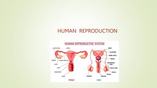
- 2. INTRODUCTION : Reproductions the process of giving birth to their young ones which are identical to their parents, reproduction in human is sexual reproduction, which involves internal fertilization by sexual intercourse. The human reproductive event include formation of gametes(gametogenesis), i.e, sperms in male and ovum in females, transfer of sperms into the female genital tract (insemination) and fusion of male and female gametes (fertilization) leading to formation of zygote. There are remarkable differences between the reproductive events in the male and in the female, e.g – sperm formation continues even in old men, but formation of ovum ceases in women around the age of 50 year. The human reproductive system mainly consists of : The male reproductive system, and the female reproductive system
- 3. MALE REPRODUCTIVE SYSTEM The reproductive system is positioned in the pelvic region and comprises a pair of testes in addition to the accessory glands, ducts, and the external genitalia. A pouch-like structure known as scrotum encloses the testes located outside the abdominal cavity. Scrotum helpd in maintaining the low temperature of the testes (2-2.5 degree c lower than the normal internal body temp), which is necessary for the spermatogenesis. Each testis has close to 250 testicular lobules (compartments). These lobules comprise 1-3 seminiferous tubules where in the sperms are produced. The lining of these tubules consists of two types of cell – male germ cells and sertoli cells. The exterior of these tubules consist of spaces containing blood vessels and leydig cells(produce testosterone). Male sex accessory ducts includes rete testis(carries sperm from the seminiferous tubules to the efferent duct), vasa efferntia(arise from rete testis, connects the seminiferous tubules to the ductus efferentes), epididymis(convoluted duct behind the testis along which sperm passes) and vas deferens (transport sperm from the epididymis) The urethra opens externally to the urethral meatus The male external genitalia, the penis is covered by foreskin which is a loose fold of skin.
- 4. Accessory Glands of male 1) Seminal vesicles : these are paired, tubular, coiled glands situated between the bladder and the rectum. They secrete viscous fluid which constitutes the main part of the ejaculate. Seminal fluid contains fructose, citric acid, inositol(help with mental condition such as panic, depression) and prostaglandins (group of lipids or hormone). 2) Prostate gland : the prostate is a walnut-sized gland located between the bladder and the penis. Prostate secretes fluid that nourishes and protects sperm. The alkaline secretions of prostate gland help the sperm to become active. 3)Bulbourethral glands or Cowper’s gland : the bulbourethral glands are a pair of pea shaped exocrine glands located posterolateral to the membranous urethra. Provide mucus protein that lubricate the urethra.
- 5. Semen : semen is a mixture of sperm and seminal fluid, which is the liquid portion of semen that consists of secretions of the seminiferous tubules, seminal vesicles, prostate gland, and bulbourethral glands. The average volume of semen in an ejaculation is 2.5 – 5 ml, with a sperm count of 200 to 300 million sperms. Path of sperm through the male body : Rete testis Vasa efferentia Epididymis Vas deferens Ejaculatory duct Urethra
- 6. Delivery of Sperm : The urethra passes through the penis, an erectile copulatory organ that deposites the semen in the female reproductive tract.
- 7. Female reproductive system The female reproductive system consists of a pair of ovaries, a duct system consisting of a pair of fallopian tubes(oviduct), a uterus, cervix and vagina. A pair of mammary glands are accessory genital glands. 1)OVARIES : The ovary is the primary female sex organ. It produces ova and secretes the female sex hormones, estrogens and progesterone which are responsible for the development of secondary female sex charecters. Human ovaries are small about 2 to 4 cm in length and is connected to the pelvic wall and uterus by ligaments. Location : Ovaries are located near kidneys and remain attached to the lower abdominal cavity through mesovarium. 2) Fallopian Tubes (Oviducts): these are one pair of long (10 to 12 cm), ciliated, muscular and tubular structures which extend from the periphery of each ovary to the uterus. Each oviduct is suspended by mesosalpinx and is differentiated into three part : i) Infundibulum : the part of oviduct closer to the ovary is the funnel shaped infundibulum. It opens into the abdominal cavity by an aperture called ostium.
- 8. ii) Ampulla : The infundibulum leads to a wider part of the oviduct called ampulla. iii) Isthmus : it is the last and narrow part having narrow lumen that links to the uterus. Fertilization occurs at the junction of ampulla and isthmus. 3) Uterus : it is a large hollow, muscular, highly vascular and inverted pear shaped structure present in the pelvis between the bladder and rectum. Uterus is also known as the womb. The cervical cavity is known as the cervical canal which goes onto form the birth canal along with the vagina. 4) Vagina : it is long (8.5 cm), fibro-muscular tube. It extends backward in front of rectum and anal canal from cervix to the vestibule. It is a highly vascular tube lined internally by mucus membrane which is raised into transverse folds called vaginal rugae. Vaginal orifice is covered partially by a membranous diaphragm called hymen. However , it can also be broken by a sudden fall, insertion of a viginal tampon, active participation in some sports like horseback riding, cycling etc. In some women the hymen persists even after coitus, in fact, the presence or absence of hymen is not reliable indicator of virginity or sexual experience. Vagina act both as copulation canal (as it receives the sperms from penis during copulation) and as birth canal along with cervix.
- 9. Female external Genitalia Female external genitalia comprises – monus pubis, labia majora, labia minora, clitoris and hymen. Mons pubis is a cushion of fatty tissue covered by skin and pubic hair. The labia majora are fleshy folds of skin, which extend down from mons pubis and surround the vaginal opening. The labia minora are paired folds of tissue in the form of lips under the labia majora. The opening of vagina is often covered partially by a membrane called hymen. The clitoris is a tiny finger-like structure which lies at the upper junction of the two labia minora above the urethral opening. It is formed by two erectile bodies and is covered by skin fold called prepuce.
- 10. Mammary gland/Breasts The mammary glands are paired structure that contain glandular tissue and variable amount of fat. These remain undeveloped till puberty. At puberty, these start developing under the influence of oestrogen and progesterone hormones. The glandular tissue of each breast is divided into 15-20 mammary lobes containing cluster of cells called alveoli. The cells of alveoli secrete milk, which is stored in the cavities of alveoli. The alveoli opens into mammary tubules. Several mammary ducts join to form a wider mammary ampulla which is connected to lactiferous duct through which milk is sucked out.
- 11. Gametogenesis The primary sex organs – the testis in the males and the ovaries in the female, produce gametes i.e, sperms and ovum respectively, by the process called gametogenesis. Gametogenesis is the process of division of diploid cells to produce new haploid cells. Spermatogenesis : sperm formation Oogenesis : ovum formation 1) SPERMATOGENESIS : In male, immature germ cells are produced in the testes. At puberty, in male, these immature germ cells or spermatogonia are converted into sperms by the process of spermatogenesis. Spermatogonia are diploid cells which undergo mitotic division and their number increases. Primary spermatocytes undergo meiosis and produce haploid cells, secondary spermatocytes. These secondary spermatocytes undergo the second meiotic division to produce immature sperms or spermatids. These spermatids undergo spermiogenesis to transform into sperms. Various hormones like gonadotropin (GnRH), Luteinising hormone (LH), Follicle stimulating hormone(FSH) and androgens are involved in stimulating the spermatogenesis.
- 12. 2) Oogenesis : In females, the oogonia are converted to the mature ovum. This process is called oogenesis. In the female ovary, millions of oogonia or mother cells are formed during fetal development. These mother cells undergo the meiotic cell division, the meiotic division rests at the prophase – 1 and lead to the production of primary oocytes. Primary oocytes are embedded within the primary follicles on the outer layer. Primary follicles get surrounded by more granulosa cell layer and forms secondary follicles. Secondary follicles then turn into the tertiary follicle. At the stage of female puberty, the primary oocytes present in the tertiarty follicles complete meiosis and form secondary oocytes (haploid) and the polar body by unequal division. The tertiary follicle undergoes some structural and functional changes and produces mature Graafian follicle. Secondary oocyte undergoes second meiotic division to form an ovum. Ovum is released from the Graafian follicle during the menstrual cycle. The release of an ovum from the Graafian follicle is called ovulation. Ovulation is controlled by the female reproductive hormone which is stimulated by the pituitary gland.
- 13. Spermatogenesis vs Oogenesis Spermatogenesis The production of sperms from spermatogonia is known as spermatogenesis. Occurs in testes All stages are completed in testes Produce motile gametes Oogenesis The production of eggs from oogonia is known as oogenesis Occurs inside the ovary The major part of oogenesis occurs inside the ovary. The last few stages occur in the oviduct. Produce non-motile gametes
- 14. Section view of ovary oogenesis Structure of a sperm Spermatogenesis
- 15. Menstrual cycle The reproductive cycle in the female primates (e.g- monkey, apes, and human beings) is called menstrual cycle. The first menstruation begins at puberty and is called menarche. In human females, menstruation is repeated at an average interval of about 28/29 days, and the cycle of events from one menstruation till next one, is called the menstrual cycle. One ovum is released (ovulation) during the middle of each menstrual cycle of 28 days. PHASE OF MENSTRUAL CYCLE : i) Menstrual phase ii) Follicular phase iii) Ovulation iv) Luteal/secretory phase
- 16. i) Menstrual phase : menstrual flow occurs in this phase and it lasts for 3 to 4 days. This flow results due to the breakdown of endometrial lining of the uterus and its body vessels which forms a liquid and flows out through the vagina. Menstruation usually occurs about 14 days after ovulation , if the released ovum is not fertilized. Lack of menstruation may be indicative of pregnancy. ii) Follicular phase : in this phase follicles in the ovary mature from primary follicle to a fully mature graafian follicle. It ends with ovulation. Estrogen rises as an egg prepares to be released. iii) Ovulation : LH includes ovulation which usually occurs on 14th day in the 28 days cycle. The Graafian follicleand secondary oocyte (ovum) is released. iv)Luteal phase : it occurs after ovulation (when your ovaries release an egg) and before menstrual start. During this time, the lining of yours uterus normally gets thicker to prepare for a possible pregnancy. During pregnancy all events of the menstrual cycle stop and there is no menstruation. In human beings, menstrual cycles ceases around 50 yr of age, that is termed as menopause.
- 18. Fertilisation and Implantation During copulation semen (contain sperm cell) is released by the penis into the vagina of female, called insemination. A human sperm can live for many weeks in male genital duct. Once ejaculated in the semen, it lives only for 24 to 48 hr outside the body. The motile sperms swim rapidly, pass through the cervix, enter into the uterus and final reach the ampullary region of the fallopian tube. The ovum released by the ovary is also transported to the ampullary region where fertilization takes place. The process of fusion of a sperm with the ovum is called fertilization. During fertilization, sperm comes in contact with zona pellucida layer of ovum. This includes changes in the membrane that prevents the entry of more sperms. Thus, only one sperm can fertilize an ovum. Secretion of the acrosome help the sperm to enter the cytoplasm of ovum through zona pellucida and plasma membrane. Sperm entry stimulates 2nd meiotic division of secondary oocyte.
- 19. Sperm + ovum fertilization Zygote (diploid) (haploid) (haploid) Sperms surrounding the ovum
- 20. Who determines the sex of the baby? Mother or Father? Human female – XX ; Human male – XY Human female gametes (ova) : X X [ only 1 type] Human male gametes (sperm) : X Y [ Two type] If male gamete containing X-chromosome fertilize female gamete, female baby [XX] If male gamete containing Y- chromosome fertilizes female gamete, male baby [XY] Thus, sex of the baby is determined by father and not by the mother. CLEAVAGE : It is a series of rapid mitotic divisions of zygote immediately after fertilization to form a hollow, multicellular structure called blastula. Cleavage occurs in the fallopian tube during the conduction of zygotetowards uterus. Cleavage converts zygote into a mass of cell called morula (8-16 blastomeres)
- 21. Blastulation (Formation of Blastula – Blastocyst) : the process of rearrangement of blastomeres and formation of central fluid filled cavity in the morula is called blastulation. Site : it occurs in upper part of uterus. Blastomeres in blastocyst are arranged into trophoblast (outer layer) and inner group of cell attached to trophoblast are called inner cell mass. Trophoblast layer gets attached to endometrium and inner cell mass differentiates as the embryo. After attachment, Implantation : the process of attachment of the blastocyst (mammalian blastula) on the endometrium of the uterus is called implantation. This leads to pregnancy. Transport of ovum, fertilization and passage of growing embryo through fallopian tube
- 22. Pregnancy and Embryonic Development PREGNANCY : The condition of carrying one or more embryos in the uterus is called pregnancy. Human pregnancy lasts for average 266 days from fertilization of the egg or 280 days from the start of the last menstrual cycle. Human pregnancy can be divided for convience into three trimesters of about three months each. i) The first trimester : After the 1st month of pregnancy , the embryo’s heart is formed. After the end of 2nd month, foetus develops limbs and digits By the end of 12 weeks (First trimester), most of the major organ systems are formed. E.g – limbs and external genital organs are well-developed. ii)Second trimester : 5th month First movment of foetus. Appearance of hair on the head By the end of 24 weeks (2nd trimester), body is covered with fine hair, Eye lids separate, Eye lashes are formed.
- 23. iii) Third (final) trimester : After the end of nine months of pregnancy , the foetus is fully developed and the baby is ready for delivery. Foetus with the uterus Delivery of the baby
- 24. Placentation A temporary structural and functional unit formed between foetus and maternal tissues is called placenta. The intimate connection established between thr foetal membrane and the uterine wall is known as the placenta. The placenta has two parts. i) Chorionic villi ii) Part of uterine wall Chorionic villi : The part of placenta contributed by the foetus is called chorionic villi. It is also called the foetal placenta. Function: i) Placenta helps in the nutrition of the embryo as the nutrients like amino acids, monosugar, vitamins, etc, pass from the maternal blood into foetal blood through placenta. ii) it also help in respiration of the embryo as Oxygen of the maternal blood and co2 of the foetal blood diffuse through placenta into the foetal blood and maternal blood respectively. It also helps in excretion of the embryo as nitrogenous wastes of foetal blood like urea pass into marernal blood through placenta. It also act as endocrine gland as it secretes certain hormones like estrogens, progesterone, human chorionic gonadotrophin (HCG) and human placenta loactogen (HPL)
- 25. After implantation, inner cell mass (embryo) differentiates into : Ectoderm (outer layer) Endoderm (inner layer) Give rise to all tissues in adults Mesoderm (middle layer between ectoderm and endoderm) PARTURITION AND LACTATION : PARTURITION : Parturition is the process of expelling fully formed young ones or baby from the mother’s uterus after the gestation period. The foetal ejection reflex is caused by the fully developed foetus and hormones from placenta. The foetal ejection causes the release of oxygen from posterior pituitary which causes stronger contraction of uterine muscles. The stronger contraction stimulates further release of oxytocin and with continued powerful contraction, the child is expelled from the uterus through the birth canal.
- 26. LACTATION : Production and release of milk after birth in mammals. Mammary glands of female undergo differentiation during pregnancy and start producing milk by the end of pregnancy. SIGNIFICANCE OF LACTATION : i) Fulfil nutritional requirements of the baby. ii)Protection from infection . iii)Helps mother in feeding new born. iv)Colostrum : Released after birth for 2-3 days, of lactation. Provides antibodies to the baby to develop resistance.